Estudios estructurales de relaxasas involucradas en el ... · 1-β1, donde se localiza la segunda...
53
Estudios estructurales de relaxasas involucradas en el proceso de conjugación bacteriana Silvia Russi Barcelona, 2009
Transcript of Estudios estructurales de relaxasas involucradas en el ... · 1-β1, donde se localiza la segunda...
Estudios estructurales de relaxasas
involucradas en
el proceso de conjugación bacteriana
Silvia Russi Barcelona, 2009

119
5. CONCLUSIONES

120
121
5. Conclusiones
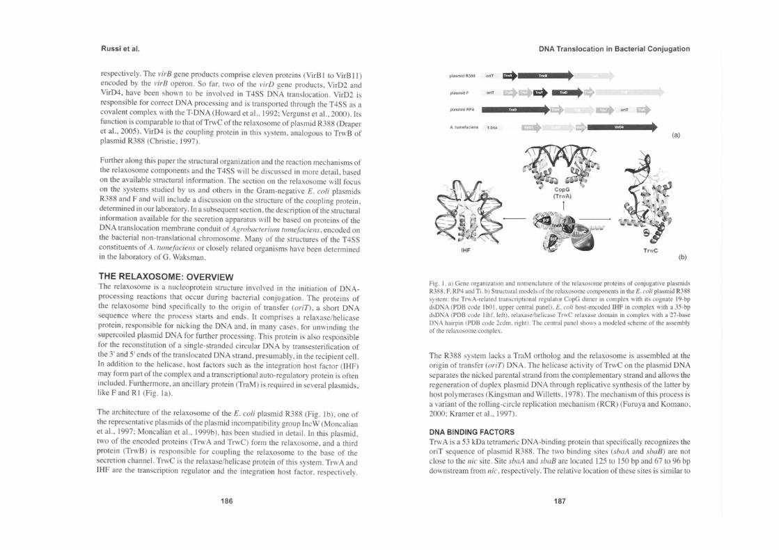
5.1. La relaxasa TrwC del plásmido conjugativo R388
El análisis de la unión del complejo TrwC-ADN25 a diferentes metales
divalentes (Zn2+, Cu2+, Ni2+, Mn2+, Mg2+, Ca2+) por difracción anómala de rayos X,
junto con los ensayos bioquímicos, permitió establecer que el metal fisiológicamente
empleado por la proteína TrwC en la actividad catalítica es el Zn2+.
El metal desarrolla un papel clave en el mecanismo enzimático, en el que no solo
interviene en la coordinación de la tríada de histidinas localizada en el sitio activo de la
proteína, sino que además su unión al grupo fosfato lo polariza, y debilita el enlace
fosfo-diéster a cortar.
En los complejos TrwC-ADN25-M2+ el lazo α1-β1, donde se localiza la segunda
tirosina catalítica Tyr26, pudo ser completamente trazado. Si bien la posición de este
residuo es bastante lejana del sitio activo, no puede descartarse su participación en el
segundo corte de la cadena de ADN. Es posible que ocurran arreglos conformacionales,
o incluso que la proteína actúe como dímero, para permitir que el corte se realice. La
información estructural no permite establecer sin ambigüedad el rol exacto que juega
este residuo en el mecanismo catalítico.
La estructura del complejo TrwC-ADN27 en la que está presente el enlace fosfo-
diéster a cortar permitió la localización del mismo respecto al sitio activo, ayudando a
comprender el papel del metal en la reacción de corte de la cadena de ADN.
En base a la información estructural obtenida para los complejos TrwC-ADN25-
M2+ y el complejo TrwC-ADN27, es posible proponer un mecanismo a nivel molecular,
del primer paso de la reacción de corte de la cadena de ADN. La Tyr18, activada por el
Asp85, realiza el ataque nucleofílico al grupo fosfato, siendo ayudada por el ión
metálico, que como se ha indicado, lo polariza favoreciendo el ataque.
La posición de grupos sulfato en la estructura TrwC-ADN25, que mimetizan la
122
posición de las bases de la cadena de ADN, junto con lo observado en la estructura del
complejo TrwC-ADN27, sugieren la existencia de dos posibles rutas de salida para la
cadena de ADN cuya bifurcación se produce cerca del sitio de unión al metal. Una
hipótesis plausible es que la proteína utiliza ambos caminos en su mecanismo.
5.2. La relaxasa MobM del plásmido movilizable pMV158
Se logró determinar la estructura del dominio N-terminal de la relaxasa MobM
en complejo con ADN, encontrándose en ella los elementos estructurales conservados
de la familia de las relaxasas: el núcleo central formado por cinco hebras beta
flanqueadas por ambos lados por hélices alfa.
Las interacciones de la cadena de ADN con la proteína son múltiples, dada la
extensa superficie de interacción que involucra (un 30% de la superficie total accesible
de la proteína). Entre ellas, destacan el reconocimiento de la proteína a la cadena de
ADN mediante el lazo α2-β3. Dos giros betas consecutivos de dicho lazo orientan dos
residuos arginina (Arg71 y Arg74) hacia el estrecho surco menor de la doble cadena de
ADN, penetrando en él y estableciendo enlaces de hidrógeno que permiten el
reconocimiento específico del ADN.
La arquitectura del sitio activo es semejante a la observada en la estructura de
los complejos TrwC-ADN, con la característica tríada de histidinas encargada de
coordinar el ión metálico. Las His126 e His133 adoptan conformaciones diferentes que
en la estructura de la TrwC producto de la ausencia del metal en el sitio activo.
La identidad del residuo nucleofílico es ambigua. Los resultados sugieren que en
la MobM la Tyr122 podría cumplir este papel catalítico, aunque se encuentra más
alejada del grupo fosfato a cortar que en el caso de las relaxasas de las bacterias Gram-
negativas TrwC o TraI. La tirosina Tyr44, por su parte, se localiza en una posición
homóloga a la Tyr26 de la TrwC y podría cumplir un papel similar al de esta. La His22
podría ser también un residuo esencial.
123
El extremo 3’ de la cadena de ADN no entra al sitio activo y en su lugar se
coloca el lazo de la horquilla de una cadena de ADN vecina. La posición relativa de
dicha cadena parece estar mimetizando la conformación IR-2 en la que el sitio de corte
estaría en el lazo de la horquilla del ADN.
Si bien no puede establecerse con certeza la razón por la cual el extremo 3’ de la
cadena de ADN no entra al sitio activo, la comparación de la estructura obtenida con la
de la relaxasa TrwC sugiere que el constructo de MobM con el que se trabajó carece de
los elementos de secuencia secundaria indispensables para guiar al ADN hacia el sitio
de corte, indicando que las hélices o “dedos” en la parte C-terminal del dominio serían
imprescindibles para el correcto posicionamiento del substrato.

124

125
6. BIBLIOGRAFÍA

126
127
6. Bibliografía
Atmakuri, K., Cascales, E., and Christie, P.J. (2004). Energetic components VirD4,
VirB11 and VirB4 mediate early DNA transfer reactions required for bacterial type IV
secretion. Mol Microbiol 54, 1199-1211.
Backert, S., and Meyer, T.F. (2006). Type IV secretion systems and their effectors in
bacterial pathogenesis. Curr Opin Microbiol 9, 207-217.
Boer, D.R., Ruíz-Masó, J.A., López-Blanco, J.R., Blanco, A.G., Vives-Llàcer, M.,
Chacón, P., Uson, I., Gomis-Rüth, F.X., Espinosa, M., Llorca, O., et al. (2009). Plasmid
replication initiator RepB forms a hexamer reminiscent of ring helicases and has mobile
nuclease domains. EMBO J 28, 1666-1678.
Boer, R., Russi, S., Guasch, A., Lucas, M., Blanco, A.G., Perez-Luque, R., Coll, M.,
and de la Cruz, F. (2006). Unveiling the molecular mechanism of a conjugative
relaxase: The structure of TrwC complexed with a 27-mer DNA comprising the
recognition hairpin and the cleavage site. J Mol Biol 358, 857-869.
Bradford, M.M. (1976). A rapid and sensitive method for the quantitation of microgram
quantities of protein utilizing the principle of protein-dye binding. Anal Biochem 72,
248-254.
Brunger, A.T., Adams, P.D., Clore, G.M., DeLano, W.L., Gros, P., Grosse-Kunstleve,
R.W., Jiang, J.S., Kuszewski, J., Nilges, M., Pannu, N.S., et al. (1998). Crystallography
& NMR system: A new software suite for macromolecular structure determination.
Acta Crystallogr D Biol Crystallogr 54 ( Pt 5), 905-921.
Burdett, V. (1980). Identification of tetracycline-resistant R-plasmids in Streptococcus
agalactiae (group B). Antimicrob Agents Chemother 18, 753-760.
Burns, D.L. (2003). Type IV transporters of pathogenic bacteria. Curr Opin Microbiol
6, 29-34.
128
Byrd, D.R., and Matson, S.W. (1997). Nicking by transesterification: the reaction
catalysed by a relaxase. Mol Microbiol 25, 1011-1022.
Cabezon, E., and de la Cruz, F. (2006). TrwB: an F(1)-ATPase-like molecular motor
involved in DNA transport during bacterial conjugation. Res Microbiol 157, 299-305.
Cabezon, E., Sastre, J.I., and de la Cruz, F. (1997). Genetic evidence of a coupling role
for the TraG protein family in bacterial conjugation. Mol Gen Genet 254, 400-406.
Campos-Olivas, R., Louis, J.M., Clerot, D., Gronenborn, B., and Gronenborn, A.M.
(2002). The structure of a replication initiator unites diverse aspects of nucleic acid
metabolism. Proc Natl Acad Sci U S A 99, 10310-10315.
Cascales, E., and Christie, P.J. (2003). The versatile bacterial type IV secretion systems.
Nat Rev Microbiol 1, 137-149.
Collaborative Computational Project, N. (1994). The CCP4 suite: programs for protein
crystallography. Acta Crystallogr D Biol Crystallogr 50, 760-763.
Cowtan, K. (2002). Generic representation and evaluation of properties as a function of
position in reciprocal space. J Appl Cryst 35, 655-663.
Chen, I., Christie, P.J., and Dubnau, D. (2005). The ins and outs of DNA transfer in
bacteria. Science 310, 1456-1460.
Christie, P.J. (2001). Type IV secretion: intercellular transfer of macromolecules by
systems ancestrally related to conjugation machines. Mol Microbiol 40, 294-305.
Christie, P.J. (2004). Type IV secretion: the Agrobacterium VirB/D4 and related
conjugation systems. Biochim Biophys Acta 1694, 219-234.
Christie, P.J., Atmakuri, K., Krishnamoorthy, V., Jakubowski, S., and Cascales, E.
(2005). Biogenesis, architecture, and function of bacterial type IV secretion systems.
129
Annu Rev Microbiol 59, 451-485.
Christie, P.J., and Cascales, E. (2005). Structural and dynamic properties of bacterial
type IV secretion systems (review). Mol Membr Biol 22, 51-61.
Christie, P.J., and Vogel, J.P. (2000). Bacterial type IV secretion: conjugation systems
adapted to deliver effector molecules to host cells. Trends Microbiol 8, 354-360.
Datta, S., Larkin, C., and Schildbach, J.F. (2003). Structural insights into single-
stranded DNA binding and cleavage by F factor TraI. Structure 11, 1369-1379.
Davis, I.W., Leaver-Fay, A., Chen, V.B., Block, J.N., Kapral, G.J., Wang, X., Murray,
L.W., Arendall, W.B., 3rd, Snoeyink, J., Richardson, J.S., et al. (2007). MolProbity: all-
atom contacts and structure validation for proteins and nucleic acids. Nucleic Acids Res
35, W375-383.
de Antonio, C., Farias, M.E., García de Lacoba, M., and Espinosa, M. (2004). Features
of the Plasmid pMV158-encoded MobM, a Protein Involved in its Mobilization. . J Mol
Biol 335, 733-743.
de la Cruz, F., and Davies, J. (2000). Horizontal gene transfer and the origin of species:
lessons from bacteria. Trends Microbiol 8, 128-133.
Ding, Z., Atmakuri, K., and Christie, P.J. (2003). The outs and ins of bacterial type IV
secretion substrates. Trends Microbiol 11, 527-535.
Draper, O., Cesar, C.E., Machon, C., de la Cruz, F., and Llosa, M. (2005). Site-specific
recombinase and integrase activities of a conjugative relaxase in recipient cells. Proc
Natl Acad Sci U S A 102, 16385-16390.
Dreiseikelmann, B. (1994). Translocation of DNA across bacterial membranes.
Microbiol Rev 58, 293-316.
Durrenberger, M.B., Villiger, W., and Bachi, T. (1991). Conjugational junctions:
130
morphology of specific contacts in conjugating Escherichia coli bacteria. J Struct Biol
107, 146-156.
Emsley, P., and Cowtan, K. (2004). Coot: model-building tools for molecular graphics.
Acta Crystallogr D Biol Crystallogr 60, 2126-2132.
Enemark, E.J., Stenlund, A., and Joshua-Tor, L. (2002). Crystal structures of two
intermediates in the assembly of the papillomavirus replication initiation complex.
Embo J 21, 1487-1496.
Evans, P.R. (1993). Data reduction. In Proceedings of CCP4 Study Weekend. Data
Collection & Processing, 114-122.
Farias, M.E., Grohmann, E., and Espinosa, M. (1999). Expression of the mobM gene of
the streptococcal plasmid pMV158 in Lactococcus lactis subsp. lactis. FEMS Microbiol
Lett 176, 403-410.
Firth, N., Ippen-Ihler, K., and Skurray, R.A. (1996). Structure and function of the F
factor and mechanism of conjugation. In Escherichia Coli and Salmonella: Cellular and
Molecular Biology, N. FC, ed. (American Sociaty of Microbiology), pp. 2377-2401.
Francia, M.V., Varsaki, A., Garcillan-Barcia, M.P., Latorre, A., Drainas, C., and de la
Cruz, F. (2004). A classification scheme for mobilization regions of bacterial plasmids.
FEMS Microbiol Rev 28, 79-100.
Furuya, N., and Komano, T. (2000). Initiation and termination of DNA transfer during
conjugation of IncI1 plasmid R64: roles of two sets of inverted repeat sequences within
oriT in termination of R64 transfer. J Bacteriol 182, 3191-3196.
Garcillan-Barcia, M.P., Jurado, P., Gonzalez-Perez, B., Moncalian, G., Fernandez, L.A.,
and de la Cruz, F. (2007). Conjugative transfer can be inhibited by blocking relaxase
activity within recipient cells with intrabodies. Molecular microbiology 63, 404-416.
Gomis-Ruth, F.X., Sola, M., Acebo, P., Parraga, A., Guasch, A., Eritja, R., Gonzalez,
131
A., Espinosa, M., del Solar, G., and Coll, M. (1998). The structure of plasmid-encoded
transcriptional repressor CopG unliganded and bound to its operator. Embo J 17, 7404-
7415.
Gomis-Ruth, F.X., Sola, M., de la Cruz, F., and Coll, M. (2004). Coupling factors in
macromolecular type-IV secretion machineries. Curr Pharm Des 10, 1551-1565.
Grandoso, G., Avila, P., Cayon, A., Hernando, M.A., Llosa, M., and de la Cruz, F.
(2000). Two active-site tyrosyl residues of protein TrwC act sequentially at the origin of
transfer during plasmid R388 conjugation. J Mol Biol 295, 1163-1172.
Grohmann, E., Muth, G., and Espinosa, M. (2003). Conjugative plasmid transfer in
gram-positive bacteria. Microbiol Mol Biol Rev 67, 277-301, table of contents.
Guasch, A., Lucas, M., Moncalian, G., Cabezas, M., Perez-Luque, R., Gomis-Ruth,
F.X., de la Cruz, F., and Coll, M. (2003). Recognition and processing of the origin of
transfer DNA by conjugative relaxase TrwC. Nat Struct Biol 10, 1002-1010.
Guzmán, L.M., and Espinosa, M. (1997). The mobilization protein MobM, of the
streptoccocal plasmid pMV158, specifically cleaves supercoiled DNA at the plasmid
oriT. J Mol Biol 266, 688-702.
Hickman, A.B., Ronning, D.R., Kotin, R.M., and Dyda, F. (2002). Structural unity
among viral origin binding proteins: crystal structure of the nuclease domain of adeno-
associated virus Rep. Mol Cell 10, 327-337.
Howard, M.T., Nelson, W.C., and Matson, S.W. (1995). Stepwise assembly of a
relaxosome at the F plasmid origin of transfer. J Biol Chem 270, 28381-28386.
Kingsman, A., and Willetts, N. (1978). The requirements for conjugal DNA synthesis in
the donor strain during flac transfer. J Mol Biol 122, 287-300.
Kramer, M.G., Khan, S.A., and Espinosa, M. (1997). Plasmid rolling circle replication:
identification of the RNA polymerase-directed primer RNA and requirement for DNA
132
polymerase I for lagging strand synthesis. Embo J 16, 5784-5795.
Lanka, E., and Wilkins, B.M. (1995). DNA processing reactions in bacterial
conjugation. Annu Rev Biochem 64, 141-169.
Laskowski, R.A., MacArthur, M.W., Moss, D.S., and Thornton, J.M. (1993).
PROCHECK: a program to check the stereochemical quality of protein structures. J
Appl Cryst 26, 283-291.
Lederberg, J., and Tatum, E. (1946). Gene recombination in E. coli. Nature 158, 558.
Leslie, A.G.W. (1992). Recent changes to the MOSFLM package for processing film
and image plate data Joint CCP4 + ESF-EAMCB Newsletter on Protein Crystallography
26.
Lupas, A., Van Dyke, M., and Stock, J. (1991). Predicting coiled coils from protein
sequences. Science 252, 1162-1164.
Luscombe, N.M., Laskowski, R.A., and Thornton, J.M. (1997). NUCPLOT: a program
to generate schematic diagrams of protein-nucleic acid interactions. Nucleic Acids Res
25, 4940-4945.
Llosa, M., and de la Cruz, F. (2005). Bacterial conjugation: a potential tool for genomic
engineering. Res Microbiol 156, 1-6.
Llosa, M., Gomis-Ruth, F.X., Coll, M., and de la Cruz Fd, F. (2002). Bacterial
conjugation: a two-step mechanism for DNA transport. Mol Microbiol 45, 1-8.
Llosa, M., Grandoso, G., Hernando, M.A., and de la Cruz, F. (1996). Functional
domains in protein TrwC of plasmid R388: dissected DNA strand transferase and DNA
helicase activities reconstitute protein function. J Mol Biol 264, 56-67.
Llosa, M., and O'Callaghan, D. (2004). Euroconference on the Biology of Type IV
Secretion Processes: bacterial gates into the outer world. Mol Microbiol 53, 1-8.
133
Marsden, R.L., McGuffin, L.J., and Jones, D.T. (2002). Rapid protein domain
assignment from amino acid sequence using predicted secondary structure. Protein Sci
11, 2814-2824.
Matthews, B.W. (1968). Solvent content of protein crystals. J Mol Biol 33, 491-497.
Meinke, G., Bullock, P.A., and Bohm, A. (2006). Crystal structure of the simian virus
40 large T-antigen origin-binding domain. J Virol 80, 4304-4312.
Moncalian, G., Cabezon, E., Alkorta, I., Valle, M., Moro, F., Valpuesta, J.M., Goni,
F.M., and de La Cruz, F. (1999a). Characterization of ATP and DNA binding activities
of TrwB, the coupling protein essential in plasmid R388 conjugation. J Biol Chem 274,
36117-36124.
Moncalian, G., and de la Cruz, F. (2004). DNA binding properties of protein TrwA, a
possible structural variant of the Arc repressor superfamily. Biochim Biophys Acta
1701, 15-23.
Moncalian, G., Grandoso, G., Llosa, M., and de la Cruz, F. (1997). oriT-processing and
regulatory roles of TrwA protein in plasmid R388 conjugation. J Mol Biol 270, 188-
200.
Moncalian, G., Valle, M., Valpuesta, J.M., and de la Cruz, F. (1999b). IHF protein
inhibits cleavage but not assembly of plasmid R388 relaxosomes. Mol Microbiol 31,
1643-1652.
Moscoso, M., Eritja, R., and Espinosa, M. (1997). Initiation of replication of plasmid
pMV158: mechanisms of DNA strand-transfer reactions mediated by the initiator RepB
protein. J Mol Biol 268, 840-856.
Murray, J., and Garman, E. (2002). Investigation of possible free-radical scavengers and
metrics for radiation damage in protein cryocrystallography. J Synchrotron Radiat 9,
347-354.
134
Murshudov, G.N., Vagin, A.A., and Dodson, E.J. (1997). Refinement of
macromolecular structures by the maximum-likelihood method. Acta Crystallogr D Biol
Crystallogr 53, 240-255.
Murzin, A.G., Brenner, S.E., Hubbard, T., and Chothia, C. (1995). SCOP: a structural
classification of proteins database for the investigation of sequences and structures. J
Mol Biol 247, 536-540.
Ochman, H., Lawrence, J.G., and Groisman, E.A. (2000). Lateral gene transfer and the
nature of bacterial innovation. Nature 405, 299-304.
Priebe, S.D., and Lacks, S.A. (1989). Region of the streptococcal plasmid pMV158
required for conjugative mobilization. J Bacteriol 171, 4778-4784.
Ramachandran, G.N., and Sasisekharan, V. (1968). Conformation of polypeptides and
proteins. Adv Protein Chem 23, 283-438.
Read, R.J. (2001). Pushing the boundaries of molecular replacement with maximum
likelihood. Acta Crystallogr D Biol Crystallogr 57, 1373-1382.
Rice, P.A., Yang, S., Mizuuchi, K., and Nash, H.A. (1996). Crystal structure of an IHF-
DNA complex: a protein-induced DNA U-turn. Cell 87, 1295-1306.
Rossmann, M.G., and Blow, D.M. (1962). The detection of sub-units within the
crystallographic asymmetric unit. Acta Crystallogr 15, 24-31.
Roussel, A., and Cambilleau, C. (1989). Turbo-Frodo. In Silicon Graphics geometry
partners directory (Mountain View, CA, Silicon Graphics), pp. 77-79.
Santini, J.M., and Stanisich, V.A. (1998). Both the fipA gene of pKM101 and the pifC
gene of F inhibit conjugal transfer of RP1 by an effect on traG. J Bacteriol 180, 4093-
4101.
135
Scott, J.R., and Churchward, G.G. (1995). Conjugative transposition. Annu Rev
Microbiol 49, 367-397.
Schneider, T.R., and Sheldrick, G.M. (2002). Substructure solution with SHELXD.
Acta Crystallogr D Biol Crystallogr 58, 1772-1779.
Schroder, G., and Lanka, E. (2005). The mating pair formation system of conjugative
plasmids-A versatile secretion machinery for transfer of proteins and DNA. Plasmid 54,
1-25.
Seubert, A., Hiestand, R., de la Cruz, F., and Dehio, C. (2003). A bacterial conjugation
machinery recruited for pathogenesis. Mol Microbiol 49, 1253-1266.
Sexton, J.A., and Vogel, J.P. (2002). Type IVB secretion by intracellular pathogens.
Traffic 3, 178-185.
Sheldrick, G. (2002). Macromolecular phasing with SHELXE. Z Krist 217, 644-650.
Silverman, P.M. (1997). Towards a structural biology of bacterial conjugation. Mol
Microbiol 23, 423-429.
Szpirer, C.Y., Faelen, M., and Couturier, M. (2001). Mobilization function of the
pBHR1 plasmid, a derivative of the broad-host-range plasmid pBBR1. J Bacteriol 183,
2101-2110.
Terwilliger, T. (2004). SOLVE and RESOLVE: automated structure solution, density
modification and model building. J Synchrotron Radiat 11, 49-52.
Ton-That, H., and Schneewind, O. (2003). Assembly of pili on the surface of
Corynebacterium diphtheriae. Mol Microbiol 50, 1429-1438.
Uson, I., and Sheldrick, G.M. (1999). Advances in direct methods for protein
crystallography. Curr Opin Struct Biol 9, 643-648.
136
Vagin, A., and Teplyakov, A. (1997). MOLREP: an automated program for molecular
replacement. J Appl Cryst 30, 1022-1025.
Walker, J.E., Saraste, M., Runswick, M.J., and Gay, N.J. (1982). Distantly related
sequences in the alpha- and beta-subunits of ATP synthase, myosin, kinases and other
ATP-requiring enzymes and a common nucleotide binding fold. Embo J 1, 945-951.
Waters, V.L. (2001). Conjugation between bacterial and mammalian cells. Nature
genetics 29, 375-376.
Weik, M., Ravelli, R.B., Kryger, G., McSweeney, S., Raves, M.L., Harel, M., Gros, P.,
Silman, I., Kroon, J., and Sussman, J.L. (2000). Specific chemical and structural
damage to proteins produced by synchrotron radiation. Proc Natl Acad Sci U S A 97,
623-628.
Wilkins, B., and Lanka, E. (1993). DNA processing and replication during plasmid
transfer between Gram-negative bacteria (New York, Plenum Press).
Ziegelin, G., Furste, J.P., and Lanka, E. (1989). TraJ protein of plasmid RP4 binds to a
19-base pair invert sequence repetition within the transfer origin. J Biol Chem 264,
11989-11994.

137
7. PUBLICACIONES

138
139
7. Publicaciones
Los resultados derivados de este proyecto de tesis están incluidos en las
siguientes publicaciones.
Artículos publicados:
Unveiling the Molecular Mechanism of a Conjugative Relaxase: The Structure of TrwC
Complexed with a 27-mer DNA Comprising the Recognition Hairpin and the Cleavage
Site, R. Boer*, S. Russi*, A. Guasch, M. Lucas, A.G. Blanco, R. Pérez-Luque, M. Coll1
& F. de la Cruz. J. Mol. Biol., 2006, Vol. 358, 857-869. *Ambos autores contribuyeron por
igual al trabajo
Capítulos de libros:
Molecular machinery for DNA translocation in bacterial conjugation, S. Russi, R. Boer
& M. Coll, 2008, PLASMIDS: current research and future trends, edited by G. Lipps
(Caister Academic Press).
Artículos en preparación:
Crystal structure of the N-terminal relaxase domain of the mobilization protein MobM,
from plasmid pMV158, in complex with its cognate DNA. Russi et al., 2009.

140
Unveiling the Molecular Mechanism of a ConjugativeRelaxase: The Structure of TrwC Complexed witha 27-mer DNA Comprising the Recognition Hairpinand the Cleavage Site
Roeland Boer1†, Silvia Russi1†, Alicia Guasch1,3, Marıa Lucas2
Alexandre G. Blanco1, Rosa Perez-Luque1, Miquel Coll1*and Fernando de la Cruz2
1Institut de Biologia Molecularde Barcelona (CSIC) andInstitut de Recerca BiomedicaParc Cientıfic de BarcelonaJosep Samitier 1-5, 08028Barcelona, Spain
2Departamento de BiologıaMolecular (Laboratorio asociadoal CIB, CSIC), Universidad deCantabria, Herrera Oria, s/n39011 Santander, Spain
3Plataforma Automatitzada deCristal$lografia (PCB-CSIC)Parc Cientific de BarcelonaJosep Samitier 1-5, 08028Barcelona, Spain
TrwC is a DNA strand transferase that catalyzes the initial and final stagesof conjugative DNA transfer. We have solved the crystal structure of theN-terminal relaxase domain of TrwC in complex with a 27 base-long DNAoligonucleotide that contains both the recognition hairpin and the scissilephosphate. In addition, a series of ternary structures of protein–DNAcomplexes with different divalent cations at the active site have beensolved. Systematic anomalous difference analysis allowed us to determineunambiguously the nature of the metal bound. Zn2C, Ni2C and Cu2C werefound to bind the histidine-triad metal binding site. Comparison of thestructures of the different complexes suggests two pathways for the DNAto exit the active pocket. They are probably used at different steps of theconjugative DNA-processing reaction. The structural information allowsus to propose (i) an enzyme mechanism where the scissile phosphate ispolarized by the metal ion facilitating the nucleophilic attack of thecatalytic tyrosine, and (ii) a probable sequence of events during conjugativeDNA processing that explains the biological function of the relaxase.
q 2006 Elsevier Ltd. All rights reserved.
Keywords: X-ray structure; relaxase; bacterial conjugation; DNA transfer;DNA–protein complex*Corresponding author
Introduction
Bacterial conjugation is a process by which aDNA molecule is transferred from a donor to arecipient bacterium. The mechanism is efficient, andallows bacteria to acquire new adaptive traits suchas antibiotic resistance. Thus, it is considered animportant mechanism in bacterial evolution.1,2 Theunderlying biochemical process can be operation-ally divided into two steps, DNA processing andDNA transport, each of which is carried out by aspecific set of proteins encoded by the tra genes of agiven conjugative plasmid. Conjugative DNAprocessing starts by cleavage of a specific phospho-diester bond (the nic site) in the donor supercoiledDNA by a plasmid-specific relaxase. The resulting
nucleoprotein complex, the relaxosome, contactsthe transport site, where a multi-protein DNAtransport apparatus effects the transfer process ofthe cleaved DNA strand to the recipient cell.Presumably, the relaxase religates the transferredDNA strand, and finally host proteins replicateboth single strands in donor and recipient bacteriato regenerate the double-stranded conjugativeplasmid.3,4
Most conjugative systems contain phylogeneti-cally related relaxases, according to their aminoacid sequences.5 Remarkably conspicuous is ahistidine triad that has been intimately involvedin the catalytic mechanism. A few selected relaxaseshave been analyzed at a biochemical level. Thepurified proteins can specifically cleave oligo-nucleotides containing their respective nic sitesequences so that the 5 0 phosphoryl end of thecleaved product becomes covalently bound to thehydroxyl group of a specific tyrosyl residue ofthe protein. Relaxases can then transfer the bound
0022-2836/$ - see front matter q 2006 Elsevier Ltd. All rights reserved.
† R.B. and S.R. contributed equally to this work.E-mail address of the corresponding author:
doi:10.1016/j.jmb.2006.02.018 J. Mol. Biol. (2006) 358, 857–869
DNA to an appropriate acceptor oligonucleotide bya second DNA strand-transfer reaction, so that ahybrid oligonucleotide is released from theenzyme.6 The P-family relaxase TraI of plasmidRP4 is one of the best analyzed with respectto the biochemical details of these reactions. Byusing oligonucleotides bound to a solid support,Pansegrau & Lanka7 showed that the TraI oligo-nucleotide adduct was incapable of carrying out thestrand transfer reaction when challenged with asecond oligonucleotide containing nic. Theinterpretation was that a second TraI molecule isrequired to complete the reaction, presumably byproviding a new Tyr to catalyze the second strandtransfer reaction.
The F-family relaxase TrwC, from the IncWplasmid R388, is a large protein composed of anN-terminal domain with DNA-relaxase activityand a C-terminal domain with DNA helicaseactivity.8 It can cleave a supercoiled plasmidDNA containing oriT in vitro in the absence ofaccessory proteins.9 TrwC contains two active-sitetyrosyl residues (instead of the single one inP-family relaxases) that play different functionalroles in the DNA processing reactions. Thus, it wasproposed that conjugative DNA processing ofplasmid R388 occurs by a variant of the flip-flopmechanism used in fX-174 replication10 in whichthe two active tyrosine residues catalyze theinitiation and termination steps in conjugativeDNA replication.11
The atomic structures of two F-family relaxases,R388 TrwC12 and F plasmid TraI,13 have beenreported. The TrwC structure shows a complex ofthe protein with the nicDNA up to the cleavage site.This complex was shown both as a binary DNA–protein complex and as a ternary complex with aZn2C in the three-histidine pocket. Although thesestructures provided valuable information withrespect to the DNA and metal binding sites, theydid not reveal the identity of the metal ion and didnot provide a complete view of the reactionmechanism catalyzed by relaxases. In particular,both the exit path for the DNA and the position ofthe scissile phosphate with respect to the catalyticresidues were unclear because the DNA used forthe DNA–TrwC complex12 did not include anyresidue downstream of the cleavage site nor thescissile phosphate (A26, according to our naming).Recently, the structure of TraI complexed with a10 bp oligonucleotide, including the scissile bondbut not the full recognition hairpin, has beenreported.14 Here we present additional structuralinformation of TrwC with the description of arelaxase ternary complex with a 27-mer DNAoligonucleotide encompassing both the recognitionhairpin and the cleavage site. Also, we present adetailed structural analysis of the binding ofdifferent metal ions. Finally, we provide a structurewhere the loop a1-b1 that includes the secondcatalytic residue Tyr26 is fully traced, in contrast toprevious structures where it was disordered. As aresult we clarify crucial aspects of the mechanism
for the successive DNA-strand transfer reactionscatalyzed by relaxases.
Results
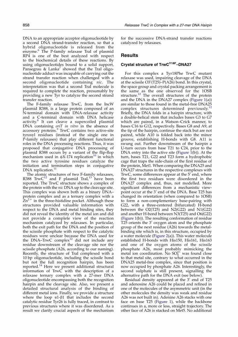
Crystal structure of TrwCY18F–DNA27
For this complex a Tyr18Phe TrwC mutantrelaxase was used, impairing cleavage of the DNAat the scissile O3 0(T25)–P(A26) bond. In this crystal,the space group and crystal packing arrangement isthe same as the one observed for the 1OSBstructure.12 The overall structures of the proteinand the DNA in the DNA27 complex (Figure 1(a))are similar to those found in the metal-free DNA25complex structures determined previously.12
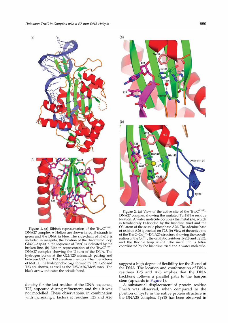
Briefly, the DNA folds in a hairpin structure, witha double-helical stem that includes bases G3 to G7which are paired, in a Watson–Crick manner, tobases C16 to G12, respectively. Bases G8 and A9, atthe tip of the hairpin, continue the stack but are notpaired, while A10 is folded back into the minorgroove, establishing H-bonds with G8. A11 isswung out. Further downstream of the hairpin aU-turn occurs from base T21 to C24, prior to theDNA entry into the active site (Figure 1(b)). At theturn, bases T21, G22 and T23 form a hydrophobiccage that traps the side-chain of the first residue ofthe protein, Met1. When comparing the DNA25 andDNA27 structures in the respective complexes withTrwC, some differences appear at the 5 0 end, wherethe first two residues were disordered in theDNA27 complex and, thus, not modelled. Moresignificant differences from a mechanistic view-point occur at the 3 0 end of the DNA. Base T25 haschanged its orientation (when compared to 1OSB)to form a non-complementary base-pairing withG22, with a three-centered (bifurcated) H-bondbetween the O2(T25) and N2(G22) and N1(G22)and another H-bond between N3(T25) and O6(G22)(Figure 1(b)). The resulting conformation of residueT25 orients the 3 0 oxygen atom and the phosphategroup of the next residue (A26) towards the metal-binding site which is, in this structure, occupied bya water molecule (Figure 2(a)). This water moleculeestablished H-bonds with His150, His161, His163and one of the oxygen atoms of the scissilephosphate A26, most probably mimicking themetal ion coordination. No SO2K
4 was found closeto that metal site, contrary to what occurred in theDNA25 metal-free complex, since that position isnow occupied by phosphate A26. Interestingly, thesecond sulphate is still present, signalling thealternative path for the DNA exit (see below).
Residual density appeared at the 3 0 end of T25and adenosine A26 could be placed and refined inone of the molecules of the asymmetric unit (in theother molecules the density was weak and residueA26 was not built in). Adenine A26 stacks with oneface on base T25 (Figure 1), while the backbonecontinues in a, more or less, straight trajectory. Theother face of A26 is stacked on Met5. No additional
858 Relaxase TrwC in Complex with a 27-mer DNA Hairpin
density for the last residue of the DNA sequence,T27, appeared during refinement, and thus it wasnot modelled. These observations, in combinationwith increasing B factors at residues T25 and A26
suggest a high degree of flexibility for the 3 0 end ofthe DNA. The location and conformation of DNAresidues T25 and A26 implies that the DNAbackbone follows a parallel path to the hairpinstem (upwards in Figure 1).A substantial displacement of protein residue
Phe18 was observed, when compared to theposition of Tyr18 in the native protein structure inthe DNA25 complex. Tyr18 has been observed in
Figure 2. (a) View of the active site of the TrwCY18F–DNA27 complex showing the mutated Tyr18Phe residuelocation. Awater molecule occupies the metal site, whichis tetrahedraly H-bonded by the histidine triad and theO3 0 atom of the scissile phosphate A26. The adenine baseof residue A26 is stacked on T25. (b) View of the active siteof the TrwC–Cu2C–DNA25 structure showing the coordi-nation of the Cu2C, the catalytic residues Tyr18 and Tyr26,and the flexible loop a1–b1. The metal ion is tetra-coordinated by the histidine triad and a water molecule.
Figure 1. (a) Ribbon representation of the TrwCY18F–DNA27 complex. a-Helices are shown in red, b-strands ingreen and the DNA in blue. The side-chain of Phe18 isincluded in magenta, the location of the disordered loopGlu20–Asp30 in the sequence of TrwC is indicated by thebroken line. (b) Ribbon representation of the TrwCY18F–DNA27 complex showing the U-turn of the DNA. Thehydrogen bonds at the G22:T25 mismatch pairing andbetween G22 and T23 are shown as dots. The interactionsof Met1 at the hydrophobic cage formed by T21, G22 andT23 are shown, as well as the T25/A26/Met5 stack. Theblack arrow indicates the scissile bond.
Relaxase TrwC in Complex with a 27-mer DNA Hairpin 859
slightly different positions in different crystals (seebelow and Figure 5(a)), but the Tyr18Phe mutationfurther frees the side-chain, since the hydrogenbond of the Tyr18 hydroxyl group to Asp85 is notpossible anymore.
Crystal structures with different metal ions
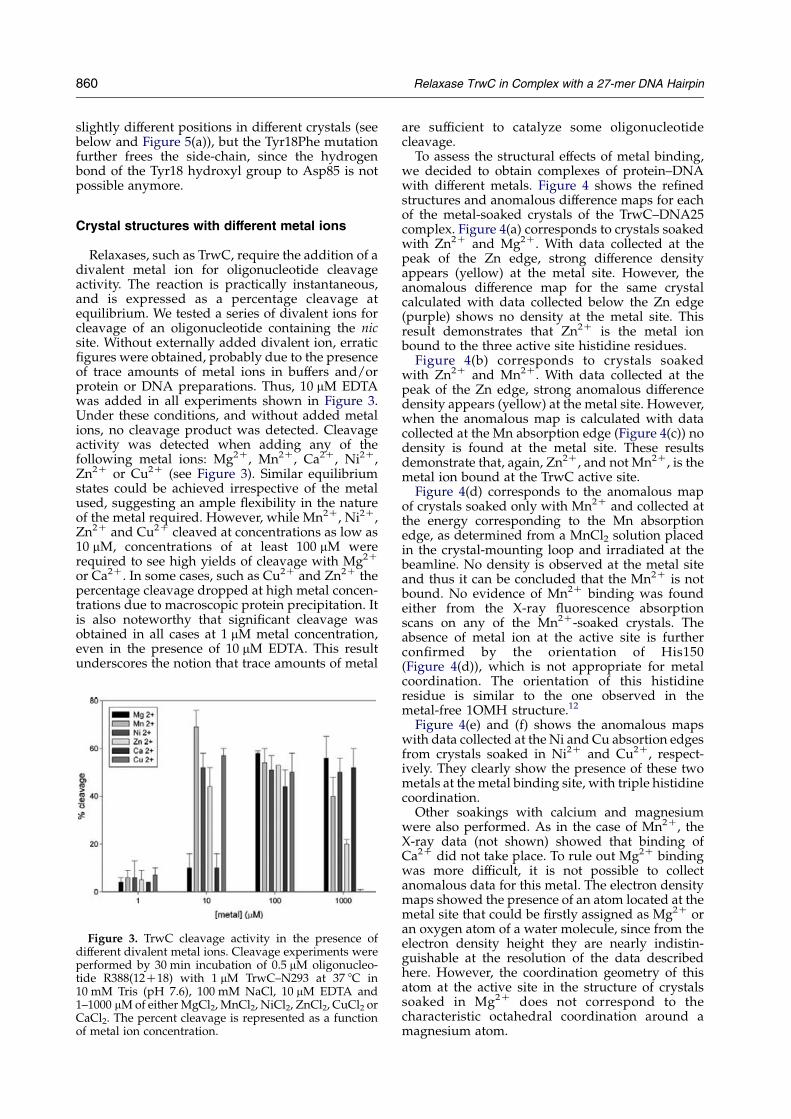
Relaxases, such as TrwC, require the addition of adivalent metal ion for oligonucleotide cleavageactivity. The reaction is practically instantaneous,and is expressed as a percentage cleavage atequilibrium. We tested a series of divalent ions forcleavage of an oligonucleotide containing the nicsite. Without externally added divalent ion, erraticfigures were obtained, probably due to the presenceof trace amounts of metal ions in buffers and/orprotein or DNA preparations. Thus, 10 mM EDTAwas added in all experiments shown in Figure 3.Under these conditions, and without added metalions, no cleavage product was detected. Cleavageactivity was detected when adding any of thefollowing metal ions: Mg2C, Mn2C, Ca2C, Ni2C,Zn2C or Cu2C (see Figure 3). Similar equilibriumstates could be achieved irrespective of the metalused, suggesting an ample flexibility in the natureof the metal required. However, while Mn2C, Ni2C,Zn2C and Cu2C cleaved at concentrations as low as10 mM, concentrations of at least 100 mM wererequired to see high yields of cleavage with Mg2C
or Ca2C. In some cases, such as Cu2C and Zn2C thepercentage cleavage dropped at high metal concen-trations due to macroscopic protein precipitation. Itis also noteworthy that significant cleavage wasobtained in all cases at 1 mM metal concentration,even in the presence of 10 mM EDTA. This resultunderscores the notion that trace amounts of metal
are sufficient to catalyze some oligonucleotidecleavage.
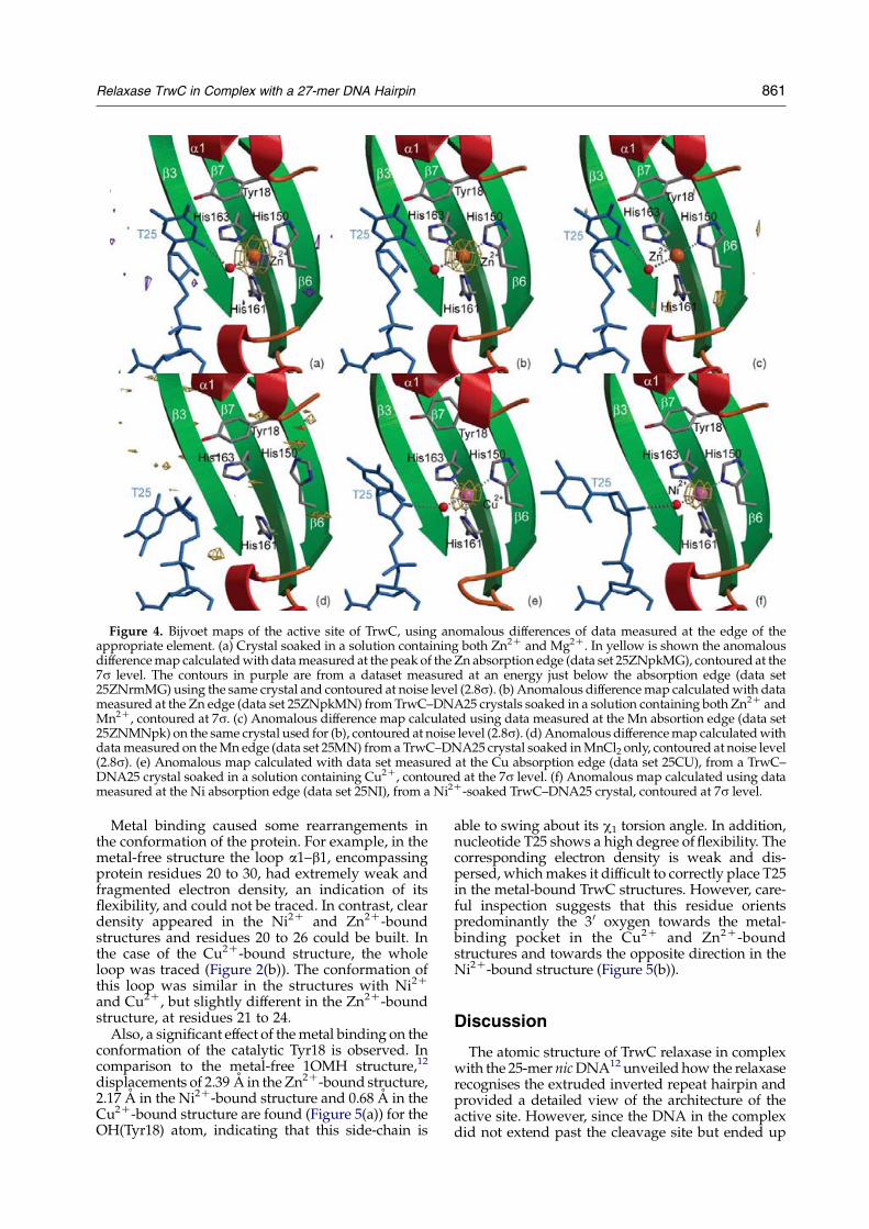
To assess the structural effects of metal binding,we decided to obtain complexes of protein–DNAwith different metals. Figure 4 shows the refinedstructures and anomalous difference maps for eachof the metal-soaked crystals of the TrwC–DNA25complex. Figure 4(a) corresponds to crystals soakedwith Zn2C and Mg2C. With data collected at thepeak of the Zn edge, strong difference densityappears (yellow) at the metal site. However, theanomalous difference map for the same crystalcalculated with data collected below the Zn edge(purple) shows no density at the metal site. Thisresult demonstrates that Zn2C is the metal ionbound to the three active site histidine residues.
Figure 4(b) corresponds to crystals soakedwith Zn2C and Mn2C. With data collected at thepeak of the Zn edge, strong anomalous differencedensity appears (yellow) at the metal site. However,when the anomalous map is calculated with datacollected at the Mn absorption edge (Figure 4(c)) nodensity is found at the metal site. These resultsdemonstrate that, again, Zn2C, and not Mn2C, is themetal ion bound at the TrwC active site.
Figure 4(d) corresponds to the anomalous mapof crystals soaked only with Mn2C and collected atthe energy corresponding to the Mn absorptionedge, as determined from a MnCl2 solution placedin the crystal-mounting loop and irradiated at thebeamline. No density is observed at the metal siteand thus it can be concluded that the Mn2C is notbound. No evidence of Mn2C binding was foundeither from the X-ray fluorescence absorptionscans on any of the Mn2C-soaked crystals. Theabsence of metal ion at the active site is furtherconfirmed by the orientation of His150(Figure 4(d)), which is not appropriate for metalcoordination. The orientation of this histidineresidue is similar to the one observed in themetal-free 1OMH structure.12
Figure 4(e) and (f) shows the anomalous mapswith data collected at the Ni and Cu absortion edgesfrom crystals soaked in Ni2C and Cu2C, respect-ively. They clearly show the presence of these twometals at the metal binding site, with triple histidinecoordination.
Other soakings with calcium and magnesiumwere also performed. As in the case of Mn2C, theX-ray data (not shown) showed that binding ofCa2C did not take place. To rule out Mg2C bindingwas more difficult, it is not possible to collectanomalous data for this metal. The electron densitymaps showed the presence of an atom located at themetal site that could be firstly assigned as Mg2C oran oxygen atom of a water molecule, since from theelectron density height they are nearly indistin-guishable at the resolution of the data describedhere. However, the coordination geometry of thisatom at the active site in the structure of crystalssoaked in Mg2C does not correspond to thecharacteristic octahedral coordination around amagnesium atom.
Figure 3. TrwC cleavage activity in the presence ofdifferent divalent metal ions. Cleavage experiments wereperformed by 30 min incubation of 0.5 mM oligonucleo-tide R388(12C18) with 1 mM TrwC–N293 at 37 8C in10 mM Tris (pH 7.6), 100 mM NaCl, 10 mM EDTA and1–1000 mMof either MgCl2, MnCl2, NiCl2, ZnCl2, CuCl2 orCaCl2. The percent cleavage is represented as a functionof metal ion concentration.
860 Relaxase TrwC in Complex with a 27-mer DNA Hairpin
Metal binding caused some rearrangements inthe conformation of the protein. For example, in themetal-free structure the loop a1–b1, encompassingprotein residues 20 to 30, had extremely weak andfragmented electron density, an indication of itsflexibility, and could not be traced. In contrast, cleardensity appeared in the Ni2C and Zn2C-boundstructures and residues 20 to 26 could be built. Inthe case of the Cu2C-bound structure, the wholeloop was traced (Figure 2(b)). The conformation ofthis loop was similar in the structures with Ni2C
and Cu2C, but slightly different in the Zn2C-boundstructure, at residues 21 to 24.
Also, a significant effect of themetal binding on theconformation of the catalytic Tyr18 is observed. Incomparison to the metal-free 1OMH structure,12
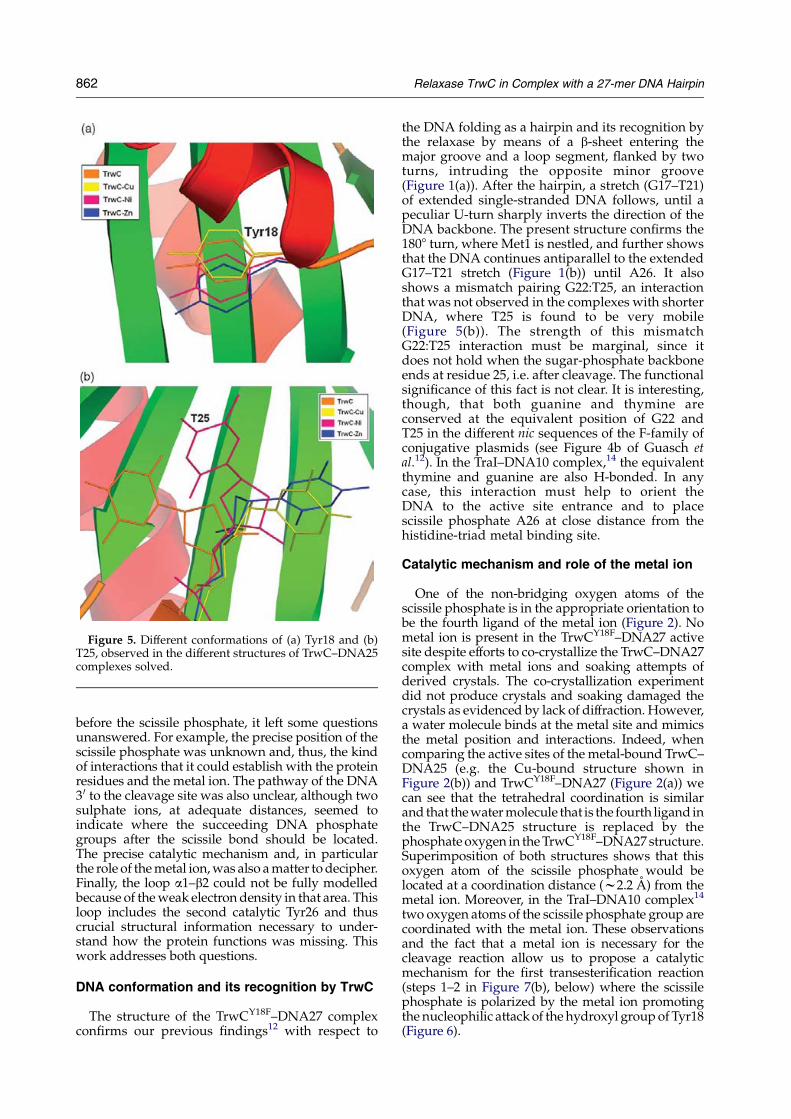
displacements of 2.39 A in theZn2C-bound structure,2.17 A in the Ni2C-bound structure and 0.68 A in theCu2C-bound structure are found (Figure 5(a)) for theOH(Tyr18) atom, indicating that this side-chain is
able to swing about its c1 torsion angle. In addition,nucleotide T25 shows a high degree offlexibility. Thecorresponding electron density is weak and dis-persed, whichmakes it difficult to correctly place T25in the metal-bound TrwC structures. However, care-ful inspection suggests that this residue orientspredominantly the 30 oxygen towards the metal-binding pocket in the Cu2C and Zn2C-boundstructures and towards the opposite direction in theNi2C-bound structure (Figure 5(b)).
Discussion
The atomic structure of TrwC relaxase in complexwith the 25-mer nicDNA12 unveiled how the relaxaserecognises the extruded inverted repeat hairpin andprovided a detailed view of the architecture of theactive site. However, since the DNA in the complexdid not extend past the cleavage site but ended up
Figure 4. Bijvoet maps of the active site of TrwC, using anomalous differences of data measured at the edge of theappropriate element. (a) Crystal soaked in a solution containing both Zn2C and Mg2C. In yellow is shown the anomalousdifferencemap calculatedwith datameasured at the peak of the Zn absorption edge (data set 25ZNpkMG), contoured at the7s level. The contours in purple are from a dataset measured at an energy just below the absorption edge (data set25ZNrmMG) using the same crystal and contoured at noise level (2.8s). (b) Anomalous differencemap calculatedwith datameasured at the Zn edge (data set 25ZNpkMN) fromTrwC–DNA25 crystals soaked in a solution containing both Zn2C andMn2C, contoured at 7s. (c) Anomalous difference map calculated using data measured at the Mn absortion edge (data set25ZNMNpk) on the same crystal used for (b), contoured at noise level (2.8s). (d) Anomalous differencemap calculatedwithdatameasured on theMn edge (data set 25MN) froma TrwC–DNA25 crystal soaked inMnCl2 only, contoured at noise level(2.8s). (e) Anomalous map calculated with data set measured at the Cu absorption edge (data set 25CU), from a TrwC–DNA25 crystal soaked in a solution containing Cu2C, contoured at the 7s level. (f) Anomalous map calculated using datameasured at the Ni absorption edge (data set 25NI), from a Ni2C-soaked TrwC–DNA25 crystal, contoured at 7s level.
Relaxase TrwC in Complex with a 27-mer DNA Hairpin 861
before the scissile phosphate, it left some questionsunanswered. For example, the precise position of thescissile phosphate was unknown and, thus, the kindof interactions that it could establish with the proteinresidues and the metal ion. The pathway of the DNA30 to the cleavage site was also unclear, although twosulphate ions, at adequate distances, seemed toindicate where the succeeding DNA phosphategroups after the scissile bond should be located.The precise catalytic mechanism and, in particularthe role of themetal ion,was also amatter todecipher.Finally, the loop a1–b2 could not be fully modelledbecause of theweak electrondensity in that area. Thisloop includes the second catalytic Tyr26 and thuscrucial structural information necessary to under-stand how the protein functions was missing. Thiswork addresses both questions.
DNA conformation and its recognition by TrwC
The structure of the TrwCY18F–DNA27 complexconfirms our previous findings12 with respect to
the DNA folding as a hairpin and its recognition bythe relaxase by means of a b-sheet entering themajor groove and a loop segment, flanked by twoturns, intruding the opposite minor groove(Figure 1(a)). After the hairpin, a stretch (G17–T21)of extended single-stranded DNA follows, until apeculiar U-turn sharply inverts the direction of theDNA backbone. The present structure confirms the1808 turn, where Met1 is nestled, and further showsthat the DNA continues antiparallel to the extendedG17–T21 stretch (Figure 1(b)) until A26. It alsoshows a mismatch pairing G22:T25, an interactionthat was not observed in the complexes with shorterDNA, where T25 is found to be very mobile(Figure 5(b)). The strength of this mismatchG22:T25 interaction must be marginal, since itdoes not hold when the sugar-phosphate backboneends at residue 25, i.e. after cleavage. The functionalsignificance of this fact is not clear. It is interesting,though, that both guanine and thymine areconserved at the equivalent position of G22 andT25 in the different nic sequences of the F-family ofconjugative plasmids (see Figure 4b of Guasch etal.12). In the TraI–DNA10 complex,14 the equivalentthymine and guanine are also H-bonded. In anycase, this interaction must help to orient theDNA to the active site entrance and to placescissile phosphate A26 at close distance from thehistidine-triad metal binding site.
Catalytic mechanism and role of the metal ion
One of the non-bridging oxygen atoms of thescissile phosphate is in the appropriate orientation tobe the fourth ligand of the metal ion (Figure 2). Nometal ion is present in the TrwCY18F–DNA27 activesite despite efforts to co-crystallize the TrwC–DNA27complex with metal ions and soaking attempts ofderived crystals. The co-crystallization experimentdid not produce crystals and soaking damaged thecrystals as evidenced by lack of diffraction. However,a water molecule binds at the metal site and mimicsthe metal position and interactions. Indeed, whencomparing the active sites of themetal-bound TrwC–DNA25 (e.g. the Cu-bound structure shown inFigure 2(b)) and TrwCY18F–DNA27 (Figure 2(a)) wecan see that the tetrahedral coordination is similarand that thewatermolecule that is the fourth ligand inthe TrwC–DNA25 structure is replaced by thephosphateoxygen in theTrwCY18F–DNA27structure.Superimposition of both structures shows that thisoxygen atom of the scissile phosphate would belocated at a coordination distance (w2.2 A) from themetal ion. Moreover, in the TraI–DNA10 complex14
two oxygen atoms of the scissile phosphate group arecoordinated with the metal ion. These observationsand the fact that a metal ion is necessary for thecleavage reaction allow us to propose a catalyticmechanism for the first transesterification reaction(steps 1–2 in Figure 7(b), below) where the scissilephosphate is polarized by the metal ion promotingthe nucleophilic attack of the hydroxyl groupofTyr18(Figure 6).
Figure 5. Different conformations of (a) Tyr18 and (b)T25, observed in the different structures of TrwC–DNA25complexes solved.
862 Relaxase TrwC in Complex with a 27-mer DNA Hairpin
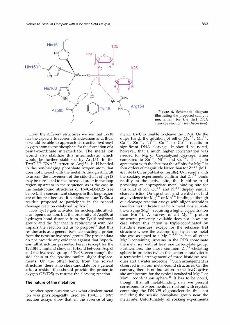
From the different structures we see that Tyr18has the capacity to reorient its side-chain and, thus,it would be able to approach its reactive hydroxyloxygen atom to the phosphate for the formation of apenta-coordinate intermediate. The metal ionwould also stabilize this intermediate, whichwould be further stabilized by Arg154. In theTrwCY18F–DNA27 structure Arg154 is H-bondedto the non-bridging phosphate oxygen atom thatdoes not interact with the metal. Although difficultto assess, the movement of the side-chain of Tyr18may be correlated to the increased order in the loopregion upstream in the sequence, as is the case inthe metal-bound structures of TrwC–DNA25 (seebelow). The concomitant changes in this loop regionare of interest because it contains residue Tyr26, aresidue proposed to participate in the secondcleavage reaction catalyzed by TrwC.
How Tyr18 gets activated for nucleophilic attackis an open question, but the proximity of Asp85, athydrogen bond distance from the Tyr18 hydroxylgroup, and the fact that its replacement with Alaimpairs the reaction led us to propose12 that thisresidue acts as a general base, abstracting a protonfrom the tyrosine hydroxyl group. The present datado not provide any evidence against that hypoth-esis: all structures presented herein (except for theTyr18Phe mutant) show an H-bond between Asp85and the hydroxyl group of Tyr18, even though theside-chain of the tyrosine suffers slight displace-ments. On the other hand, from the solvedstructures, there is no clear candidate for a generalacid, a residue that should provide the proton tooxygen O3 0(T25) to resume the cleaving reaction.
The nature of the metal ion
Another open question was what divalent metalion was physiologically used by TrwC. In vitroreaction assays show that, in the absence of any
metal, TrwC is unable to cleave the DNA. On theother hand, the addition of either Mg2C, Mn2C,Ca2C, Zn2C, Ni2C, Cu2C or Co2C results insignificant DNA cleavage. It should be noted,however, that a much higher concentration wasneeded for Mg or Ca-catalyzed cleavage, whencompared to Zn2C, Ni2C and Cu2C. This is inagreement with the fact that the affinity for Mg2C isfour orders of magnitude lower than for Zn2C (M.L.& F. de la C., unpublished results). Our results withthe soaking experiments confirm that Zn2C bindsreadily to the active site, the histidine triadproviding an appropriate metal binding site forthis kind of ion. Cu2C and Ni2C display similarcharacteristics. On the other hand we did not findany evidence for Mg2C or Mn2C binding, althoughour cleavage reaction assays with oligonucleotides(see Results) indicate that both metal ions activatethe enzyme (Mg2C requiring a higher concentrationthan Mn2C). A survey of all Mg2C proteinstructures presently available does not show anycase where this cation is triple-coordinated byhistidine residues, except for the relaxase TraIstructure where the electron density at the metalsite was assigned to a Mg2C.4,13 In fact, all otherMg2C-containing proteins in the PDB coordinatethe metal ion with at least one carboxylate group.Furthermore, the most common Zn2C-chelatingsphere in proteins (when this cation is catalytic) isa tetrahedral arrangement of three histidine resi-dues and a water molecule.15 Such arrangement isobserved in all our metal-bound structures. On thecontrary, there is no indication in the TrwC activesite architecture for the typical octahedral Mg2C orMn2C coordination sphere.16 It has to be noted,though, that all metal-binding data we presentcorrespond to experiments carried out with crystalscontaining the DNA25 oligonucleotide, thus notincluding the scissile phosphate group near themetal site. Unfortunately, all soaking experiments
Figure 6. Schematic diagramillustrating the proposed catalyticmechanism for the first DNAcleavage reaction (see Discussion).
Relaxase TrwC in Complex with a 27-mer DNA Hairpin 863
with the DNA27 oligonucleotide resulted in crystalsthat either lacked any metal at the active site (shorttime soaking) or did not diffract. Therefore, we cannot rule out the binding of Mg2C or Mn2C to theTrwC active site, since the presence of phosphateA26 could provide a better environment for thesemetals to bind.
Two exit paths for the DNA
One important feature of the TrwCY18F–DNA27complex is the fact that the DNA downstream of thecleavage site does not follow one of the paths thatcould be inferred from our previous work with themetal-free TrwC–DNA25 complex (PDB code1OMH).12 From that structure, it was apparentthat the deep active site crevice continued after themetal binding site. Two sulphate ions, one close tothe metal binding site and another 7 A further away(labelled SO and SB in Figure 7(a)) suggestedfavorable locations for DNA phosphate groupsafter residue 25. Our prediction has been provencorrect for the first position, since in the presentTrwCY18F–DNA27 structure the scissile phosphate(A26) occupies the first sulphate SO position. Butafter that the DNA does not follow the crevice.Instead, it continues “upwards” (Figure 1), anti-parallel to the single-stranded DNA stretch thatextends between the hairpin and the U-turn. Sincethe crevice is almost closed over the active site byresidues Arg14 and Arg154, the DNA seems to go,in the TrwCY18F–DNA27 structure, towards a cul-de-sac. However, closer inspection indicates that anexit path for the DNA is possible, as shown bymodel building. Indeed, a third sulphate ion wasobserved in that path, bound to Arg14 and Arg154(sulphate SA and path A in Figure 7(a)). Inagreement, in TraI–DNA10 complex,14 the DNAfollows a similar path A. Still, we may speculatethat an alternative functional path exists (path B)following the active site crevice as suggested bysulphate SB. This sulphate ion is present in both theTrwC–DNA2512 structure and in the TrwCY18F–DNA27 structure reported here, close to conservedresidues Glu151 (an Asp in some relaxases of thefamily) and Thr152 (Figure 7(a)).
An attractive hypothesis is that both paths areused by the enzyme. In this hypothesis, theinitiation and termination reactions will occur asschematized in Figure 7(b). We propose that, for theinitiation reaction, the DNA follows path A. Thisconformation might be the preferred one in theconstrained binding to supercoiled DNA, whereonly Tyr18 can cleave the DNA.11 After the firstcleavage, Tyr18 forms a covalent complex with theDNA 3 0 to the cleavage site. The DNA moietycontaining the 3 0-OH leaves the active site and isextended by rolling-circle replication. Meanwhile,the relaxase is shot to the recipient cell, where ittracks on the incoming DNA by means of its 5 0/3 0
DNA helicase activity. When the reformed nic siteDNA enters the recipient cell, it is recognized againand bound by the relaxase following path B
(Figure 7(b), blueCgreen strand). It has to be sobecause path A is still occupied by the 3 0 moiety ofthe DNA covalently attached to Tyr18. Besides, it isknown that TrwC recognizes only specific bases 5 0
to the cleavage site.12 Since Tyr18 can swing(Figure 5(a)), it might well get slightly away fromthe active site to leave room for the new DNA(Figure 7(b), blueCgreen strand).
Role of the second catalytic tyrosine
The second cleavage reaction could be catalyzedby Tyr26, located at the tip of the a1–b2 loop asshown in Figure 2(b). After second cleavage, a new3 0-OH appears at the protein active site and thereligation reaction with the 5 0 end phosphate couldtake place by reversal of the initial cleavagereaction, thus re-circularizing the transferred DNAstrand, as shown in Figure 7(b). We know from thedifferent structures that a 3 0-free T25 can rotate byan angle of 1808 (Figure 5(b)). This ability couldfacilitate the adjustment of the 3 0-OH position forattacking the phospho-tyrosine linkage.
In the different structures loop a1–b2 appearseither fully disordered from residue 20 to 30 and notmodelled (TrwC–DNA25, TrwCY18F–DNA27), par-tially ordered and partially modelled (TrwC–Ni2C–DNA25) or fully modelled (TrwC–Cu2C–DNA25).Still, in this later case, the temperature factors arerather high indicating a considerable flexibility.Where Tyr26 could be built in (Figure 2(b)) itappears to be at 17 A distance from themetal site, itshydroxyl group pointing out in the oppositedirection and making a hydrogen bond with themain-chain of Ala96, a residue of helix a4. A longmovement, with the loop a1–b2 folding backtowards the active site, must take place if theTyr26 has to come in close proximity to the scissilephosphate. We cannot exclude, though, that thissecond cleavage is performed by a second TrwCmolecule. In favor of this hypothesis is the fact thatTyr26 is located at the surface of the protein.However, in order to reach the active site of anotherrelaxase molecule, the loop a1–b2 must furtherextend and stick out of the protein surface becausethe active site of the protein is located in a ratherdeep crevice. An example of such trans-attackmechanism is IS608 transposase. IS608 transposasebelongs to the family of rolling-circle transposases,which contain catalytic Tyr residues and histidinetriad motifs in their active sites. They are thusrelated to the superfamily of rolling-circle replica-tion initiation proteins and conjugative relaxases.17
The atomic structure of IS608 transposase18 showsthat the IS608 active Tyr is located in an a-helix thatreaches the scissile bond of the target DNA locatedat the histidine triad catalytic site of the adjacentprotein monomer.
Provided one relaxase molecule has been shot tothe recipient, as proposed in the shoot and pumpconjugation model3 and recently confirmedexperimentally,19 there are two possible locationsfor termination reaction, the donor or the recipient
864 Relaxase TrwC in Complex with a 27-mer DNA Hairpin
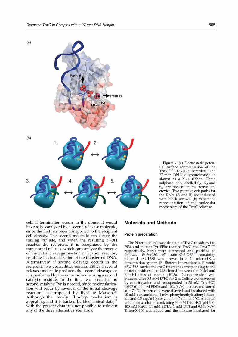
cell. If termination occurs in the donor, it wouldhave to be catalyzed by a second relaxase molecule,since the first has been transported to the recipientcell already. The second molecule can cleave thetrailing nic site, and when the resulting 3 0-OHreaches the recipient, it is recognized by thetransported relaxase which can catalyze the reverseof the initial cleavage reaction or ligation reaction,resulting in circularization of the transferred DNA.Alternatively, if second cleavage occurs in therecipient, two possibilities remain. Either a secondrelaxase molecule produces the second cleavage orit is performed by the samemolecule using a secondcatalytic residue. In the first two scenarios nosecond catalytic Tyr is needed, since re-circulariza-tion will occur by reversal of the initial cleavagereaction, as proposed by Byrd & Matson.20
Although the two-Tyr flip-flop mechanism isappealing, and it is backed by biochemical data,11
with the present data it is not possible to rule outany of the three alternative scenarios.
Materials and Methods
Protein preparation
The N-terminal relaxase domain of TrwC (residues 1 to293), and mutant Tyr18Phe (named TrwC and TrwCY18F,respectively, here) were expressed and purified asfollows.21 Escherichia coli strain C43-DE322 containingplasmid pSU1588 was grown in a 2 l micro-DCUfermentation system (B. Biotech International). PlasmidpSU1588 carries the trwC fragment corresponding to theprotein residues 1 to 293 cloned between the NdeI andBamHI sites of vector pET3a. Overexpression wasinduced with 0.5 mM IPTG for 2 h. Cells were harvestedby centrifugation and resuspended in 50 mM Tris–HCl(pH 7.6), 10 mM EDTA and 10% (v/v) sucrose, and storedat K70 8C. Frozen cells were thawed and incubated with10 mM benzamidine, 1 mM phenylmethylsulfonyl fluor-ide and 0.5 mg/ml lysozyme for 45 min at 0 8C. An equalvolume of a solution containing 50 mMTris–HCl (pH 7.6),400 mM NaCl, 0.1 mM EDTA, 1 mM DTT and 0.5% (v/v)Triton-X-100 was added and the mixture incubated for
Figure 7. (a) Electrostatic poten-tial surface representation of theTrwCY18F–DNA27 complex. The27-mer DNA oligonucleotide isshown as a blue ribbon. Threesulphate ions, labelled SA, SO andSB, are present in the active sitecrevice. Two putative exit paths forthe DNA (A and B) are indicatedwith black arrows. (b) Schematicrepresentation of the molecularmechanism of the TrwC relaxase.
Relaxase TrwC in Complex with a 27-mer DNA Hairpin 865
another 5 min on ice. The lysate was centrifuged at145,000g for 30 min at 4 8C. Supernatants were applied toa P11-phosphocellulose column equilibrated in buffer A(50 mM Tris–HCl (pH 7.6), 200 mM NaCl, 0.1 mM EDTA)and eluted at 600 mM NaCl. Fractions containing N293–TrwC were pooled, diluted to 200 mM NaCl, loaded to aMonoS column (Amersham), and eluted with a linearNaCl gradient (200 mM–600 mM NaCl) in bufferA. Finally, a gel filtration step was carried out in aSuperdex75HR column (Amersham) equilibrated in50 mM Tris–HCl (pH 7.6), 500 mM NaCl, 0.1 mM EDTA.TrwC–N293 eluted as a monomer of about 40 kDa. Itsconcentration was estimated by UVabsorbance at 280 nmusing an extinction coefficient of 31,500 MK1 cmK1, whichwas calculated as described by Edelhoch et al.23 No loss ofactivity was observed after six months storage at K70 8C.Purification of N293–TrwC labelled with seleno-meth-
ionine (SeMet) was performed using B834(DE3) strainand New Minimal Medium supplemented with SeMet.24
The purification was performed according to thedescribed procedure. For subsequent crystallizationtrials, both protein variants were concentrated to2–6 mg/ml and stored at K20 8C in 50 mM Hepes/NaOH (pH 7.6), 450 mM NaCl.
Oligonucleotide cleavage reactions
Reaction mixtures contained 0.5 mMfluorescein-labeledoligonucleotide R388(12C18) (TGCGTATTGTCT/ATAGCCCAGATTTAAGGA), 1 mM TrwC–N293 in clea-vage buffer (10 mM Tris (pH 7.6), 100 mM NaCl, 10 mMEDTA) plus or minus a variety of divalent metal salts(MgCl2, MnCl2, NiCl2, ZnCl2, CuCl2 and CaCl2). Metalion concentrations ranged from 1 mM to 10 mM. Afterincubation for 30 min at 37 8C, samples were treatedwith 0.6 mg/ml proteinase K and 0.05% (w/v) SDS for20 min at 37 8C. Reaction products were separated andquantified by capillary electrophoresis, using the CEOligonucleotide Analysis Kit (BioRad) in the capillarysystem BioFocusw2000 (BioRad). The capillary usedwas a BioCAP Oligonucleotide Analysis Capillary(30 cm!75 mm i.d.!375 mm o.d.). Samples were intro-duced in the capillary by pressure (200 psi/s). Electro-phoresis was carried out at 12 kV at 40 8C. A laser-induced detector was employed for fluorescencedetection. Peak information (migration time, peak areaand height) was obtained using the CE IntegratorSoftware (BioRad).
Crystallization and structure determinationof TrwCY18F–DNA27 complex
In order to prepare the protein–DNA complex, asolution 0.23 mM SeMet TrwCY18F, 450 mM NaCl,50 mM Hepes (pH 7.6) was mixed with a solution of0.23 mM of annealed DNA oligonucleotide, encompass-ing the nic sequence 25 nucleotides upstreamand two nucleotides downstream from the cleavagesite (DNA27Z5 0-GCGCACCGAAAGGTGCGTATTGTCT-3 0), dissolved in the same buffer and salt. Theresulting TrwCY18F–DNA27 complex was purified bysize-exclusion chromatography and concentrated to6.9 mg/ml of protein. Crystals were obtained by vapordiffusion, at 4 8C, from hanging drops prepared bymixing2 ml of the concentrated TrwC solution and 1 ml of theprecipitant solution containing 32% (w/v) PEG MME2000, 0.3 M ammonium sulphate, 0.1 M sodium acetate(pH 4.6). Crystals appeared after seven days and
continued to grow to a maximum size after 27 days.A complete dataset was measured at beamline ID14-4, atESRF (Grenoble). Data collection is summarized inTable 1. The TrwC–DNA25 (see below for definition ofDNA25) metal-free structure previously published12
(PDB code 1OSB), modified to correctly represent theTyr18Phe mutation, was used as a starting model formolecular replacement. Refinement was carried out withREFMAC5.2, part of the CCP4 program suite.25 Initialrefinement indicated a change in the position of the baseT25 of the DNA strand. Subsequently, this residue andwater molecules in and around the active site wereremoved, after which refinement was restarted. The DNAresidue T25 could be correctly placed upon inspection ofthe 2FoKFc and FoKFc Fourier maps. Another cycle ofrestrained refinement and map inspection revealed theposition of DNA residue A26, which was subsequentlyadded. Residue 27, however, could not be unambiguouslylocated and was not modelled. Final refinement statisticsare shown in Table 1.
Crystallization and structure determinationof TrwC–DNA25 metal ion complexes
In order to prepare TrwC–DNA binary complexes, asolution of 0.179 mM TrwC in 450 mM NaCl, 50 mMHepes (pH 7.6), was mixed with a solution of 0.179 mM ofannealed DNA oligonucleotide, encompassing the OriTsequence 25 nucleotides upstream from the nic site(DNA25Z5 0-GCGCACCGAAAGGTGCGTATTGTCT-3 0)dissolved with similar buffer and salt. The resultingTrwC–DNA25 complex was purified by size-exclusionchromatography and concentrated to 6.8 mg/mlof protein. Crystals were grown as described.12 Thesecrystals were used to prepare different metal ioncomplexes by soaking them in metal ion solutions andX-ray diffraction data were collected at the ESRF(Grenoble), as follows: (1) crystals soaked in 5 mMZnSO4 and 10 mM MgCl2 for 24 h were measured at theK absorption edge of Zn (data set 25ZNpkMG) and 24 eVbelow this edge (data set 25ZNrmMG), on beamline ID14-4; (2) crystals soaked in 5 mM ZnSO4, 10 mM MnCl2 for24 h were measured at both the K absorption edges of Zn(data set 25ZNpkMN) and Mn (data set 25ZNMNpk), onbeamline BM16; (3) crystals soaked in 5 mM MnCl2 for24 h (data set 25MN) were measured at the K absorptionedge of Mn, on beamline BM16; (4) crystals soaked in5 mM CuCl2 for 24 h (data set 25CU) were measuredbelow the K absorption edge of Cu (0.97855 A), atbeamline ID23EH1; and (5) crystals soaked in 5 mMNiCl2 for 24 h (25NI) were measured below the Kabsorption edge of Ni, at beamline BM14. Data collectionstatistics are summarized in Table 1.Refinement against datasets 25ZNpkMG and
25ZNpkMN was performed with REFMAC5.2 programin the CCP4 program suite,25 starting from a rigid bodyrefinement using the previously deposited Zn2C-boundstructure of TrwC complexed with the same DNA25fragment12 (PDB code 1QX0), followed by a restrainedrefinement. For the 25MN structure a similar protocol wasused, starting from the deposited metal-free structure12
(PDB code 1OMH). Anomalous maps were calculatedfrom a combination of the corresponding phases and theanomalous differences associated with each dataset. Themetal-free protein structure (PDB code 1OMH) was alsoused as initial model for refinement against data of theCu2C and Ni2C-soaked crystals. Initial inspection of thedensity maps clearly indicated the presence of metal ionsat the active site. Subsequent simulated annealing and
866 Relaxase TrwC in Complex with a 27-mer DNA Hairpin
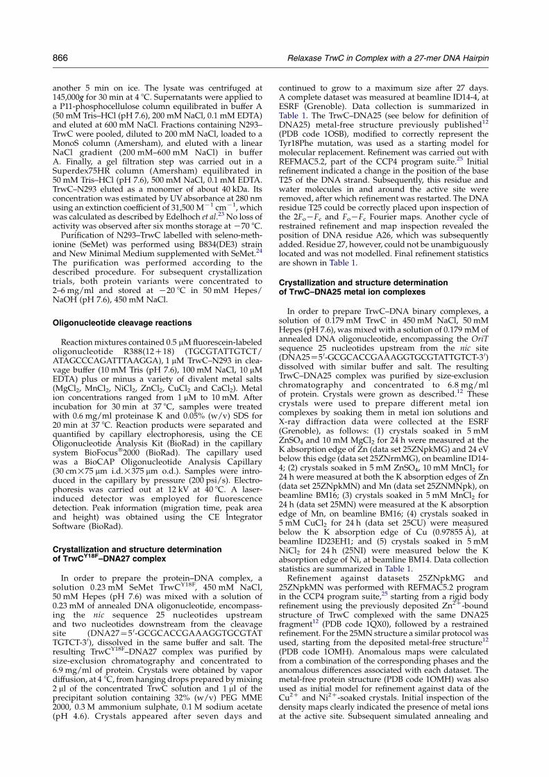
Table 1. Data collection and refinement statistics
Data seta 27NAT 25ZNpkMG 25ZNrmMG 25ZNpkMN 25ZNMNpk 25MN 25CU 25NI
A. Data collectionUnit cell (A) aZbZ148.42,
cZ75.52aZbZ90.70,cZ204.22
aZbZ90.75,cZ204.36
aZbZ90.69,cZ204.22
aZbZ90.53,cZ203.77
aZbZ90.24,cZ202.08
aZbZ91.088,cZ205.55
aZbZ92.7312,cZ208.8551
Space group P65 P6122 P6122 P6122 P6122 P6122 P6122 P6122Wavelength (A) 0.97930 1.2820 1.2852 1.2823 1.89223 1.8924 0.97855 1.00100Resolution range (A)b 25.0–2.7
(2.9–2.7)19.8–2.7(2.9–2.7)
19.8–2.7(2.9–2.7)
51.4–3.16(3.2–3.0)
45.3–2.90(3.1–2.9)
43.9–2.6(2.7–2.6)
24.7–2.5(2.6–2.5)
43.9–2.3(2.4–2.3)
No. of observed reflectionsb 165,382 (22,961) 169,473 (24,851) 146,870 (21,542) 120,010 (17,326) 130,924 (18,815) 171,348 (25,127) 386,179(56,283)
114,701 (13,061)
No. of unique reflectionsb 27,451 (3996) 143,70 (2037) 14,393 (2039) 10,633 (1496) 11,442 (1598) 15,710 (2226) 18,252 (2592) 28,857 (3506)Completenessb 99.7 (100) 99.9 (100) 99.8 (100) 100 (100) 98.9 (98.0) 99.9 (99.8) 99.9 (100) 98.8 (98.8)Rmerge
b,c 10.9 (41.3) 9.0 (22.4) 5.9 (15.6) 14.1 (42.7) 9.9 (36.1) 12.7 (45.5) 10.4 (51.6) 4.9 (14.6)I/s(I)b 5.4 (1.5) 5.7 (3.2) 9.1 (4.6) 4.8 (1.7) 7.1 (2.0) 4.6 (1.5) 5.6 (1.4) 10.2 (4.8)Multiplicityb 6.0 (5.7) 11.8 (12.2) 10.2 (10.6) 11.3 (11.6) 11.4 (11.8) 10.9 (11.3) 21.2 (21.7) 4.7 (4.1)Anomalous completenessb 99.0 (99.9) 99.9 (100) 99.9 (100) 100 (100) 99.4 (98.6) 100 (100) 100(100) 98.3 (88.9)Anomalous multiplicityb 3.1 (2.9) 6.5 (6.6) 5.6 (5.6) 6.4 (6.3) 6.4 (6.4) 5.9 (6.0) 11.7(11) 2.5 (2.2)B. RefinementProtein atomsd 2213C2213 2288 Not refinede 2288 Not refinede 2226 2314 2275DNA atomsd 492C475 512 512 512 512 512Metal atomd – Zn2C Zn2C – Cu2C Ni2C
Water O atomsd 239 178 178 256 207 162No. of reflections used in refinementf 26,050 13,597 Not refinede 10,055 Not refinede 10,614 18,200 24,598R factor (%)f 20.5 21.8 19.9 20.4 23.3 20.6Rfree (%)f 24.7 26.0 24.0 25.0 27.6 24.5rmsd from target valuesBonds (A) 0.009 0.009 0.011 0.010 0.005 0.013Angles (deg.) 1.37 1.31 1.44 1.35 1.52 1.35Chiral (A) 0.10 0.075 0.079 0.074 0.080 0.086Mean B factor (A2) 49.2 34.0 32.4 31.6 37.1 38.2
a See Materials and Methods for an explanation.b Outermost resolution shell values are in parenthesis.c RmergeZ ½ShklSijIiðhklÞK!IðhklÞO j=ShklSiIiðhklÞ�!100.d Per asymmetric unit.e Structures were not refined against data sets 25ZNrmMG and 25 NMNpk.f RfactorZ ½ShkljjFobsjKkjFcalcjj=ShkljFobsj�!100; Rfree, same as for a test set of reflections not used during refinement.
positional and overall B-factor refinement were per-formed using CNS.26 The correction of the model,through visual inspection of the electron density mapsusing TURBO–FRODO,27 was alternated with solventmolecule addition and refinement, including atomicB-factor refinement, using CNS. Final refinement statisticsare shown in Table 1. Anomalous difference Fourier maps(Bijvoet maps) were calculated from various datasets asindicated in the legend to Figure 4. All pictures wereprepared using PyMol†, Bobscript extention of Mol-script28 and the Raster3D package.29
Protein Data Bank accession codes
Atomic coordinates have been deposited with the RCSBPDB under accession codes 2CDM (TrwCY18F–DNA27),1S6M (TrwC-Ni2C–DNA25) and 1ZM5 (TrwC-Cu2C–DNA25).
Acknowledgements
This study was supported by the Ministerio deEducacion y Ciencia of Spain (grants BIO2002-03964, GEN2003-20642 and BFU2005-06758/BMCto M.C.; grant BMC2002-00379 to F.C.) and by theGeneralitat de Catalunya (grant 2001SGR-346 toM.C.). Synchrotron data collection was supportedby the ESRF and the EU. S.R. is the recipient of apredoctoral fellowship from the Generalitat deCatalunya. M.L. acknowledges a fellowship fromthe Fundacion Marques de Valdecilla IFIMAV.
References
1. Ochman, H., Lawrence, J. G. & Groisman, E. A. (2000).Lateral gene transfer and the nature of bacterialinnovation. Nature, 405, 299–304.
2. de la Cruz, F. & Davies, J. (2000). Horizontal genetransfer and the origin of species: lessons frombacteria. Trends Microbiol. 8, 128–133.
3. Llosa,M., Gomis-Ruth, F. X., Coll,M.&de la Cruz Fd, F.(2002). Bacterial conjugation: a two-stepmechanism forDNA transport. Mol. Microbiol. 45, 1–8.
4. Zechner, E. L., de la Cruz, F., Eisenbrandt, T., Grahn,A. M., Koraimann, G., Lanka, E. et al. (2000).Conjugative DNA transfer processes. In The Horizon-tal Gene Pool: Bacterial Plasmids and Gene Spread(Thomas, C. M., ed.), pp. 87–174, Harwood AcademicPublishers, London.
5. Francia, M. V., Varsaki, A., Garcillan-Barcia, M. P.,Latorre, A., Drainas, C. & de la Cruz, F. (2004). Aclassification scheme for mobilization regions ofbacterial plasmids. FEMS Microbiol. Rev. 28, 79–100.
6. Lanka, E. & Wilkins, B. M. (1995). DNA processingreactions in bacterial conjugation. Annu. Rev. Biochem.64, 141–169.
7. Pansegrau, W. & Lanka, E. (1996). Mechanisms ofinitiation and termination reactions in conjugativeDNA processing. Independence of tight substrate
binding and catalytic activity of relaxase (TraI) ofIncPalpha plasmid RP4. J. Biol. Chem. 271,13068–13076.
8. Llosa, M., Grandoso, G., Hernando, M. A. & de laCruz, F. (1996). Functional domains in protein TrwCof plasmid R388: dissected DNA strand transferaseand DNA helicase activities reconstitute proteinfunction. J. Mol. Biol. 264, 56–67.
9. Llosa,M.,Grandoso,G.&de laCruz, F. (1995).Nickingactivity of TrwC directed against the origin of transferof the IncW plasmid R388. J. Mol. Biol. 246, 54–62.
10. Hanai, R. & Wang, J. C. (1993). The mechanism ofsequence-specific DNA cleavage and strand transferby phi X174 gene A* protein. J. Biol. Chem. 268,23830–23836.
11. Grandoso, G., Avila, P., Cayon, A., Hernando, M. A.,Llosa, M. & de la Cruz, F. (2000). Two active-sitetyrosyl residues of protein TrwC act sequentially atthe origin of transfer during plasmid R388 conju-gation. J. Mol. Biol. 295, 1163–1172.
12. Guasch, A., Lucas, M., Moncalian, G., Cabezas, M.,Perez-Luque, R., Gomis-Ruth, F. X. et al. (2003).Recognition and processing of the origin of transferDNA by conjugative relaxase TrwC. Nature Struct.Biol. 10, 1002–1010.
13. Datta, S., Larkin, C. & Schildbach, J. F. (2003).Structural insights into single-stranded DNA bindingand cleavage by F factor TraI. Structure (Camb), 11,1369–1379.
14. Larkin, C., Datta, S., Harley, M. J., Anderson, B. J.,Ebie, A., Hargreaves, V. & Schildbach, J. F. (2005).Inter- and intramolecular determinants of the speci-ficity of single-stranded DNA binding and cleavageby the f factor relaxase. Structure (Camb), 13,1533–1544.
15. Dudev, T. & Lim, C. (2003). Principles governing Mg,Ca, and Zn binding and selectivity in proteins. Chem.Rev. 103, 773–788.
16. Bock, C.W., Katz, A. K.,Markham, G.D.&Glusker, J. P.(1999). Manganese as a replacement for magnesiumand zinc: functional comparison of the divalent ions.J. Am. Chem. Soc. 121, 7360–7372.
17. Garcillan-Barcia, M. P., Bernales, I., Mendiola, M. V. &de la Cruz Fd, F. (2005). IS91 rolling-circle trans-position. In Mobile DNA II (Craig, N. L., Craigie, R.,Gellert, M. & Lambowitz, A., eds), pp. 891–904, ASMPress, Washington, DC.
18. Ronning, D. R., Guynet, C., Ton-Hoang, B., Perez,Z. N., Ghirlando, R., Chandler, M. & Dyda, F. (2005).Active site sharing and sub-terminal hairpin recog-nition in a new class of DNA transportases. Mol. Cell,20, 143–154.
19. Draper, O., Cesar, C., Machon, C., de la Cruz, F. &Llosa, M. (2005). Site-specific recombinase andintegrase activities of a conjugative relaxase inrecipient cells. Proc. Natl Acad. Sci. USA, 102,16385–16390.
20. Byrd, D. R. & Matson, S. W. (1997). Nicking bytransesterification: the reaction catalysed by a relax-ase. Mol. Microbiol. 25, 1011–1022.
21. Grandoso, G., Llosa, M., Zabala, J. C. & de la Cruz, F.(1994). Purification and biochemical characterization ofTrwC, the helicase involved in plasmid R388 conjugalDNA transfer. Eur. J. Biochem. 226, 403–412.
22. Miroux, B. & Walker, J. E. (1996). Over-productionof proteins in Escherichia coli: mutant hosts thatallow synthesis of some membrane proteins andglobular proteins at high levels. J. Mol. Biol. 260,289–298.† http:/www.pymol.org
868 Relaxase TrwC in Complex with a 27-mer DNA Hairpin
23. Edelhoch, H. (1967). Spectroscopic determination oftryptophan and tyrosine in proteins. Biochemistry, 6,1948–1954.
24. Budisa, N., Steipe, B., Demange, P., Eckerskorn, C.,Kellermann, J. & Huber, R. (1995). High-levelbiosynthetic substitution of methionine in proteinsby its analogs 2-aminohexanoic acid, selenomethio-nine, telluromethionine and ethionine in Escherichiacoli. Eur J Biochem. 230, 788–796.
25. Collaborative Computational Project, Number 4.(1994). The CCP4 suite: programs for proteincrystallography. Acta Crystallog. sect. D, 50, 760–763.
26. Brunger, A. T., Adams, P. D., Clore, G. M., DeLano,W. L., Gros, P., Grosse-Kunstleve, R. W. et al. (1998).
Crystallography &NMR system: a new software suitefor macromolecular structure determination. ActaCrystallog. sect. D, 54, 905–921.
27. Roussel, A. & Cambilleau, C. (1989). Turbo-Frodo. InSilicon Graphics Geometry Partners Directory, pp. 77–79.Silicon Graphics, Mountain View, CA.
28. Esnouf, R. M. (1999). Further additions to MolScriptversion 1.4, including reading and contouring ofelectron-density maps. Acta Crystallog. sect. D, 55,938–940.
29. Merritt, E. & Bacon, D. (1997). Raster3D: photorealis-tic molecular graphics. In Methods in Enzymology(Carter, C., Jr & Sweet, R., eds), vol. 277, pp. 505–524,Elsevier, San Diego.
Edited by R. Huber
(Received 27 October 2005; received in revised form 3 February 2006; accepted 8 February 2006)Available online 28 February 2006
Relaxase TrwC in Complex with a 27-mer DNA Hairpin 869