FHAISOL MAT AMIN
40
UNIVERSITI PUTRA MALAYSIA COMPARATIVE MORPHOLOGY OF THE GASTROINTESTINAL TRACT OF WHITE EDIBLE BIRD'S NEST SWIFTLET (Aerodramus fuciphagus [Thunberg]) AND HOUSE SWIFT (Apus nipalensis [Hodgson]) FHAISOL MAT AMIN FPV 2014 14
Transcript of FHAISOL MAT AMIN
UNIVERSITI PUTRA MALAYSIA
COMPARATIVE MORPHOLOGY OF THE GASTROINTESTINAL TRACT OF WHITE EDIBLE BIRD'S NEST SWIFTLET
(Aerodramus fuciphagus [Thunberg]) AND HOUSE SWIFT (Apus nipalensis [Hodgson])
FHAISOL MAT AMIN
FPV 2014 14
© COPYRIG
HT UPM
COMPARATIVE MORPHOLOGY OF THE
GASTROINTESTINAL TRACT OF
WHITE EDIBLE BIRD'S NEST SWIFTLET
(Aerodramus fuciphagus [Thunberg]) AND
HOUSE SWIFT (Apus nipalensis [Hodgson])
FHAISOL MAT AMIN
MASTER OF VETERINARY SCIENCE
UNIVERSITI PUTRA MALAYSIA
2014
© COPYRIG
HT UPM
COMPARATIVE MORPHOLOGY OF THE
GASTROINTESTINAL TRACT OF WHITE EDIBLE BIRD'S
NEST SWIFTLET (Aerodramus fuciphagus [Thunberg])
AND HOUSE SWIFT (Apus nipalensis [Hodgson])
By
FHAISOL MAT AMIN
Thesis submitted to the School of Graduate Studies, University Putra
Malaysia
in Fulfilment of the Requirements for the Degree of
Master of Veterinary Science.
January 2014

© COPYRIG
HT UPM
COPYRIGHT
All material contained within the thesis, including without limitation text,
logos, icons, photographs and all other artwork, is copyright material of
Universiti Putra Malaysia unless otherwise stated. Use may be made of
any material contained within the thesis for non-commercial purposes
from the copyright holder. Commercial use of material may only be
made with the express, prior, written permission of Universiti Putra
Malaysia.
Copyright © Universiti Putra Malaysia
© COPYRIG
HT UPM
ii
Abstract of thesis presented to the Senate of Universiti Putra Malaysia in fulfillment
of the requirement for the degree of Masters of Veterinary Science.
COMPARATIVE MORPHOLOGY OF THE GASTROINTESTINAL
TRACT OF WHITE EDIBLE BIRD'S NEST SWIFTLET
(Aerodramus fuciphagus [Thunberg]) AND
HOUSE SWIFT (Apus nipalensis [Hodgson])
By
Fhaisol Mat Amin
Chairman : Intan Shameha Binti Abd Razak, Ph.D
Faculty : Veterinary Medicine
A. fuciphagus or White Edible bird’s-nest Swiftlet (EBN Swiftlet) and A. nipalensis
or House swift belong to Apodidae family. A. fuciphagus is the only bird in the world
constructing its nest using saliva, whereas A. nipalensis builds its nest using grass
and saliva as adhesive materials. The objective of this study is to evaluate the macro
and microscopic morphology of the gastrointestinal tract of six selected male A.
fuciphagus and four selected male A. nipalensis. The birds were caught in Kuala
Terengganu, Terengganu, and in FELDA Redong, Segamat, Johore, using mist net
according to the FAO standards, and were transported and immediately euthanized
by injecting pentobarbital sodium (Nembutal®) at 80 mg/kg body weight through a
brachial ulnar vein upon arrival at the Anatomy Laboratory, Faculty of Veterinary
Medicine UPM. The body weight and length and the gastrointestinal tract (GIT)
weight and length of each bird were measured, recorded and calculated into relative
GIT weight and length. These values were analyzed using Mann-Whitney U Test for
non-parametric data. The GIT specimens were then fixed in Bouin’s solution and
underwent various processes for histomorphological and histomorphometrical
evaluations. The histological evaluations include the used of H&E, Masson
trichrome, Van Gieson, Gomori trichrome. Periodic acid-Schiff (PAS), Aldehyde
fuchsin, Alcian blue pH 1.0 and pH 2.5, Aldehyde fuchsin-Alcian blue (AF-AB) and
Periodic acid-Schicff-Alcian blue (PAS-AB) were used to determine and classify the
type of mucins. Histomorphometric evaluations were also conducted on koilin
thickness, villus height, and crypt depth, thickness of the tunica muscularis externa
and goblet cell of the intestines. It was observed that the tongue had a sharp
burficated apex, the esophagus was an elongated tubular structure with absence of
crop and it continuously connected to the proventriculus, ventriculus and small
intestine. The ventriculus was formed by a thick and strong muscle, whereas the
intestines lacked both a cecum and Meckel’s diverticulum. All the recorded weight
and length of the GIT and its glandular organs of A.fuciphagus were found to be
smaller (P≤0.05) than those of A.nipalensis, except for the esophagus and pancreas.
However, when calculated based on the relative weight and length, all the GIT and

© COPYRIG
HT UPM
iii
its glandular organs of A.fuciphagus were found to be significantly greater (P≤0.05)
than those of A.nipalensis, except for the esophagus and pancreas. The tissue
arrangement of the GIT in both Apodidae species consisted of mucosa at the
innermost layer, with absent of the muscularis mucosa. It was followed by a
submucosa rich in glandular structure, the tunica muscularis externa, comprised of
inner longitudinal and outer circular muscle, and finally the serosa as the outermost
layer. The tongue of both species of birds was comprised of a keratinized squamous
epithelium, which was found to be thicker in the dorsal than ventral region. The
submucosa layer consisted of a massive glandular structure, absent in the apical
region of the tongue. The mucins were detected in the glands of the tongue and also
along the GIT of A. fuciphagus and A. nipalensis. The lingual and esophageal glands
of A. fuciphagus consist of acid carboxylated mucins, but A. nipalensis had a
mixture of carboxylated and sulfated mucins. Along the GIT, the type of mucins was
considerably similar. In turn, the thickness of koilin, height of the villus, crypt depth,
thickness of the tunica muscularis externa and goblet cells in the intestinal segments
were found to be significantly different (P≤0.05) in both A. fuciphagus and A.
nipalensis. As a conclusion, the significant differences (P≤0.05) in the morphology
of the GIT of A. fuciphagus and A. nipalensis are related to both the type of diet and
the nest building ability.

© COPYRIG
HT UPM
iv
Abstrak tesis yang dikemukakan kepada Senat Universiti Putra Malaysia Sebagai
memenuhi keperluan ijazah Sarjana Sains Veterinar
PERBANDINGAN MORFOLOGI TRAKUS GASTROUSUS BURUNG
WALIT SARANG PUTIH (Aerodramus fuciphagus [Thunberg]) DAN
BURUNG LAYANG – LAYANG RUMAH (Apus nipalensis [Hodgson])
Oleh
FHAISOL MAT AMIN
Pengerusi : Dr Intan Shameha Binti Abd Razak, Ph.D
Fakulti : Fakulti Perubatan Veterinar, UPM
A.fuciphagus atau Burung Walit Sarang Putih (EBN walit ) dan A. nipalensis atau
Layang – Layang Rumah tergolong dalam keluarga Apodidae. A. fuciphagus
merupakan satu-satunya burung di dunia yang membuat sarang dengan
menggunakan air liur manakala A.nipalensis membina sarangnya menggunakan
rumput dan air liur sebagai bahan pelekat. Objektif kajian ini adalah untuk menilai
morfologi makro dan mikroskopik saluran gastrousus ke atas enam ekor jantan A.
fuciphagus dan empat ekor A. nipalensis terpilih. Burung – burung itu ditangkap di
Kuala Terengganu, Terengganu dan di FELDA Redong, Segamat, Johor
menggunakan pukat mengikut piawaian FAO yang kemudiannya dibawa dan
dimatikan dengan menyuntik sodium pentobarbital (Nembutal®) pada kadar
80mg/kg berat badan melalui vena ulnar brakial sejurus tiba di Makmal Anatomi,
Fakulti Perubatan Veterinar UPM. Berat dan panjang badan; berat dan panjang
saluran gastrousus setiap burung diukur dan direkodkan. Ia kemudiannya dikira
berdasarkan kepada berat dan panjang relatif. Data-data ini dianalisis dengan
menggunakan Ujian Mann -Whitney U untuk data bukan parametrik . Spesimen GIT
kemudiannya ditetapkan ke dalam larutan Bouin dan menjalani pelbagai proses
untuk penilaian histomorfologi dan histomorfometrik. Penilaian histologi termasuk
pewarnaan ‘H&E’, ‘Masson trichrome’, ‘Van Gieson’, ‘Gomori trichrome’ telah
digunakan. Pewarnaan ‘Periodic acid-Schiff’ (PAS ), aldehid fuksin , alcian biru pH
1.0 dan pH 2.5, Aldehid fuksin - 'Alcian blue' ( AF- AB) dan ‘Periodic acid-Schiff’ –
‘Alcian blue’ (PAS- AB ) telah digunakan untuk menentu dan mengkelaskan jenis
mucins. Penilaian histomorfometik juga telah dijalankan ke atas ketebalan koilin,
ketinggian vilus, kedalaman krip, ketebalan maskularis eksterna dan sel goblet usus.
Diperhatikan bahawa lidah mempunyai hujung bercabang yang tajam, esofagus
berbentuk tiub panjang dengan ketiadaan tembolok dan ia bersambung ke
proventrikulus, ventrikulus dan usus kecil. Ventrikulus terbentuk daripada otot yang
tebal dan kuat, usus pula didapati tiada sekum dan ‘Meckel’s diverticulum’. Berat
sebenar semua organ trakus gastrousus dan organ kelenjar di dalam A.fuciphagus
yang direkodkan didapati rendah dengan ketara (P≤0.05) berbanding A.nipalensis
kecuali esofagus dan pancreas. Walau bagaimanapun, apabila perkiraan dibuat
berdasarkan berat dan panjang relatif, Walau bagaimanapun, apabila ia telah dikira
© COPYRIG
HT UPM
v
berdasarkan berat relatif dan panjang, semua organs trakus gastrousus dan organ
kelenjar di dalam A.fuciphagus didapati lebih tinggi dengan ketara (P≤0.05) daripada
A.nipalensis kecuali esofagus dan pankreas. Susunan tisu GIT dalam kedua-dua
spesies Apodidae ini terdiri daripada mukosa di lapisan terdalam dengan tidak
mukosa maskularis. Ia diikuti oleh submukosa yang kaya dengan struktur kelenjar;
selaput muskularis eksterna terdiri daripada otot bulat membujur dalaman dan otot
melintang luar dan akhirnya lapisan serosa di bahagian paling luar. Lidah kedua-dua
spesies burung ini terdiri daripada epitelium skuamus berkeratin yang didapati tebal
pada bahagian dorsal berbanding bahagian ventral. Lapisan submukosa pula terdiri
daripada struktur kelenjar yang banyak tetapi tiada pada bahagian apeks lidah.
Mucins dikesan di kelenjar lidah dan juga di sepanjang trakus gastrousus
A.fuciphagus dan A.nipalensis. Kelenjar lidah esophagus A.fuciphagus terdiri
daripada mucins berasid berkarboksilat tetapi dalam A.nipalensis adalah campuran
mucins berkarboksilat dan bersulfat. Dalam GIT lain, jenis mucins adalah sama.
Ketebalan koilin , ketinggian vilus , kedalaman krip, ketebalan muskularis eksterna
dan sel goblet dalam usus didapati berbeza dengan ketara (P≤0.05) dalam kedua-dua
spesies A.fuciphagus dan A.nipalensis yang dikaji. Sebagai kesimpulannya,
perbezaan yang ketara dalam morfologi trakus gastrousus A.fuciphagus dan
A.nipalensis dipengaruhi oleh jenis diet dan juga keupayaan membina sarang.

© COPYRIG
HT UPM
vi
ACKNOWLEDGEMENTS
In the name of Allah, the most Benovolent and the most Merciful. I am thankful for
giving me strength which enabled me to complete this study.
I deeply express my gratitude to my supervisor Dr Intan Shameha binti Abd. Razak
for giving me an opportunity to complete my thesis and devoted her time for
invaluable guidance, advice, supervision and support throughout the course of study.
I also would like to express my sincere gratitude to my co-supervisors, Professor Dr.
Md Zuki bin Zakaria @Abu Bakar and Associate Professor Dr. Azhar bin Kasim for
investing time time and knowledge for valueable comments and recommendation in
my study.
I also express my grateful to my sponsor, Malaysian Agricultural Research and
Development Institute (MARDI) for providing me two years of sponsorship to
complete my study in Universiti Putra Malaysia.
My acknowledgments also extended to those who helping and providing facilities
especially to Dr Rueben Sharma of Parasite laboratory, Mr Saifulzaman of Serology
laboratory, Mrs Jamilah of Histopathology laboratory, Dr Mehdi Ebrahimi for
assistance of data analysis, Dr Ong Kang Woei and Mr Marwan for assistance of
sample processing and to all member of the faculty at Faculty of Veterinary
Medicine, UPM for everything they have done for me in completing my study.
© COPYRIG
HT UPM
vii
I certify that a Thesis Examination Committee has met on 7th
January 2014 to
conduct the final examination of Fhaisol Bin Mat Amin on his thesis entitled
"Comparative morphology of the gastrointestinal tract of the White Edible Bird's
nest Swiftlet (Aerodramus fuciphagus [Thunberg]) and House Swift (Apus nipalensis
[Hodgson])" in accordance with the Universities and University Colleges Act 1971
and the Constitution of the Universiti Putra Malaysia [P.U.(A) 106] 15 March 1998.
The Committee recommends that the student be awarded the Master of Veterinary
Science.
Members of the Thesis Examination Committee were as follows:
Mohamad Ali Rajion, PhD
Professor
Faculty of Veterinary Medicine
Universiti Putra Malaysia
(Chairman)
Noordin Mohamad Mustaffa, PhD
Professor
Faculty of Veterinary Medicine
Universiti Putra Malaysia
(Internal Examiner I)
Jalila Abu, PhD
Assoc. Professor
Faculty of Veterinary Medicine
Universiti Putra Malaysia
(Internal Examiner II)
Srihadi Agungpriyono, PhD
Assoc. Professor
Bogor Agriculture University
(External Examiner)
NORITAH OMAR, PhD
Associate Professor and Deputy Dean
School of Graduate Studies
Universiti Putra Malaysia
Date:

© COPYRIG
HT UPM
viii
This thesis was submitted to the senate of Universiti Putra Malaysia and has been
accepted as fulfilment of the requirement for the degree of Master of Veterinary
Science.
The members of the Supervisory Committee were as follow:
Intan Shameha Binti Abdul Razak, Ph.D
Senior Lecturer
Faculty of Veterinary Medicine
Universiti Putra Malaysia
(Chairman)
Md Zuki Bin Zakaria @ Abu Bakar, Ph.D
Professor
Faculty of Veterinary Medicine
Universiti Putra Malaysia
(Member)
Azhar Bin Kasim, Ph.D
Associate Professor,
Faculty of Agriculture
Universiti Putra Malaysia
(Member)
_______________________
BUJANG BIN KIM HUAT, PhD
Professor and Dean
School of Graduate Studies
Universiti Putra Malaysia
Date:
© COPYRIG
HT UPM
ix
DECLARATION
Declaration by graduate student
I hereby confirm that:
This thesis is my original work;
Quotations, illustrations and citations have been duly referenced;
This thesis has not been submitted previously or concurrently for any other
degree at any other institutions;
Intellectual property from the thesis and copyright of thesis are fully-owned
by Universiti Putra Malaysia, as according to the Universiti Putra Malaysia
(Research) Rules 2012;
Written permission must be obtained from supervisor and the office of
Deputy Vice-Chancellor (Research and Innovation) before thesis is published
(in the form of written, printed or in electronic form) including books,
journals, modules, proceedings, popular writings, seminar papers,
manuscripts, posters, reports, lecture notes, learning modules or any other
materials as stated in the Universiti Putra Malaysia (Research) Rules 2012;
There is no plagiarism or data falsification/fabrication in the thesis, and
scholarly integrity is upheld as according to the Universiti Putra Malaysia
(Graduate Studies) Rules 2003 (Revision 2012-2013) and the Universiti Putra
Malaysia (Research) Rules 2012. The thesis has undergone plagiarism
detection software.
Signature :
_____________________
Date : 7th January 2014
Name and Matric
No
FHAISOL BIN MAT AMIN
(GS 29998)

© COPYRIG
HT UPM
x
Declaration by Member of Supervisory Committee
This is to confirm that:
The research conducted and the writing of this thesis was under our
supervision;
Supervision responsibilities as stated in the Universiti Putra Malaysia
(Graduate Studies) Rules 2003 (Revision 2012-2013) are adhered to.
Signature: ______________________
Signature: ____________________
Name of
Chairman of
Supervisory
Committee:
__________________
Name of
Member of
Supervisory
Committee:
_________________
Signature: ______________________
Signature: ____________________
Name of
Member of
Supervisory
Committee:
_________________
Name of
Member of
Supervisory
Committee:
_________________

© COPYRIG
HT UPM
xi
TABLE OF CONTENTS
Page
DEDICATION i
ABSTRACT ii
ABSTRAK iv
ACKNOWLEDGEMENT vi
APPROVAL SHEET vii
DECLARATION ix
LIST OF TABLES xiv
LIST OF FIGURES xv
LIST OF ABBREVIATIONS
xvii
CHAPTER
1 INTRODUCTION
1
2 LITERATURE REVIEW
2.1 A. fuciphagus 4
2.1.1 Taxonomical Descriptions 4
2.1.2 Morphological Characteristics 4
2.1.3 Ecology and Habitat 5
2.1.4 Diet 5
2.1.5 Echolocation 6
2.1.6 Breeding Biology 6
2.2 A. nipalensis 7
2.2.1 Taxonomical Descriptions 7
2.2.2 Ecology and Habitat 7
2.2.3 Diet 7
2.2.4 Breeding Biology 8
2.3 Diet And Dietary Pattern in Avian Groups 8
2.3.1 Nomenclature of the Avian Group Based on Dietary
Pattern
8
2.4 Nutritional Strategies and Adaptations 9
2.5 Gross Morphology of Avian Gastrointestinal Tract 10
2.5.1 Mouth and Tongue 11
2.5.2 Esophagus 13
2.5.3 Proventriculus and Ventriculus 13
2.5.4 Small Intestines 14
2.5.5 Large Intestines 15
2.5.6 Pancreas 16
2.5.7 Liver 16
2.6 Histomorphology of the Avian Gastrointestinal Tract 17
2.6.1 Tongue 17
2.6.2 Esophagus 17
2.6.3 Proventriculus and Ventriculus 18
2.6.4 Intestines 19
2.6.5 Pancreas 20
2.6.6 Liver
21

© COPYRIG
HT UPM
xii
3 GROSS MORPHOLOGICAL EVALUATIONS OF THE
GASTROINTESTINAL TRACT OF THE EDIBLE-BIRD’S NEST
SWIFTLET (Aerodramus fuciphagus [Thunberg]) AND HOUSE
SWIFT (Apus nipalensis [Hodgson])
3.1 Introduction 22
3.2 Materials and Methods 22
3.2.1 Animals 22
3.2.1.1 A. fuciphagus 22
3.2.1.2 A. nipalensis 23
3.2.2 Transportation of Birds 24
3.2.3 Euthanasia 24
3.2.4 Animal Care and Use Committee (ACUC) 25
3.2.5 Post Mortem and General Gross Examination 25
3.2.6 Statistical Analysis 25
3.3 Results 25
3.3.1 Body Weight and General Gross Morphology 25
3.3.2 Tongue 27
3.3.3 Esophagus 28
3.3.4 Proventriculus and Ventriculus 29
3.3.5 Intestines 31
3.3.6 Liver 33
3.3.7 Pancreas 34
3.4 Discussion 34
3.5 Conclusion
37
4 HISTOLOGICAL AND HISTOCHEMICAL EVALUATION OF
THE GASTROINTESTINAL TRACT OF WHITE EDIBLE
BIRD’S-NEST SWIFTLET (Aerodramus fuciphagus [Thunberg])
AND HOUSE SWIFT (Apus nipalensis [Hodgson])
4.1 Introduction 38
4.2 Materials And Methods 39
4.2.1 Animals 39
4.2.2 Collection and Samples Processing 39
4.2.3 Samples for Histological and Histochemical Evaluation 41
4.2.4 General Histological Observations 41
4.2.5 Histochemical Evaluations 42
4.3 Results 42
4.3.1 Tongue 42
4.3.2 Esophagus 48
4.3.3 Proventriculus and Ventriculus 52
4.3.4 Intestines 61
4.3.5 Pancreas 62
4.3.6 Liver 64
4.4 Discussion 65
4.5 Conclusion 69

© COPYRIG
HT UPM
xiii
5 HISTOMORPHOMETRIC EVALUATIONS OF THE
VENTRICULUS AND INTESTINES OF EDIBLE-BIRD’S NEST
SWIFTLET(Aerodramus fuciphagus [Thunberg]) AND HOUSE
SWIFT (Apus nipalensis [Hodgson])
5.1 Introduction 70
5.2 Materials and Methods 71
5.2.1 Animals 71
5.2.2 Transportation of Birds 71
5.2.3 Euthanasia 71
5.2.4 Sampling and Tissue Processing for
Histomorphometrical Evaluations
71
5.2.5 Measurements of the Thickness of the Koilin in the
Ventriculus, Villi Height, Crypt Depth, Thickness of
Muscularis External and Goblet Cell Count of the
Intestines
72
5.2.6 Data Analysis 73
5.3 Results 74
5.3.1 Koilin Thickness of Ventriculus 74
5.3.2 Height of the Intestinal Villi 74
5.3.3 Crypt Depth 75
5.3.4 The Thickness of Tunica Muscularis Externa 76
5.3.5 Goblet Cell Count 76
5.4 Discussion 77
5.5 Conclusion
79
6 GENERAL DISCUSSIONS, CONCLUSIONS AND
RECOMMENDATIONS FOR FUTURE RESEARCH
6.1 General Discussions 80
6.2 Conclusions 82
6.3 Limitation of the Study 82
6.4 Future Recommendations 82
REFERENCES 84
APPENDICES 101
BIODATA OF THE STUDENT 110
LIST OF PUBLICATIONS 111
© COPYRIG
HT UPM
xiv
LIST OF TABLES
Table Page
2.1 Subspecies and distribution of A. fuciphagus 5
2.2 The avian nomenclature based on on their diet 8
3.1 Morphometric data (weight, length, relative weight and relative
length) of the esophagus of A. fuciphagus and A. nipalensis.
28
3.2 Morphometric data (weight, length, relative weight and relative
length) of proventriculus and ventriculus of A. fuciphagus and
A. nipalensis.
30
3.3 Morphometric data (weight, length, relative weight and relative
length) of the doudenum and remaining intestinal regions of
A. fuciphagus and A. nipalensis.
33
3.4 Morphometric data (weight and relative weight) of the liver of
A. fuciphagus and A. nipalensis.
34
3.5 Morphometric date (weight and relative weight) of the pancreas of
A. fuciphagus and A. nipalensis.
34
4.1 Histochemical reactions of the lingual glands in A. fuciphagus and
A. nipalensis
48
5.1 Koilin thickness of the gizzard in A. fuciphagus and A. nipalensis 74
5.2 The villus height in intestinal segment in A. fuciphagus and
A. nipalensis
75
5.3 The crypt depth in all intestinal segments of A. fuciphagus and
A. nipalensis
75
5.4 The thickness of tunica muscularis externa in A. fuciphagus and
A. nipalensis.
76
5.5 The goblet cell count/area in A. fuciphagus and A. nipalensis 77
© COPYRIG
HT UPM
xv
LIST OF FIGURES
Figure
Page
1.1 Phylogenetic tree of Apodidae family 2
1.2 Photographs of similarities and differences between A. fuciphagus
and A. nipalensis
2
2.1 The morphology of the tongue of several birds use their tongue for
food collection
12
3.1 Photograph showing the method of trapping A. fuciphagus and
A.nipalensis
23
3.2 Photograph showing the method of euthanasia 24
3.3 Dorsal view of A. fuciphagus and A. nipalensis 26
3.4 Ventral view of A. fuciphagus and A. nipalensis 26
3.5 Photographs showing the gross appearance of the tongue in
A. fuciphagus and A. nipalensis
27
3.6 Photographs showing the oral cavity of A. fuciphagus and
A. nipalensis
28
3.7 Photographs showing the proventriculus, ventriculus and attached
GIT of A. fuciphagus
29
3.8 Photographs showing the proventriculus, ventriculus and it internal
surface of A. nipalensis
30
3.9 The relative weight and length of the proventriculus, ventriculus of
A. fuciphagus and A. nipalensis
31
3.10 Photographs showing the GIT with attached esophagus,
proventriculus, ventriculus and intestines of A. fuciphagus and
A. nipalensis
31
3.11 Photographs showing the gross morphology of the intestines of
A. fuciphagus
32
3.12 Photographs showing the liver and pancreas of A. fuciphagus and
A. nipalensis
33
4.1 Schematic diagram of identified segments for histological
sampling for esophagus and intestines.
40
4.2 The identified section used for histological evaluation 40
4.3 Longitudinal section of A. fuciphagus tongue stained with
Hematoxylin and Eosin (H&E).
43
4.4 Longitudinal section of A. nipalensis tongue stained with
Hematoxylin and Eosin (H&E).
44
4.5 Photomicrograph of the lingual glands of A. fuciphagus and
A. nipalensis stained with Alcian blue pH 2.5 (ABpH2.5)
45
4.6 Photomicrographs showing the presence of acid and neutral
mucins in the tongue of A. fuciphagus and A. nipalensis.
46
4.7 Photomicrographs showing the lingual glands of A. fuciphagus and
A. nipalensis stained with Aldehyde fuchsin-Alcian blue
47
4.8 Photomicrographs showing the cross section of the esophagus of
A. fuciphagus stained with H&E and Masson trichrome stain.
49
4.9 Photomicrographs showing the cross section of the esophagus of
A. nipalensis stained with H&E and Masson trichrome stain.
50
© COPYRIG
HT UPM
xvi
4.10 Photomicrographs showing the cross section of the esophagus of
A. fuciphagus and A. nipalensis stained with Aldehyde fuchsin-
Alcian blue
51
4.11 Photomicrographs showing the cross section of the proventriculus
of A. fuciphagus and A. nipalensis stained with H&E
53
4.12 Photomicrographs showing the cross section of the proventriculus
of A. fuciphagus and A. nipalensis stained with Masson trichrome
stain.
54
4.13 Photomicrographs showing the alveoli of the proventricular gland
of A. fuciphagus and A. nipalensis stained with Masson trichrome
stain.
55
4.14 Photomicrographs showing the papillae of the mucosa of the
proventricular gland in A. fuciphagus and A. nipalensis react to
PAS
56
4.15 Photomicrographs showing the proventricular glands
A. fuciphagus and A. nipalensis react to Aldehyde fuchsin-Alcian
blue.
57
4.16 Photomicrographs showing the koilin and mucosal layer of the
gizzard in A. fuciphagus and A. nipalensis stained with H&E
58
4.17 Photomicrographs showing the cross section of the gizzard in A.
fuciphagus and A. nipalensis stained with Masson trichrome stain.
59
4.18 Photomicrographs reactions of the mucosal papillae of gizzard in
A. fuciphagus and A. nipalensis stained with Alcian blue pH2.5
60
4.19 Photomicrographs showing the intestines of A. fuciphagus and
A. nipalensis stained with Masson trichrome stain and PAS
61
4.20 Photomicrographs showing the intestines of A. fuciphagus and
A. nipalensis react to Aldehyde fuchsin-Alcian blue
62
4.21 Photomicrographs showing the pancreas of A. fuciphagus and
A. nipalensis stained with H&E
63
4.22 Photomicrograph showing the cross section of the liver of
A. fuciphagus stained with H&E
64
4.23 Photomicrographs showing the liver of A. fuciphagus and
A. nipalensis stained with PAS
65
5.1 Schematic diagram of systematic randomization for
histomorphometric sampling
71
5.2 Schematic diagram of sampling area for histomorphometrical
evaluation
72
5.3 Photomicrographs showing the measurement of villus height and
the thickness of tunica muscularis externa of intestine in
A. fuciphagus and A. nipalensis
73

© COPYRIG
HT UPM
xvii
LIST OF ABBREVIATIONS
AB-PAS Alcian blue- Periodic acid-Schiff stain
AB-PAS Alcian blue- Periodic acid-Schiff stain
AF-AB Aldehyde fuchsin-Alcian blue stain
ANOVA Analysis of Variance
AD Anno Domini
cm Centimeter oC Degree Celsius
ETP Economic Transformation Program
EBN Edible bird’s-nest
FAO Food and Agricultural Organization
GIT Gastrointestinal tract
g Gram
H&E Haematoxylin and Eosin
HCL Hydrochloric acid
kg Kilogram
µm Microgram
ml Milimeter
mg/kg Milligram per kilogram
% Percentage
PEMANDU Performance Management Delivery Unit
PAS Periodic acid-Schiff stain
RM Ringgit Malaysia
SE Standard error
SPSS Statistical Package for the Social Sciences
UPM Universiti Putra Malaysia
© COPYRIG
HT UPM
1
CHAPTER 1
INTRODUCTION
Toward the goal of becoming a high-income country by 2020, Malaysia has
recognized swiftlet farming as one of the major projects under the Economic
Transformation Program (ETP) (Pemandu, 2011). In the 2011 budget, the Government
of Malaysia has allocated a total of RM 135 million to promote the participation of
farmers in the high-value agriculture cluster (swiftlet farming, herbs, ornamental fish,
seaweed and aquaculture) (Razak, 2010). Currently, there are about 50,000 active
premises of swiftlet houses in Malaysia, mainly in Sabah and Sarawak, making
Malaysia the second largest producer in the world of Edible Bird's-nest (EBN) after
Indonesia (Hobbs, 2004), with 10% of world market and approximately 375 metric
tons annually (Kadir, 2012). In 2001, Hong Kong and North America were the world’s
largest importer and consumer of the processed nests, respectively (Goh et al., 2001).
In 2020, EBN are expected to generate approximately RM 4.5 billion for Gross
National Income (GNI) with 20,800 jobs created and an additional increased revenue
from RM 0.5 billion to 4.5 billion. (Pemandu, 2011).
All members of Apodidae family are able to produce saliva in feeding and as nest
cement (Camfield, 2004). However, due to their medicinal value, three species from
Apodidae, namely A. fuciphagus (white edible bird's-nest swiftlet), A. maxima (black
edible bird's-nest swiftlet) and A. unicolor (Indian swiftlet), were reported to be highly
exploited for human consumption, especially by the Chinese community (Nguyen and
Voisin, 1993; Lau and Melville, 1994). A. fuciphagus constructs its nests using their
saliva, occasionally in combination with feathers (Lee et al., 1996). In turn, around
10% of the nest of A. maxima is normally composed of feathers, while the nest of A.
unicolor contains saliva, vegetation and feathers (Kang and Lee, 1991; Kang et al,
1991; Lau and Melville, 1994).
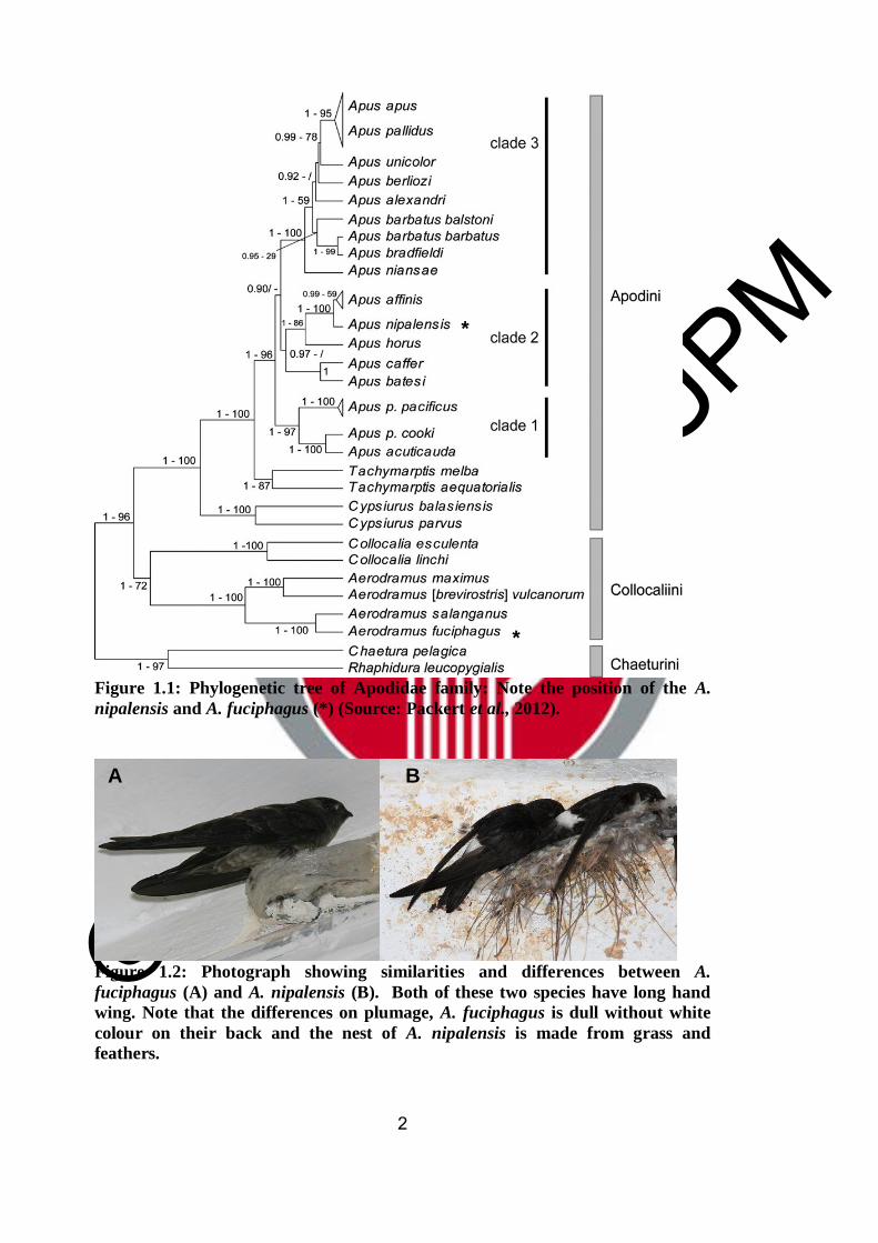
Both the House Swift (A. nipalensis) and the White Edible Bird's-nest Swiftlet (A.
fuciphagus) belong to the order Apodiformes, suborder Apodi and family Apodidae
(Figure 1.1) (Chantler and Driessen, 1995; 2000; Packert et al., 2012; Bird Life
International, 2013). These two species are aerial insectivorous birds where the insects
their diet (Zhou, 2002). Unlike the White edible bird's-nest, the House Swift, also
known as Malay House Swift (A. nipalensis), is categorized as the least concerned
bird (Camfield, 2004; BirdLife Int. 2013) and is a native bird of the country in South
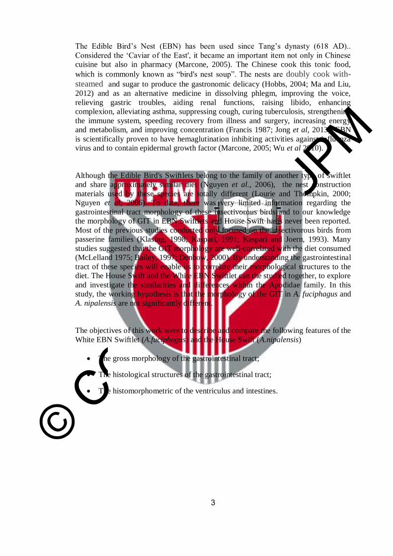
East Asia and East Asia (BirdLife Int. 2013). There are also some similarities and
differences between A. fuciphagus and A. nipalensis. For instance, both of these two
species have long hand-wing., A. fuciphagus has dull plumage without white color on
their back and the nest of A. nipalensis is made from grass and feathers (Figure 1.2).
© COPYRIG
HT UPM
2
Figure 1.1: Phylogenetic tree of Apodidae family: Note the position of the A.
nipalensis and A. fuciphagus (*) (Source: Packert et al., 2012).
Figure 1.2: Photograph showing similarities and differences between A.
fuciphagus (A) and A. nipalensis (B). Both of these two species have long hand
wing. Note that the differences on plumage, A. fuciphagus is dull without white
colour on their back and the nest of A. nipalensis is made from grass and
feathers.
*
*
A B
© COPYRIG
HT UPM
3
The Edible Bird’s Nest (EBN) has been used since Tang’s dynasty (618 AD)..
Considered the ‘Caviar of the East', it became an important item not only in Chinese
cuisine but also in pharmacy (Marcone, 2005). The Chinese cook this tonic food,
which is commonly known as “bird's nest soup”. The nests are doubly cook with-
steamed and sugar to produce the gastronomic delicacy (Hobbs, 2004; Ma and Liu,
2012) and as an alternative medicine in dissolving phlegm, improving the voice,
relieving gastric troubles, aiding renal functions, raising libido, enhancing
complexion, alleviating asthma, suppressing cough, curing tuberculosis, strengthening
the immune system, speeding recovery from illness and surgery, increasing energy
and metabolism, and improving concentration (Francis 1987; Jong et al, 2013). EBN
is scientifically proven to have hemaglutination inhibiting activities against influenza
virus and to contain epidermal growth factor (Marcone, 2005; Wu et al 2010).
Although the Edible Bird's Swiftlets belong to the family of another type of swiftlet
and share approximately similar diet (Nguyen et al., 2006), the nest construction
materials used by these species are totally different (Lourie and Thompkin, 2000;
Nguyen et al, 2006). To date, there was very limited information regarding the
gastrointestinal tract morphology of these insectivorous birds and to our knowledge
the morphology of GIT in EBN Swiftlets and House Swift hasis never been reported.
Most of the previous studies conducted only focused on the insectivorous birds from
passerine families (Klasing, 1998; Kaspari, 1991; Kaspari and Joern, 1993). Many
studies suggested that the GIT morphology are well-correlated with the diet consumed
(McLelland 1975; Bailey, 1997; Denbow, 2000). By understanding the gastrointestinal
tract of these species will enable us to correlate their morphological structures to the
diet. The House Swift and the White EBN Swiftlet can the studied together, to explore
and investigate the similarities and differences within the Apodidae family. In this
study, the working hypothesis is that the morphology of the GIT in A. fuciphagus and
A. nipalensis are not significantly different.
The objectives of this work were to describe and compare the following features of the
White EBN Swiftlet (A.fuciphagus) and the House Swift (A.nipalensis)
The gross morphology of the gastrointestinal tract;
The histological structures of the gastrointestinal tract;
The histomorphometric of the ventriculus and intestines.

© COPYRIG
HT UPM
84
REFERENCES
Adnyane, I. K. M., Zuki, A.B., Noordin, M.M., and Agungpriyono, S. (2011).
Morphological study of the lingual papillae in the barking deer, Muntiacus
muntjak. Anatomia, Histologia, Embryologia, 40(1), 73-77
Akester A.R. (1986). Structure of the glandular layer and koilin membrane in the
gizzard of the adult domestic fowl (Gallus gallus domesticus). Journal of
Anatomy, 147, 1–25.
Aitken, R. N. C. (1954). A histochemical study ofthe stomach and intestine of the
chicken. Journal of Anatomy, 92(1932), 453 – 470.
Al-Mansour, M.I. and Jarrar, B.M. (2007) Morphological, histological and
histochemical study of the lingual salivary glands of the Little Egret, Egretta
garzetta. Saudi Journal of Biological Sciences, 14: 75–81
Ankney, C.D and Scott, S.M (1998) Size of digestive organs in breeding Brown-
headed cowbird, Molothrus ater relative to diet. Canadian. Zoology. 66: 1254-
1257
Aptekmann, K.P., Baraldi Arton, S.M., Stefanini, M.A. and Orsi, M.A. (2001).
Morphometric analysis of the intestine of domestic quails (Coturnix coturnix
japonica) treated with different levels of dietary calcium. Anatomia, Histologia,
Embryologia, 30(5), 277–80.
Arthitvong, S., Makmee, N. and Suprasert, A. (1999). Histochemical detection of
glycoconj- ugates in the anterior lingual salivary glands of the domestic fowl.
Kasetsart Journal of Natural Science, 250, 243–250.
Bacha, J. W. and Bacha, M. L. (2000) Color Atlas of Veterinary Histology, 2nd edn.
Philadelphia, PA: Lippincott Williams and Wilkins
Bailey, T. A., Mensah-Brown, E. P., Samour, J. H., Naldo, J., Lawrence, P. and
Garner, A. (1997). Comparative morphology of the alimentary tract and its
glandular derivatives of captive bustards. Journal of Anatomy, 191(3), 387–98.
Bancroft, J.D. and Gamble, M. (2008). Theory and Practice of Histological
Techniques (6th ed.). Edinburgh, UK: Churchill Livingstone.
Baranylova, E. and Holman, J. (1976) Morphological changes in the intestinal wall in
fed and fasted chickens in the first week after hatching. Acta Veterinaria Brno,
45: 151–158.
Baumel, J.J., King, A.S., Breazile, J.E., Evans, H.E., and Berge, J. C. (Ed.). (1993).
Baumel Handbook of Avian Anatomy: Nomina Anatomica Avium. 2nd
ed.Massachusetts: Cambridge, MA. Nuttal Ornithology Club. (pp. 1–401)
BirdLife International. (2013) Species factsheet :Apus nipalensis. Downloaded from
http://www.birdlife.org on 31/1/2013
© COPYRIG
HT UPM
85
Bjerkness, M. and Cheng, H. (2005) Gastrointestinal stem cell II. Intestinal Stem
Cells. American Journal Physiology Gastrointestinal Liver Physiology. 289:
G381-G387
Bock, P. (1978) Pancreatic duct glands. I. Staining reation of acid glycoprotien secret.
Acta Histochemica. 61:118-126
Bouwens, L., Knook, D.L. and Wisse, E. (1986) Local proliferation and extrahepatic
recruitment of Kupffer cells in partial-body irradiated rats. Journal of Leucocyte
Biology 39:687-697
Boonzaier, J., Van der Merwe, E. L., Bennett, N. C. and Kotzé, S. H. (2013). A
comparative histochemical study of the distribution of mucins in the
gastrointestinal tracts of three insectivorous mammals. Acta Histochemica,
115(6), 549–56.
Brooke, R.K. (1970) Taxonomic and evolutionary notes on the subfamilies, tribes,
genera and subgenera of swifts (Aves: Apodidae). Durban Mus. Novit. 9, 13–24.
Brooke, R.K. (1972). Generic limits in old world Apodidae and Hirundinidae.
Bulletin of British. Ornithologists Club 92, 53–57.
Burns, R. B. (1982). Histology and Immunohistology of Peyer’s patches in the
domestic fowl (Gallus domesticus). Research of Veterinary Science. 32: 359-367
Camfield, A. (2004). Apodidae (Online) Animal Diversity Web. University of
Michigan Museum of Zoology. Retrieved March 26, 2012, from
http://animaldiversity.ummz.edu/site/accounts/information/Apodidae.html
Caspary, W.F. (1992) Physiology and pathophysiology of intestinal absorption.
American Journal of Clinical Nutrition. 55: 299-308
Catroxo, M. H. B., Lima, M. A. I., and Cappellaro, C.E.M.P.D.M. (1997).
Histological aspect of the stomach (Proventriculus and Gizarrd) of the red-capped
cardinal (Paroaria gularis gularis, Linnaeus, 1766). Revista Chilena de
Anatomía, 15(1)
Caviedes-Vidal, E., Afik, D., Martinez del Rio, C., and Karasov, W. H. (2000).
Dietary modulation of intestinal enzymes of the house sparrow (Passer
domesticus): testing an adaptive hypothesis. Comparative Biochemistry and
Physiology. Part A, Molecular and Integrative Physiology, 125(1), 11–24.
Chantler, P. and Driessens, G. (1995). Swifts : A guide to the Swifts and Treeswift of
the World. East Sussex : Pica Press.
Chantler, P., and Driessens, G. (2000). Swifts: A Guide to the Swifts and Treeswifts the
World. In: Del Hoyo J, Elliott A, Sargatal, J. (ed) (2nd Edn., Vol. 118). Sussex:
Pica Press.

© COPYRIG
HT UPM
86
Cherry, J.A., Nir, I., Jones, D.E., Dunnington, E.A., Nitsan, Z. and Siegel, P.B. (1987)
Growth associated traits in parental and F1 populations of chickens under
different feeding program 1. ad libitum feeding. Poultry Science 66:1-9
Chihtung, K. (1980). Food Analysis of the House Swift (Apus affinis subfurcatus) Zoological Research 1 (2): 247 -255 (In Chinese).
Chikilian, M. and De Speroni, N. B. (1996). Comparative study of the digestive
system of three species of Tinamou. Crypturellus tataupa,
Nothoproctacinerascens and Nothura maculosa (Aves: Tinamidae). Journal of
Morphology, (228), 77–88.
Clench, M. H., and Mathias, J. R. (1995). The Avian Cecum: A review. Wilson
Bulletin, 107(l), 93–121.
Collen, T.D., Olaf, J.W. and Micheal, J.L. (2000) Anatomical and nutritional
adaptation of the Speckled Mousedird (Colius striatus) The Auk 117(3):791-794
Cooper, J.E. and Harrison, G. J. (1994). Dermatology. In L. Ritchie BW, Harrison, GJ,
Harrison (Ed.), Avian Medicine: Principles and Application. (pp. 607–639). Lake
Worth, FL: Winger Publishing.
Cranbrook, Earl of. and Medway, L. (1965). Lack of ultrasonic frequencies in the calls
of swiftlet. Ibis, 107, 258.
Creamer, B. (1967) The turnover of the epithelium of small intestine. British Medical
Bulletin. 23. 226-230
Creamer, B., G. Shorter, and J. Bamforth. (1961). The turnover and shedding of
epithelial cells. I. The turnover in the gastrointestinal tract. Gut 2:110–118.
Danford, B.R., Knabe, D.A. and Haensly W.E. (1989) Effect of soybean on
microscopic anatomy of small intestine in early-weaned pig. Journal of Animal
Science. 67. 1855-1863
De Conto, C., Oevermann, A., Burgener, I.A., Doherr, M.G. and Blum, J.W.(2010).
Gastrointestinal tract mucosal histomorphometry and epithelial cell proliferation
and apoptosis in neonatal and adult dogs. Journal of Animal Science, 88(7),
2255–64.
De Graff, R.M., Tilghman, N.G. and Anderson, S.H. (1985) Foraging guilds of North
American birds. Environmental Management.9:492-536
Devine, P.L. and McKenzie, I. F. (1992). Mucins: structure, function, and associations
with malignancy. Bioessay, 14(9), 619–625.
Denbow, D. M. (2000). Gastrointestinal Anatomy and Physiology. In G. C. Whittow
(Ed.), Sturkie’s Avian Physiology.(pp299-325) 5th Edition. San Diego,
California: Academic Press.
Dibner, J. J. and Richards, J. D. (2004). The Digestive System: Challenges and
Opportunities. The Journal of Applied Poultry Research. 13, 86– 93.

© COPYRIG
HT UPM
87
Duke, G. E. (1982). Gastrointestinal motility and its regulation. Poultry. Science. 61,
1245–1256.
Dolinsky V.W., Gilham D, Alam, M., Vance, D.E., Lehner, R. (2004). Triglycerol
hydrolase: role in intracellular lipid metabolism. Cellular and Molecular Life
Science, 61(13), 1633–1651.
Duritis, I., and Mugurevics, A. (2011). Morphometric parameters of the small and
large intestine of the ostrich (Struthio camelus Var . Domesticus ) from day 38 of
embrionic development to the age of 60 days. Proceeding of the Latvia
University of Agriculture, 26(321), 84–93.
Eglitis I. and Knouff R.A. (1962). An histological and histochemical analysis of the
inner lining and glandular epithelium of the chicken gizzard.” American Journal
of Anatomy 111.1 49–65.
El-Bakary, N.E. (2011). Surface morphology of the tongue of the hoopoe (Upupa
Epops). Journal of American Science, 7(1), 394–399.
El-Galil, A.Y., Kamel, G. and El-Magd, A. A. A. (2011). Histomorphological studies
on the stomach of the japanese quail. Asian Journal. of Poultry Science, 5(2), 56–
67.
Emad, M. A. (1987). Development of the liver in the chicken embryo. Journal of
Anatomy, (150), 181–189.
Emura, S., Okumura, T. and Chen, H. (2008) Scanning electron microscopic study of
the tongue in peregrine falcon and common kestrel. Okajimas Folia Anatomica
Japonica. 85(1):11-15
Emura, S. Okumura, T. and Chen, H. (2009): Scanning electron microscopic study of
the tongue in the Japanese pygmy woodpecker (Dendrocopes kizuki). Okajimas
Folia Anatomica Japonica 86(1): 31-35.
Erdogan, S., Sagsoz, H., Akbalik, M. E. (2012). Anatomical and histological structure
of the tongue and histochemical characteristics of the lingual salivary glands in
the Chukar partridge. British Poultry Science, 53(3), 307–315.
Ferraris, R.P., Villenas, S.A. and Diamond, J. (1992) Regulation of brush-border
enzyme activities and enterocyte migration rates in mouse small intestine.
American Journal of Physiology, 262: G1047–1059.
Fernando, F.N, Claudio, V., Victoria, M and Lopez-Calleja (1996). Seasonal changes
in diet, digestive morphology and digestive efficiency in the Rufous-Collared
Sparrow (Zonotrichia capensis) in Central Chile. The Condor, 98, 873–876.
Filipe, M. (1979). Mucins in the human gastrointestinal epithelium: A review.
Investigative Cell Pathology, (2), 195–216.
FAO. (2007). Wild Birds and Avian Influenza: an introduction to applied field
research and disease sampling techniques.In: D. Whitworth, S.H. Newman, T.
© COPYRIG
HT UPM
88
Mundkur and P. Harris (ed). FAO Animal Production and Health Manual, No. 5.
Rome.
FAO (2011) Total world production of agricultural commodities. FAOSTAT.
http://faostat.fao.org/site/339/default.aspx
Forstner, J.F. (1978) Intestinal mucins in health and disease. Digestion, 17, 234–263.
Francis, C. M. (1987). The management of edible bird's nest caves in Sabah.
Sandakan: Wildlife Section, Sabah Forest Department.
Furlan, R.L., Carvalho, N.C., Malheiros E.B. and Macari, M. (2001). Effect of early
quantitative feed restriction and environmental temperature on viscera growth
and compensatory gain of broiler chickens. Arquivo Brasileiro de Medicina
Veterinária e- Zootecnia 53(4): 1-7
Gabella, G. (1985). Anatomy and embryology structure of the musculature of the
chicken small intestine. Anatomy and Embryology, (171), 139–149.
Geyra, A., Uni, Z. and Sklan, D. (2001) Enterocyte dynamics and mucosal
development in the post-hatch chick. Poultry Science 80:776-782
Getty, R. (1975) In Sisson and Grossmans. The anatomy of the domestic animals. Vol.
2 5th Ed. London. W.B. Saunders.
Gionfriddo, J.P. and Best, L.B. (1996) Grit use pattern in North American birds : The
influence of diet, body size and gender. Wilson Bulletin 108:685-696
Goh, D. L. M., Chua, K.-Y., Chew, F.T., Seow, T. K., Ou, K. L., Yi, F. C. and Lee, B.
W. (2001). Immunochemical characterization of edible bird’s nest allergens.
Journal of Allergy and Clinical Immunology, 107(6), 1082–1088.
Goralski, A., Sawicki, W. and Blaton, O. (1975) Non-random distribution of goblet
cells around the circumference of colonic crypts. Cell Tissue Research, 160: 551-
556
Garcia-del-Rey, E., Collins, C. T. and Volpone, N. W. (2010). Food composition of
the endemic Plain Swift Apus unicolor in the Canary Islands ( Macaronesia ).
Ardea, 98(2), 211–215.
Guillemette, M. (1994) Digestive role constrains in wintering common eiders
(Somateria mollissima) : Implications for flying capabilities. Auk III: 900-909
Gulmez, N. (2003). Are glands present in the goose pancreatic ducts? Journal of the
Pancreas, 4, 125–8.
Gussekloo, S. W. S. (2006). Feeding structures in birds In V. Bels (ed.), Feeding in
domestic vertebrates: from structure to behavior (pp. 14–30). London. CAB
Publishing.
© COPYRIG
HT UPM
89
Hall, P.A., Coates, P.J., Ansari, B. and Hopwood, D. (1994) Regulation of cell
number in mammalian gastrointestinal tract : The importance of apoptosis.
Journal Cell Science. 107: 3569-3577
Hails, C.J. and Amirudin, A. (1981). Food samples and selectivity of White bellied
Swiftlet (Collocalia esculata). Ibis, 123(3), 328–333.
Hails, C.J. and Turner, A. K. (1985). The role of fat and protien during breeding in the
White-bellied Swiftlet (Collocalia esculenta). Journal of Zoology (206), 469–
484.
Hammond, K., Konarzewski, M., Torres, R. and Diamond, J.M (1994) Metabolic
celiling under a combination of peak energy demands. Physiological Zoology.
67:1479-1506
Harrison, J.G. (1964) Tongue. In Thomson (ed), A New Dictionary of Birds. London.
Nelson.
Harold, E. (1992). The gastrointestinal tract. In Clinical Anatomy A Revision and
Applied Anatomy for Clinical Students and Junior Doctors (pp. 73– 97). Victoria.
Blackwell Publishing.
Hassan, S.M., Moussa, E.A. and Cartwright, A.L. (2010) Variation by sex in
anatomical and morphological features of the tongue of Egyptian goose
(Alopochen aegyptiacus). Cells Tissues Organs 191:161-165
Herdt, T. (1993) Metabolismo/Fisiologia gastrointestinal. In Tratado de Fisiologia
Veterinaria (Cunningham J., ed) Rio de Jeneiro. Guanabara Koogan 201-222
Hill, K. J. (1971). The structure of the alimentary tract. In B. M. Bell, D.J., Freeman
(Ed.), Physiology and Biochemistry of the Domestic Fowl (pp. 1–23). London
Academic Press.
Hobbs, J. J. (2004). Problems in the harvest of edible bird's nests in Sarawak and
Sabah, Malaysian Borneo. Biodiversity and Conservation, 13(12), 2209–2226.
Hodges, R. D. (1974). The histology of the fowl. Massachusetts, Academic Press.
Homberger, D.G., Brush, A. H., and Rouge, B. (1986). Functional-morphological and
biochemical correlations of the keratinized structures in the African grey Parrot
Psittacus erithacus (Aves). Zoomorphology, (106), 103–114.
Homberger, D.G. and Meyer, R.A. (1989) Morphology of the lingual apparatus of the
domestic chicken Gallus gallus with special attention to the surface of the
Fasciae. American Journal of Anatomy 186, 217-257
Iji, P. A., Saki, A. and Tivey, D.R. (2001). Body and intestinal growth of broiler
chicks on a commercial starter diet. 1. Intestinal weight and mucosal
development. British Poultry Science, 42(4), 505–13.
© COPYRIG
HT UPM
90
Incharoen, T., Yamauchi, K., Erikawa, T. and Gotoh, H. (2010). Histology of
intestinal villi and epithelial cells in chickens fed low protein or low fat diets.
Italian Journal of Animal Science, 9(4).
Ishikawa, K., Matoba, M., Tanaka, H. and Ono, K. (1985) Anatomical study of the
insect-feeder bats, Myotis frater kaguae. Journal of Anatomy. 142 141-150
Iwasaki, S. (1992) Fine structure of the dorsal lingual epithelium of the little tern.
Sterna albifrons Pallas (Aves, Lari). Journal of Morphology 212: 13–26
Iwasaki, S. (2002). Evolution of the structure and function of the vetebrate tongue.
Journal of Anatomy, (201) 1–13.
Iwasaki, S., Asami,T. and Chiba, A. (1997) Ultrastructural study of the keratinization
of the dorsal epithelium of the tongue of Middendorff’s bean goose, Anser fabalis
mid- dendorffii (Anseres, Antidae). The Anatomical Record, 247: 149–163.
Iwasaki, S. and Kobayashi, K. (1986) Scanning electron microscopical studies on the
lingual dorsal epithelium of chickens. Acta Anatomica Nipponica. 61:83-96
Jackowiak, H. and Ludwig, M (2008) Light and scanning electron microscopic study
of the structure of the ostrich (Strutio camelus) tongue. Zoological Science
25:188-194
Jackowiak, H., Skieresz-Szewczyk, K., Kwieciński, Z., Trzcielińska-Lorych, J., and
Godynicki, S. (2010). Functional morphology of the tongue in the nutcracker
(Nucifraga caryocatactes) Zoological Science, 27(7), 589–94.
Jackowiak, H., Andrazejewki, W. and Godynicki, S. (2006). Light and scanning
electron microscopic study of the tongue in cormorant phalacrocorax carbo
(phalacrocoracidae, Aves). Zoological Science, (23), 161–167.
Jackowiak, H. and Godynicki, S. (2005). Light and scanning electron microscopic
study of the tongue in white tailed eagle (Haliaeetus albicilla, Accipitridae,
Aves). Anatomischer Anzeiger, (187), 251–259.
Jong, C. H., Tay, K. M. and Lim, C. P. (2013). Application of the fuzzy Failure Mode
and Effect Analysis methodology to edible bird nest processing. Computers and
Electronics in Agriculture, 96, 90–108.
Kadhim, K. K., Zuki, A. B. Z., Noordin, M. M., and Babjee, S. M.A. (2011).
Histomorphology of the stomach, proventriculus and ventriculus of the red jungle
fowl. Anatomia, Histologia, Embryologia, 40(3), 226–33.
Khadim, K.K. (2012). Morphological Evaluation of the Digestive System of the Red
Jungle Fowl, Village Chicken and Broiler Chicken. PhD Thesis. UPM
Kadir, I. (2012). Swiftlet nest farming: Government perspective on issues and
challenges. Edible Bird’s-Nest International Conference. 26th - 27th Nov 2012.
Putrajaya Marriot Hotel. Pg 27
© COPYRIG
HT UPM
91
Kang, N. and Lee, P.G. (1991). The Edible-nest Swiftlets Aerodramus spp. Natural
Malaysiana 16: 44-51.
Kang, N., Hails, C.J. and Sigurdsson, J. (1991). Nest construction and egg-laying in
Edible Nest swiftlet Aerodramus spp and implication for harvesting. Ibis, 133(2),
170–170.
Karasov, W.H. and McWilliams, S.R. (2005). Digestive constraints in mammalian and
avian ecology. (Pp. 87–112) In: J.M. Starck and T. Wang, (eds). Physiological
and Ecological Adaptations to Feeding in Vertebrates. Science, Enfield, NH
Kaspari, M. (1991) Prey preparation as a way that grasshopper sparrow (Amodramus
savannarum) increase the nutrient concentration of their prey. Behavioral
Ecology 2, 234-241
Kaspari, M. and Joern, A. (1993) Prey choice by three insectivorous grassland birds -
reevaluating opportunism. Oikos 68,414-430
King, A. and McLelland J. (1979) Digestive system. In King AS, McLelland J. (ed.)
Form and Function in Birds, (pp. 69–181). London: Academic Press.
Kitagawa, H., Hiratsuka, Y., Imagawa, T. and Uehara, M. (1998). Distribution of
lymphoid tissue in the caecal mucosa of chickens. Journal of Anatomy, 192, 293–
298.
Kitagawa, H., Imagawa, T., Uehara, M. (1996). The apical caecal diverticulum of the
chicken identified. Journal of Anatomy, 189, 667–672.
Klasing, C.K. (1999). Avian Gastrointestinal Anatomy and Physiology. Avian
Gastrointestinal Anatomy and Physiology (pp. 42–50). New York. Saunders
Company.
Klasing C.K. (1998). Dietary patterns In Comparative Avian Nutrition (pp. 1–8).
Wallingford, Oxon, UK: CAB International.
Konishi, M., Emlen, S.T., Ricklefs, R.E. and Wingfield, J.C. (1989) Contribution of
bird studies to biology. Science 246, 465-472
Koon, L.C. (2000). Features – Bird’s nest soup – Market demand for this expensive
gastronomic delicacy threatens the aptly name edible-nest Swiftlet with
extinction in the east. Wildlife Conservation, 103(1), 30-35
Koon, L.C. and Cranbrook, Earl of (2002). Swiftlets of Borneo – builders of edible
nests (pp. 1-171). Sabah, Malaysia : Natural History Publication (Borneo) Sdn
Bhd
Kotzé SH, Coetzee, H.L. (1994). A histocytochemical study of mucus glycoproteins or
mucins in the intestinal tract of the African elephant (Loxodonta africana).
Onderstepoort Journal of Veterinary Research., 61(2), 177–181.

© COPYRIG
HT UPM
92
Langham, N. (1980). Breeding biology of the Edible-nest swiftlet Aerodramus
fuciphagus. Ibis, 122(4), 447–461.
Langhout, D.J., Schutte, J.B., Van Leewen, P., Wiebenga, J. and Tamminga, S. (1999)
Effect of dietary high and low-methylated citrus pectin on the activity of ileal
microflora and morphology of the small intestinal wall of broiler chicks. British
Poultry Science. 40: 340-347
Langlois, I. (2003). The anatomy, physiology, and diseases of the avian proventriculus
and ventriculus. The Veterinary Clinics of North America. Exotic Animal
Practice, 6(1), 85–111.
Lasiewski, R.C. and Dawson, W. R. (1967). A re-examination of the relation between
standard metabolic rate and body weight in birds. The Condor, 69(1), 13–23.
Lau, A.S.M. and Melville, D.S (1994) International trade in swiftlet nest with special
reference to Hong Kong. Cambridge .Traffic International.
Lavin, S. R., Karasov, W. H., Ives, A. R., Middleton, K. M., and Garland, T. (2008).
Morphometrics of the avian small intestine compared with that of nonflying
mammals: a phylogenetic approach. Physiological and biochemical zoology
81(5), 526–50.
Lee, P. M., Dale, H. C., Griffiths, R., and Page, R. D. M. (1996). Does behaviour
reflect phylogeny in swiftlets (Aves: Apodidae)? Proceedings of the National
Academy of Sciences of the United States of America, 93(14), 7091–7096
Lee, P. G. and Kang, N. (1994). The reproductive strategies of edible-nest swiftlets
(Aerodramus spp .). Bulletin of The British Ornithologists’ Club, 114, 106–113.
Lein, M. (1972) A trophic comparison of avifauna. Systematic Zoology, (21), 135–
150.
Liman, N., Bayram, G., and Koçak, M. (2001). Histological and histochemical studies
on the lingual, preglottal and laryngeal salivary glands of the Japanese quail
(Coturnix coturnix japonica) at the post-hatching period. Anatomia, Histologia,
Embryologia, 30(6), 367–73.
Lourie, S. A., and Tompkins, D. M. (2000). The diets of Malaysian swiftlets. Ibis,
142(4), 596–602.
Lucas, F.A. (1896) The taxonomic value of the tongue in birds. The Auk 13(2) 109-
115
Lucas, A.M. and Dennington, E.M. (1956) Morphology of chicken liver. Poultry.
Science. 35:793-806
Liu J.W., Evans, H., Larsen, P., Pan, D., Xu, S.Z, Dong, H.C., Deng, X.B. and Wan,
B. G. (1998). Gross anatomy of the pancreatic lobes and duct in six breed of
domestic ducks and six species of wild duck in China. Anatomia, Histologia,
Embryologia, 27(6), 413–417.
© COPYRIG
HT UPM
93
Ma, F., and Liu, D. (2012). Sketch of the edible bird’s nest and its important
bioactivities. Food Research International, 48(2), 559–567.
Madkour, G.A., Hammouda, E.M. and Ibrahim, I.G. (1982) Histology of the
alimentary tract of the common Egyptian bats. Annal of Zoology 19(2) 53-73
Malewitz, T.D. and Calhoun, M. (1958). The gross and microscopic anatomy of the
digestive tract, spleen, kidney, lung and heart of the turkey. Poultry Science, 37,
388–398.
Manchi S.S. (2009). Breeding Ecology of the Edible-nest Swiftlet Aerodramus
fuciphagus and the Glossy Swiftlet Collocalia esculenta in the Andaman Islands,
PhD Thesis. India. Bharathiar University, Coimbatore.
Maneewan, B. and Yamauchi, K. (2004) Intestinal villus recovery in chicken reefed
semi purified protein, fat, fiber-free pellet diet. British Poultry Science. 45: 163-
170
Marchini, C. F. P., Silva, P. L., Nascimento, M. R. B. M. and Beletti, M. E. (2011).
Body weight , intestinal morphometry and cell proliferation of broiler chickens
submitted to cyclic heat stress. International Journal of Poultry Science, 10(6),
455–460.
Marcone, M. F. (2005). Characterization of the edible bird's nest the “Caviar of the
East”. Food Research International, 38(10),1125–1134.
Marsden, S.J. (1940). Weights and measurements of parts and organs of turkeys.
Poultry Science, 19(1), 23–28.
Marshall, A. J. and. Folley, S. J. (1956). The origin of nest cement in edible-nest
swiftlets. Proceeding of Zoological Society. 126, 383–389.
Martinez Del Rio, C., (1990). Sugar preferences in hummingbirds: the influence of
subtle chemical differences on food choice. The Condor 92, 1022–1030.
McLelland, J. (1975). Aves digestive system, In: Getty, R. (ed.), Sisson and
Grossman’s The Anatomy of the Domestic Animals, 5th ed. vol. 2 (pp. 1857–
1882.). Philadelphia, London, Toronto Saunders Company.
McLelland, J. (1989). Anatomy of the avian cecum. The Journal of experimental
zoology. Supplement, 3, 2–9.
McMinn, R.M.H. and Kugler, J. H. (1961). The glands of the bile and pancreatic
ducts: autoradiographic and histochemical studies. Journal of Anatomy. 95 1-11.
McWhorter,T.J., Caviedes-vidal, E. and Karasov, W.H. (2009) The integration of
digestion and osmoregulation in the avian gut. Biological Review 533-565
Medway, L. (1959). Echolocation among Collocalia. Nature, London, 184, 1352–
1535.

© COPYRIG
HT UPM
94
Medway, L. (1962). The relation between the reproductive cycle, moult and changes
in the sublingual salivary gland of swiftlet (Collocalia maxima, Hume).
Proceedings of the Zoological Society of London, 138, 305–315.
Micheal,R and Wojciech, P. (2011). Histology: a text and atlas: With correlated cell
and molecular biology (6th ed.). Philedelphia, USA: Lippincott William and
Wilkins.
Mitchell, P. C. (1901). On the intestinal tract of birds, with remarks on the valuation
and nomenclature of the zoological characters. Transactions of the Linnean
Society of London, Zoology, 8, 173–275.
Mitchell, M.A. and Smith, M.W. (1991). The effects of genetic selection for increased
growth rate on mucosal and muscle weights in the different regions of the small
intestine of the domestic fowl (Gallus domesticus). Comparative Biochemistry
and Physiology 99 : 251-258
Mitjans, M., Barniol, G. and Ferrer, R. (1997). Mucosal surface area in chicken small
intestine during development. Cell and Tissue Research, 290(1), 71–78.
Mitzner, S.R., Stange, J., Klammt, S., Peszynski, P., Schmidt, R. and Noldge-
Schomburg, G. (2001). Extracorporeal detoxification using the molecular
adsorbent recirculating system for critically ill patients with liver failure. Journal
of the American Society of Nephrology, 12 Suppl 1(28), S75–82.
Moniaux, N., Nollet, S., Porchet, N., Degand, P., Laine, A., and Aubert, J. (1999).
Complete sequence of the human mucin MUC4: a putative cell membrane-
associated mucin. Biochemistry Journal: 338, 325–333.
Montagne, L., Piel, C., and Lalle, J.P. (2004). Effect of diet on mucin kinetics and
composition: nutrition and health implications. Nutrition Reviews, 62(3), 105–
114.
Morse, D.H. (1975) Ecological aspects of adaptative radiation in birds. Biological
Reviews 50, 167-214
Mowry, R.W. (1956). Alcian blue techniques for histochemical study of acidic
carbohydrates. Journal of Histochemistry and Cytochemistry, (4), 403–407.
Nagy, K.A. (2001). Food requirements of wild animals: predictive equations for free-
living mammals, reptiles, and birds. Nutrition Abstracts and Reviews. B71, 21R–
31R
Nalavade, M. N. and A. T. Varute. (1977). Histochemical studies on the mucins of the
vertebrate tongues XI. Histochemical analysis of mucosubstances in the lingual
glands and taste buds of some birds. Acta. Histochemica, (60), 18–31.
Neutra, M.R., Fortsner, J. F. (1987). Gastrointestinal mucous: synthesis, secretion and
function. In L. R. Johanson (Ed.), Physiology of Gastrointesinal Tract, 2nd ed.,
(pp. 975–1009). New York: Raven Press.

© COPYRIG
HT UPM
95
Nguyen, P.Q., Voisin, J.F., and Lam, N.T. (2006). Biology of the house swift Apus
nipalensis (Hodgson) in Vietnam. Revue d’écologie, 61(4), 383–395.
Nguyen, P.Q. and Voisin, J. F. (1993). Influence of cave structure , microclimate and
nest harvesting on the breeding of the White-nest Swiftlet Collocalia fuciphaga
germani in Vietnam. Ibis, 140, 257–264.
Nickel, R., Schummer, A. and Seiferle, E. (1977). Anatomy of the domestic birds (pp.
40–61). Berlin and Hamburg. Willey & Sons
Nitsan, Z., Dunnington, E.A. and Siegel, P.B. (1991) Organs growth and digestive
enzyme levels to fifteen days of age in lines of chicken differing in body weight.
Poultry Science. 70:2040-2048
Nir, I., Nitsan, Z., Dror, Y. and Shapira, N. (1978) Influence of overfeeding on
growth, obesity, intestinal tract in young chicks of light and heavy breeds. British
Journal of Nutrition 39:27-35
Nir, I., Nitsan, Z. and Mahagna, M. (1993) Comparative growth and development of
the digestive organs and of some enzymes in broiler and egg type chicks after
hatching. British Poultry Science. 34(3):523-32.
Norberg, U.M. (1995) How long tail and changes in mass and wing shape affect the
cost for flight in animals. Functional Ecology. 9:48-54
Nudd, R.L. and Bryant, D.M. (2002) Consequences of load carrying by bird during
short flight and is found to be behavioral and energetic. American Journal
Physiology. 283: R249-R256
Ogiolda, L., Wanke, R., Rottmann, O., Hermanns, W. and Wolf, E. (1998).Intestinal
dimensions of mice divergently selected for body weight. The Anatomical Record.
250: 292-299.
Ogunkoya, Y. O., and Cook, R. D. (2009). Histomorphology of the proventriculus of
three species of Australian passerines: Lichmera indistincta, Zosterops lateralis
and Poephila guttata. Anatomia, Histologia, Embryologia, 38(4), 246–53.
Okon, E.E.(1977) Functional anatomy of the alimentary canal in the fruit bats Eidolon
helvum and the insect bats, Tadarida nigeriae. Acta Zoologica 58 83-93
O’Malley, B. (2005). Avian anatomy and physiology. In Bairbre O’Malley (Ed.),
Clinical Anatomy and Physiology of Exotic Species Structure and function of
mammals, birds, reptiles, and amphibians (pp. 97–161). Edinburgh. W.B.
Saunders.
Päckert, M., Martens, J., Wink, M., Feigl, A., and Thomas, D. (2012). Molecular
Phylogenetics and Evolution Molecular phylogeny of Old World swifts (Aves:
Apodiformes , Apodidae , Apus and Tachymarptis ) based on mitochondrial and
nuclear markers. Molecular Phylogenetics and Evolution, 63, 606–616.

© COPYRIG
HT UPM
96
Piersma, T., Koolhaas, A., and Dekinga, A. (1993). Interactions between Stomach
Structure and Diet Choice in Shorebirds. The Auk, 110(3), 552–564.
Piersma, T., Dietz, M.W., Dekinga, A., Nebel, S., Gils, J.V., Batley, P.F. and Spaans,
B. (1999). Reversible size-changes in stomach of shorebirds: when, to what
extent, and why. Acta Ornithologica, 34(2), 175–181.
Proust, J., Tchaliovska, S., and Ter Minassioan Sar, L. (1984). Mucin thin-film as a
model of the tear film rupture. Science, 98, 319.
PEMANDU. Agriculture Economic Transformation Programme: EPP2 - EBN
Swiftlet. Annual Report, Kuala Lumpur , 2011
Razak., N. (2010). The 2011 Budget Speech; Introducing The Supply Bill:
Transformation Towards A Developed and High Income Nation.
Rajabi, E., and Nabipour, A. (2009). Histological study on the oesophagus and crop in
various species of wild bird. Avian Biology Research, 2(3), 161–164.
Remington, T. E. (1989). Why do grouse have ceca? A test of the fiber digestion
theory. The Journal of experimental zoology. Supplement: published under
auspices of the American Society of Zoologists and the Division of Comparative
Physiology and Biochemistry / the Wistar Institute of Anatomy and Biology, 3,
87–94.
Rezaian, M., Shomali, T., and Branch, I. A. U. (2011). Histological changes of small
intestinal mucosa of cocks due to sunflower meal single feeding. American
Journal of Animal and Veterinary Science, 6(4), 171–175.
Ricklefs. R.E. (1996). Morphometry of the digestive tracts of some passerine birds.
The Condor, 98, 279–292.
Rocha, D.O.S. and De Lima, M. A. I. (1998). Histological aspects of the stomach of
Burrowing owl. Revista chilena de Anatomía, 16, 191–197.
Rossi, J.R., Baraldi-Artoni, S.M., Oliveira, D., Cruz, C. Da, Franzo, V. S., and Sagula,
A. (2005). Morphology of glandular stomach (Ventriculus glandularis) and
muscular stomach (Ventriculus muscularis) of the partrigde Rhynchotus
rufescens. Ciência Rural, 35(6), 1319–1324.
Rossi, J.R. Baraldi-Artoni, SM., Oliveira D., Cruz, C., Sagula, A., Pacheco, MR. and
Araujo, M. (2006). Morphology of oesophagus and crop of the partrigde
Rhynchotus rufescens ( Tiramidae ). Acta Scientarium, Biological Science, 28(2),
165–168.
Sağsöz, H., and Liman, N. (2009). Structure of the oesophagus and morphometric,
histochemical-immunohistochemical profiles of the oesophageal gland during the
post-hatching period of Japanese quails (Coturnix coturnix japonica). Anatomia,
Histologia, Embryologia, 38(5), 330–40.
© COPYRIG
HT UPM
97
Samar, M. E., Ávila, R. E., Esteban, F. J., Olmedo, L., Dettin, L., Massone, A. and
Pedrosa, J. A. (2002). Histochemical and ultrastructural study of the chicken
salivary palatine glands. Acta Histochemica, 104(2), 199–207.
Sankaran, R. (2001). The status and conservation of the Edible-nest Swiftlet
(Collocalia fuciphaga) in the Andaman and Nicobar Islands. Biological
Conservation, 97, 283–294.
Schaefer, C.M., Corsiglia, C.M., Mirales, A. and Koutsos, E.A. (2006) Turkey breeder
hen age affects growth and systemic and intestinal inflammatory responses in
female poults examines at different ages post-hatch. Poultry Science 85:1755-
1763
Scillitani, G., Zizza, S., Liquori, G. E. and Ferri, D. (2007). Lectin histochemistry of
gastrointestinal glycoconjugates in the greater horseshoe bat, Rhinolophus
ferrumequinum (Schreber, 1774). Acta Histochemica, 109(5), 347–57.
Selvan, P.S., Ushakumary. S. and Ramesh, G. (2008). Studies on the histochemistry of
proventriculus and gizzard post hatch Guinea Fowl (Numida meleagris).
International Journal of Poultry Science, 7(11), 1112–1116.
Shamoto K, Yamauchi K. (2000). Recovery responses of chick intestinal villus
morphology to different refeeding procedures. Poultry Science 79, 718–723.
Sheahan, D.G and Jervis, H. R. (1976). Comparative histochemistry of gastrointestinal
mucosubstances. American Journal of Anatomy, 146(2), 103–31.
Sherry, T.W. (1990) When are birds dietarily specialized? Distinguishing ecological
from evolutionary approaches. Studies in Avian Biology 13, 337-352
Sibley, C. G. and Monroe, B.L. (1990). Distribution and Taxonomy of the birds of the
world. Yale University Press. New Haven, USA
Sisson, S. and Grossman, J.D. (1953) The digestive system of the horse and
ruminants. In: The Anatomy of the Domestic Animal. 4th Eds. London. W.B.
Saunders.
Sklan, D. (2001). Development of the digestive tract of poultry. World’s Poultry
Science, 57, 415–428.
Smith, M.W., Mitchell, M.A. and Peacock, M.A. (1990) Effects of genetic selection
on growth rate and intestinal structure in the domestic fowl (Gallus domesticus).
Comparative Biochemistry and Physiology 97:57-63
Soler, J.J., Soler, M. and Martinez, J.G. (1993) Grit ingestion and cereal consumption
in five corvid species. Ardea 81:143-149
Spicer, S.S. and Meyer, D.B. (1960). Histochemical differentiation of acid
mucopolysaccharides by means of combined aldehyde fuchsin-alcian blue stain.
Technology Bulletin of Registered Medical Technology, (30), 53–60.
© COPYRIG
HT UPM
98
Suganuma, T., Katsuyama, T., Tsukahara M, Tatematsu, M., Sakakura, Y. and
Murata, F. (1981). Comparative histochemical study of alimentary tracts with
special reference to the mucous neck cells of the stomach. American Journal of
Anatomy. 161(2), 219–238.
Suprasert, A., Fujioka, T. and Yamada, K. (1987). The histochemistry of
glycoconjugates in the colonic epithelium of the chicken. Histochemistry, 86(5),
491–7.
Srisai, D., Juntaravimo, S., Pongkete, P., Koonjaenak, S. and Suprasert, A (2002).
Histological and histochemical studies on esophagus of the germain’s swiftlet
(Collocalia germani Oustalet, 1878). Journal of Kasetsart Veterinarians, 12(2),
16–21.
Strong, T.R., Reimer, P.R. and Braun, E. J. (1990). Morphometry of the galliform
cecum – A comparison between Gambel quail and domestic fowl. Cell and
Tissue Research, 259, 511–518.
Svihus, B. (2011). The gizzard: function, influence of diet structure and effects on
nutrient availability. World’s Poultry Science Journal, 67(02), 207–224.
Tivane, C. (2008). The morphology of the oral cavity, pharynx and esophagus of the
ostrict. MVSc Thesis. University of Pretoria, South Africa.
Toner, P.G. (1963). The fine structure of resting and active cells in the submucosal
glands of the fowl proventriculus. Journal of Anatomy, 97, 575–583.
Toner, P. G. (1964). The fine structure of gizzard gland cells in the domestic fowl.
Journal of Anatomy, 98, 77–85.
Trier, J. (1968). Morphology of the epithelium of the small intestines. In C.Code (ed)
Handbook of Physiology: Alimentary canal, Vol. III: Intestinal absorption. 1125-
1175. American Physiological Society.
Turk, D.E. (1982). The anatomy of the avian digestive tract as related to feed
utilization. Poultry Science, 61(7), 1225–1244.
Thomassen, H. ., Wiersema, A. T., De Bakker, Merijn, A. G., De Knijff., P.H., Elske,
P.G. and David, E. (2003). A new phylogeny of swiftlets (Aves: Apodidae) based
on cytochrome-b DNA. Molecular Phylogenetics and Evolution, 29(1), 86–93.
Thomassen, H. A. and Povel, G. D. E. (2006). Comparative and phylogenetic analysis
of the echo clicks and social vocalizations of swiftlets (Aves: Apodidae ).
Biological Journal of the Linnean Society, (88), 631–643.
Uni, Z., Noy, Y. and Sklan, D. (1996) Development of small intestinal function in the
poult. Poultry Science 78:215-222
Videler, J.J., Stamhuis, E.J. and Povel, G.D.E. (2004) Leading-edge vortex lifts swift.
Science 306:1960-1962
© COPYRIG
HT UPM
99
Waugh, E.E., Dzoma, B. M., Seabo, D., Aganga, A. A., Tsopito, C. M., Omphile, U. J.
and Sebolai, B. (2007). Gross adaptive morphologic changes occurring in the
gastrointestinal tract components of ostriches fed ration including or excluding
grit in Botswana. International Journal of Poultry Science, 6(4), 271–275.
Welty, J.C.(1962). The Life of Birds, Philadelphia. Saunders.
Wight, P.A.L. (1975). The occurrence of lipid in the oxynticopeptic cells of the
proventriculus of the fasting domestic fowl. Journal of Anatomy, 120(3), 485–94.
Wijtten, P. J. A. (2011). Nutrition driven small-intestinal development and
performance of weaned pigs and young broilers. PhD Thesis. Wageningen
University, The Netherland.
Wong, G.K and Cavey, M.J (1993) Development of liver in chicken embryo II.
Erythropoietic and granulopoeitic cells. The Anatomical Record. 235(1):131-43
Wu, Y.J., Chen, Y., Wang, B., Bai, L.Q., Wu, R. H. and Ge, Y.Q. (2010). Application
of SYBR green PCR and 2DGE methods to authenticate edible bird's nest food.
Food Research International, 43(8), 2020–2026
Yamauchi, K. (2007). Review of a histological intestinal approach to assessing the
intestinal function in chickens and pigs. Animal Science Journal, 78(4), 356–370.
Yamauchi, K. Yamamoto, K. and Isshiki, Y. (1995). Morphological alterations of the
intestinal villi and asorptive epithelial cells in each intestinal part in fasted
chickens. Japanese Poultry Science, 32, 241–251.
Yamauchi, K., Buwjoom, T., Koge, K. and Ebashi, T. (2006). Histological intestinal
recovery in chickens refed dietary sugar cane extract. Poultry Science, 85(4),
645–51.
Yamauchi, K.E., Lida, S. and Isshiki, Y. (1992) Post-hatching developmental changes
in the ultrastructure of the duodenal absorptive epithelial cells in 1, 10 and 60-d-
old chickens with special reference to mitochondria. British Poultry Science, 33:
475–488.
Yamauchi, K. and Tarachai, P. (2000) Changes in intestinal villi, cell area and
intracellular function in chickens. British Poultry Science 41: 416-423
Yasar, S. and Forbes, J.M. (1999) Performance and gastrointestinal response of broiler
chicken fed on cereal grain-based food soaked water. British Poultry Science.
40:65-76
Young, B., Lowe, J., Steven, A. and Heath, J. (2000). Wheather’s Functional
Histology: A text and colour atlas (5th ed.). China: Churchill Livingstone.
Yoshizawa, Shin-ichi (1978) Studies on pancreatic duct system. Gastroenterologia
Japonica 13(3):213-223

© COPYRIG
HT UPM
100
Yovchev, D., Dimitrov, R., Kostov, D. and Vladova, D. (2012) Age morphometry of
some internal organs in common pheasant (Phasianus colchicus colchicus).
Trakia Journal of Sciences 10(3): 48-52
Zavarize, K.C, , Sartori J.R, G. E. and P. A. . (2012). Morphological changes of the
intestinal mucosa of broilers and layers as affected by fasting before sample
collection. Brazilian Journal Poultry Science, 21–25.
Zhou, F. S. (2002). A study on bird communities of tropical forest at Jianfengling in
Hainan, China. PhD Thesis. Sun Yat-Sen University, Guangzhou. China.
Zabielski, R., Godlewski, M.M. and Guilloteau, P. (2008) Control of development of
gastointestinal system in neonates. Journal of Physiology and Pharmacology 59
Suppl. 1: 35-54
Zijlstra, R.T., Whang, K.Y., Easter, R.A. and Odle, J. (1996) Effect of feeding a milk
replacer to early-weaned pigs on growth, body composition, and intestinal
morphology, compared with suckled littermates. Journal of Animal Science.
74:2948-2959
Ziswiller, V. and Farners, D.S. (1972). Digestion and the digestive system In: Avian
Biology (D.S. Farner and J.R. King, Eds) Vol.2. New York and London.
Academic Press,