Gut Microbiota Modulation and Its Relationship with ... · Dinesh K. Dahiya1,Renuka2, Monica...
17
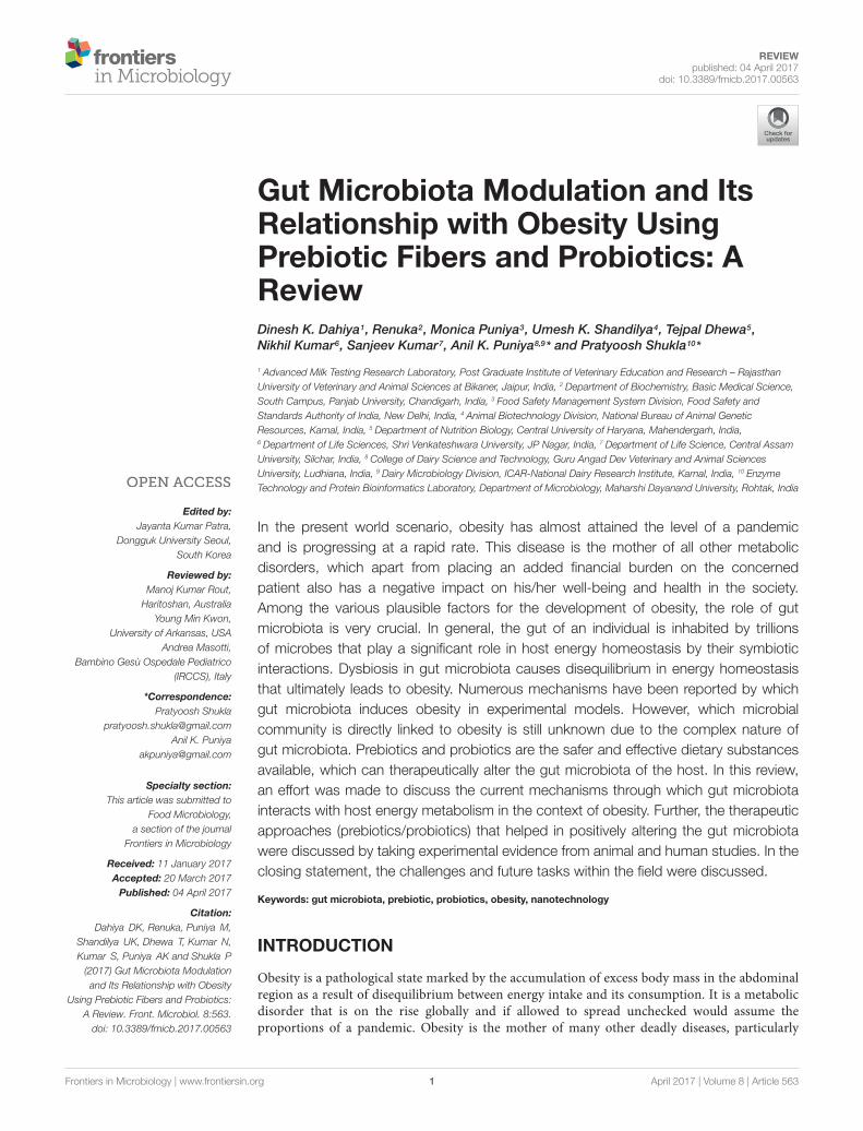
REVIEW published: 04 April 2017 doi: 10.3389/fmicb.2017.00563 Edited by: Jayanta Kumar Patra, Dongguk University Seoul, South Korea Reviewed by: Manoj Kumar Rout, Haritoshan, Australia Young Min Kwon, University of Arkansas, USA Andrea Masotti, Bambino Gesù Ospedale Pediatrico (IRCCS), Italy *Correspondence: Pratyoosh Shukla [email protected] Anil K. Puniya [email protected] Specialty section: This article was submitted to Food Microbiology, a section of the journal Frontiers in Microbiology Received: 11 January 2017 Accepted: 20 March 2017 Published: 04 April 2017 Citation: Dahiya DK, Renuka, Puniya M, Shandilya UK, Dhewa T, Kumar N, Kumar S, Puniya AK and Shukla P (2017) Gut Microbiota Modulation and Its Relationship with Obesity Using Prebiotic Fibers and Probiotics: A Review. Front. Microbiol. 8:563. doi: 10.3389/fmicb.2017.00563 Gut Microbiota Modulation and Its Relationship with Obesity Using Prebiotic Fibers and Probiotics: A Review Dinesh K. Dahiya 1 , Renuka 2 , Monica Puniya 3 , Umesh K. Shandilya 4 , Tejpal Dhewa 5 , Nikhil Kumar 6 , Sanjeev Kumar 7 , Anil K. Puniya 8,9 * and Pratyoosh Shukla 10 * 1 Advanced Milk Testing Research Laboratory, Post Graduate Institute of Veterinary Education and Research – Rajasthan University of Veterinary and Animal Sciences at Bikaner, Jaipur, India, 2 Department of Biochemistry, Basic Medical Science, South Campus, Panjab University, Chandigarh, India, 3 Food Safety Management System Division, Food Safety and Standards Authority of India, New Delhi, India, 4 Animal Biotechnology Division, National Bureau of Animal Genetic Resources, Karnal, India, 5 Department of Nutrition Biology, Central University of Haryana, Mahendergarh, India, 6 Department of Life Sciences, Shri Venkateshwara University, JP Nagar, India, 7 Department of Life Science, Central Assam University, Silchar, India, 8 College of Dairy Science and Technology, Guru Angad Dev Veterinary and Animal Sciences University, Ludhiana, India, 9 Dairy Microbiology Division, ICAR-National Dairy Research Institute, Karnal, India, 10 Enzyme Technology and Protein Bioinformatics Laboratory, Department of Microbiology, Maharshi Dayanand University, Rohtak, India In the present world scenario, obesity has almost attained the level of a pandemic and is progressing at a rapid rate. This disease is the mother of all other metabolic disorders, which apart from placing an added financial burden on the concerned patient also has a negative impact on his/her well-being and health in the society. Among the various plausible factors for the development of obesity, the role of gut microbiota is very crucial. In general, the gut of an individual is inhabited by trillions of microbes that play a significant role in host energy homeostasis by their symbiotic interactions. Dysbiosis in gut microbiota causes disequilibrium in energy homeostasis that ultimately leads to obesity. Numerous mechanisms have been reported by which gut microbiota induces obesity in experimental models. However, which microbial community is directly linked to obesity is still unknown due to the complex nature of gut microbiota. Prebiotics and probiotics are the safer and effective dietary substances available, which can therapeutically alter the gut microbiota of the host. In this review, an effort was made to discuss the current mechanisms through which gut microbiota interacts with host energy metabolism in the context of obesity. Further, the therapeutic approaches (prebiotics/probiotics) that helped in positively altering the gut microbiota were discussed by taking experimental evidence from animal and human studies. In the closing statement, the challenges and future tasks within the field were discussed. Keywords: gut microbiota, prebiotic, probiotics, obesity, nanotechnology INTRODUCTION Obesity is a pathological state marked by the accumulation of excess body mass in the abdominal region as a result of disequilibrium between energy intake and its consumption. It is a metabolic disorder that is on the rise globally and if allowed to spread unchecked would assume the proportions of a pandemic. Obesity is the mother of many other deadly diseases, particularly Frontiers in Microbiology | www.frontiersin.org 1 April 2017 | Volume 8 | Article 563
Transcript of Gut Microbiota Modulation and Its Relationship with ... · Dinesh K. Dahiya1,Renuka2, Monica...
fmicb-08-00563 March 31, 2017 Time: 16:28 # 1
REVIEWpublished: 04 April 2017
doi: 10.3389/fmicb.2017.00563
Edited by:Jayanta Kumar Patra,
Dongguk University Seoul,South Korea
Reviewed by:Manoj Kumar Rout,
Haritoshan, AustraliaYoung Min Kwon,
University of Arkansas, USAAndrea Masotti,
Bambino Gesù Ospedale Pediatrico(IRCCS), Italy
*Correspondence:Pratyoosh Shukla
[email protected] K. Puniya
Specialty section:This article was submitted to
Food Microbiology,a section of the journal
Frontiers in Microbiology
Received: 11 January 2017Accepted: 20 March 2017
Published: 04 April 2017
Citation:Dahiya DK, Renuka, Puniya M,
Shandilya UK, Dhewa T, Kumar N,Kumar S, Puniya AK and Shukla P
(2017) Gut Microbiota Modulationand Its Relationship with Obesity
Using Prebiotic Fibers and Probiotics:A Review. Front. Microbiol. 8:563.
doi: 10.3389/fmicb.2017.00563
Gut Microbiota Modulation and ItsRelationship with Obesity UsingPrebiotic Fibers and Probiotics: AReviewDinesh K. Dahiya1, Renuka2, Monica Puniya3, Umesh K. Shandilya4, Tejpal Dhewa5,Nikhil Kumar6, Sanjeev Kumar7, Anil K. Puniya8,9* and Pratyoosh Shukla10*
1 Advanced Milk Testing Research Laboratory, Post Graduate Institute of Veterinary Education and Research – RajasthanUniversity of Veterinary and Animal Sciences at Bikaner, Jaipur, India, 2 Department of Biochemistry, Basic Medical Science,South Campus, Panjab University, Chandigarh, India, 3 Food Safety Management System Division, Food Safety andStandards Authority of India, New Delhi, India, 4 Animal Biotechnology Division, National Bureau of Animal GeneticResources, Karnal, India, 5 Department of Nutrition Biology, Central University of Haryana, Mahendergarh, India,6 Department of Life Sciences, Shri Venkateshwara University, JP Nagar, India, 7 Department of Life Science, Central AssamUniversity, Silchar, India, 8 College of Dairy Science and Technology, Guru Angad Dev Veterinary and Animal SciencesUniversity, Ludhiana, India, 9 Dairy Microbiology Division, ICAR-National Dairy Research Institute, Karnal, India, 10 EnzymeTechnology and Protein Bioinformatics Laboratory, Department of Microbiology, Maharshi Dayanand University, Rohtak, India
In the present world scenario, obesity has almost attained the level of a pandemicand is progressing at a rapid rate. This disease is the mother of all other metabolicdisorders, which apart from placing an added financial burden on the concernedpatient also has a negative impact on his/her well-being and health in the society.Among the various plausible factors for the development of obesity, the role of gutmicrobiota is very crucial. In general, the gut of an individual is inhabited by trillionsof microbes that play a significant role in host energy homeostasis by their symbioticinteractions. Dysbiosis in gut microbiota causes disequilibrium in energy homeostasisthat ultimately leads to obesity. Numerous mechanisms have been reported by whichgut microbiota induces obesity in experimental models. However, which microbialcommunity is directly linked to obesity is still unknown due to the complex nature ofgut microbiota. Prebiotics and probiotics are the safer and effective dietary substancesavailable, which can therapeutically alter the gut microbiota of the host. In this review,an effort was made to discuss the current mechanisms through which gut microbiotainteracts with host energy metabolism in the context of obesity. Further, the therapeuticapproaches (prebiotics/probiotics) that helped in positively altering the gut microbiotawere discussed by taking experimental evidence from animal and human studies. In theclosing statement, the challenges and future tasks within the field were discussed.
Keywords: gut microbiota, prebiotic, probiotics, obesity, nanotechnology
INTRODUCTION
Obesity is a pathological state marked by the accumulation of excess body mass in the abdominalregion as a result of disequilibrium between energy intake and its consumption. It is a metabolicdisorder that is on the rise globally and if allowed to spread unchecked would assume theproportions of a pandemic. Obesity is the mother of many other deadly diseases, particularly
Frontiers in Microbiology | www.frontiersin.org 1 April 2017 | Volume 8 | Article 563

fmicb-08-00563 March 31, 2017 Time: 16:28 # 2
Dahiya et al. Gut Microbiota Modulation
diabetes, cardiovascular, non-alcoholic fatty liver disease(NAFLD) and some form of cancers (Kopelman, 2007;Nikolopoulou and Kadoglou, 2012; Vucenik and Stains,2012). Obesity not only affects the well-being of a person, butalso places an unwanted economic burden on the society (Wanget al., 2011; Withrow and Alter, 2011). According to a report,more than 500 million people across the world are living withthe stigma of obesity, that shows the severity of the diseaseand the challenges confronting health practitioners (Swinburnet al., 2011). Several factors such as host genetics, metabolism,lifestyle, and diet have been pinpointed as the key etiologicalagents responsible for the progression of obesity. However, thein-depth mechanisms that lead to the development of obesity areyet to be disclosed. The most recent studies have speculated thatthe gut microbiota present in the human gastrointestinal tract(GIT) have a paramount role in the onset and establishment ofobesity. The adhered gut microbiota affects the host’s nutrientsacquisition and energy homeostasis by influencing the number ofeffector molecules that finally decide the fat storage in adipocytes(Rosenbaum et al., 2015). Nonetheless, there is growing evidencethat some dietary substances, especially probiotics and prebioticscan modulate the gut microbiota of the host in a positive way andare therefore considered as important assets in the managementof obesity. Various approaches such as omics methods, systemsbiology and metabolic engineering enable us to understand andoptimize the metabolic processes (Yadav et al., 2016a,b). Themajor objectives of this review are to provide an overview of howprebiotics and probiotics modulate the gut microbiota in contextof prevention or treatment of obesity. Before we progress further,we elaborate our current understanding of how gut microbiotaare predisposed toward obesity.
RELATIONSHIP BETWEEN “GUTMICROBIOTA AND OBESITY”
Human Gut Microbiome, the“Unforeseen Organ”It is believed that the gut of a fetus during the intrauterineperiod is deprived of any bacterial communities, i.e., it isnearly sterile; however, some microbes before birth and duringparturition transit from the mother to the fetus gut andconstitute the rudimentary microbiota (Aagaard et al., 2014).The gut composition of a child varies widely during the firstfew years of life due to factors like changes in gut physiology,introduction of solid foods, use of therapeutic drugs, hostgenotype and proximity to adult microbiota (Koenig et al.,2011). During adolescence, however, the gut microbiota is nearlyconsistent and predominated by a few colonizers. Thereafter,it changes during old age when the host physiology anddietary habits change dramatically. Nevertheless, the dynamicsand structure of an individual’s gut microbiota is unique andpeople can actually be identified on the basis of microbiota“fingerprints” alone, with the help of the metagenomics approach(Franzosa et al., 2015). The gut harbors a trillion microbes,thereby constituting a complex microbial community that is
approximately comprised of 1000–1100 different bacterial speciesaltogether representing 1014–1015 microbes. This populationis 10 times the number of cells present in a eukaryotic host(Qin et al., 2010) and resemble a “world within a world.”The collective genes of these different microbial species aretermed as “microbiome,” while a combination of microbiome andhost genes is called “metagenome” (Quigley, 2011). Before theadvent of sophisticated sequencing techniques, the gut remaineda neglected organ because of the limitations of culturingmethods, but it is now considered to be a vital organ as ithelps in various metabolic functions of the host that wouldotherwise not be possible (Sommer and Bäckhed, 2013). Anearlier study inferred that the gut of an adult human being ismainly inhabited by bacteria from three major divisions, theFirmicutes (Gram-positive), Bacteroidetes (Gram-negative) andActinobacteria (Gram-positive), which together make up morethan 90% of total bacteria presented in the gut. In case of Archaea,one species Methanobrevibacter smithii predominates over others(Eckburg et al., 2005). However, obtaining an accurate picture ofthe gut is very difficult as several factors such as availability ofoxygen, diet, and physiochemical properties of the gut (e.g., pH,bile) rapidly influence its composition.
Arumugam et al. (2011) made an attempt to understandthe variation in species composition and gene pools within thehuman population from the previously available data and foundthe existence of three main distinct bacterial communities or“enterotypes” – Bacteroides, Prevotella, and Ruminococcus-basedon their abundance (Arumugam et al., 2011). Later studiesreduced the concept of three enterotypes to two – Bacteroides andPrevotella (Koren et al., 2013; Knights et al., 2014). From abovestudies, it can be inferred that gut microbiota have occupied asignificant position in human biology that interplays with themetabolic physiology and influences the health status.
Evidence that Gut Microbiota Have aRole in Obesity and DysbiosisThe pioneering evidence that linked gut microbiota to thedevelopment of obesity came from the findings of Bäckhed et al.(2004), when they transplanted the microbiota from normallygrown mice to germ free (GF) mice. The latter, consequently,gained more fat pad mass and body weight despite reductionin food consumption. Increased body weight led to insulinresistance, along with higher glucose and leptin levels in blood.The authors postulated that the transplanted microbiota helpedGF mice in harvesting excess energy from the diet. Further,they advocated that microbiota increases the expression of keytranscriptional factors to enhance lipogenesis in the liver andpromoted lipoprotein lipase (LPL) activity to store triglyceride(TG) in adipocytes (Bäckhed et al., 2004). Surprisingly, whenGF mice were maintained on a high fat diet (HFD), they wereprotected from the development of obesity. Interesting evidencein this context emerged from the effect of antibiotic experimentson body weight. Antibiotics have been used in the livestock sectorfor decades to promote the growth and body weight of animals,which indirectly indicate that role of the gut microbiota in weightmodulation. Evidence from mice has shown that early exposureto antibiotics had altered their gut microbiota, increased fat mass,
Frontiers in Microbiology | www.frontiersin.org 2 April 2017 | Volume 8 | Article 563
fmicb-08-00563 March 31, 2017 Time: 16:28 # 3
Dahiya et al. Gut Microbiota Modulation
and negatively modulated hepatic metabolism and associatedhormones, which predisposed them toward adiposity (Cho et al.,2012; Cox et al., 2014). The effect of early administration ofantibiotics on human adiposity has also been seriously reviewedover the past few years (Mueller et al., 2014; Turta and Rautava,2016; Podolsky, 2017) and there is growing consensus that theirincreased use maybe a reason for the obesity explosion we arewitnessing today.
If microbiota have a crucial role in the development of obesity,then it is obvious that the obese phenotype should have amicrobial composition distinct from lean individuals. Ley et al.(2005) during the analysis of the gut microbiota from ob/ob mice,lean ob/+ and wild-type counterparts, found that geneticallyobese mice have more of Firmicutes and less of Bacteroidetescompared with lean mice (Ley et al., 2005). These Firmicutes helpthe obese mice to draw more calories from the ingested diet,leading to obesity (Turnbaugh et al., 2006). Upon transplantationof microbiota from obese mice to GF mice, the obese phenotypeis transferred. Similar findings were observed with obese peoplewho had less of Bacteroidetes and more of Firmicutes in their gut.The proportion of Bacteroidetes increased with the initiation of alow calorie diet (Ley et al., 2006). In another study, obese childrenwere found to have more of Firmicutes and less of Bacteroidetesin their gut. In fact, they also had higher short chain fatty acids(SCFAs) that were correlated with the development of obesity(Riva et al., 2017). Overall, obese people have less microbialdiversity in comparison with lean ones (Le Chatelier et al., 2013)and dietary intervention may improve the microbial richness andassociated clinical phenotypes (Cotillard et al., 2013).
Alterations in the gut microbial population also occurred atgenus and species level, but these results were not consistent,especially in case of lactobacilli. In some findings, increase inthe population of lactobacilli was observed in obese subjectsand correlated with its pro-obesity effects (Armougom et al.,2009; Million et al., 2012b). In contrast, several studies havedocumented their anti-obesity effects as discussed elsewherein a review (Arora et al., 2013). This mystery was resolvedwith the help of a meta-analysis study which depicted thatanti-obesity activity of lactobacilli is species-specific attributeand is not a common feature of whole genera (Million et al.,2012a). Likewise, the population of bifidobacteria is negativelycorrelated with obesity, and its supplementation provided anti-obesity effects in some findings (Yin et al., 2010; An et al.,2011). In addition, Faecalibacterium prausnitzii and Akkermansiamuciniphila were also found to be significantly linked withobesity. In general, F. prausnitzii found abundant in healthyadults and its supplementation in mice have colitis preventiveeffects (Miquel et al., 2013). However, there is inconsistency inF. prausnitzii population among obese human subjects. As in onecase study their population was found to be increased in obesesubjects (Balamurugan et al., 2010) while in a recent finding,opposite results were obtained (Dao et al., 2016). Whereas,Feng et al. (2014) in reported non-significant results in theirfindings. Similarly, A. muciniphila is negatively correlated withobesity (Schneeberger et al., 2015; Remely et al., 2016) and itsadministration has weight lowering effects (Everard et al., 2013;Dao et al., 2016).
The above findings clearly indicate that gut microbiota have acrucial role in the etiology of obesity and offer an opportunity toprevent or treat obesity by its therapeutic modulation. However,it is still a matter of debate to define which “indicator” microbialgroup is responsible for causing obesity as there are manycontradictory findings with regard to the presence or absence ofa particular microbiota in obesity. The discrepancies observedin the findings might be due to genetic background of host,age, sex, gut transit time, geographical location, and the diversenature of gut microbiota. We believe that an in-depth study ofgut microbiota at functional levels, i.e., metagenomics studies,along with focus on meta-transcriptomics and meta-proteomics,would provide an improved view of the picture by correlatingthe interlinked mechanisms. The outcomes will definitely helpin understanding the known as well as unknown metabolicfunctions adhered by the gut microbiota of the host in leadingto or preventing obesity.
Gut Microbiota Link with Obesity:Mechanistic InsightGut microbiota play several crucial roles in host physiology suchas immune modulation, digestion of indigestible food materials,and production of vitamins, bile acids, bioactive compounds[conjugated linoleic acid (CLA), bacteriocins]. They are alsoknown to be involved in the degradation of toxins, carcinogens,inhibition of enteric pathogens, and maintenance of intestinalepithelia, all of which the host cannot achieve alone (Caniet al., 2013). It is proved that dysbiosis (imbalance in microbialcommunity due to pathological state) of gut microbiota leadsto the progression of several diseases in human beings suchas obesity, diabetes, NAFLD), certain form of cancers, andeven anxiety and depression (Luna and Foster, 2015; Leunget al., 2016; Perez-Chanona and Trinchieri, 2016). Therefore,understanding the relationship between host physiology andgut microbiota would pave new therapeutic opportunities. Inthe next section, we will describe the various mechanisms bywhich gut microbes influence host physiology, metabolism andenergy storage, thereby making it susceptible to obesity. Yet, theinterplay of these mechanisms and how they affect the overallmetabolic status of an individual is not fully understood.
Gut Microbiota in Energy Harvesting fromIndigestible FoodAs our digestive system is deprived of enzymes to digesthigher polysaccharides such as cellulose, xylan and pectin, uponingestion, they reach the distal gut where these are fermentedby the action of microbiota lying there. Actual digestiondepends upon the type of microbial composition. Bacteroidesare the dominating anaerobes there, which digest thesepolysaccharides, and in this context the starch hydrolytic systemof Bacteroides thetaiotaomicron has been studied extensively.The simple sugars released after the fermentation of complexpolysaccharides were influxed into glycolysis to generateATP (adenosine triphosphate). Further hydrolysis of thesebiological molecules, which are produced by different microbialfermentation pathways, lead to the generation of more ATPs andsimple carbon molecules. Of the SCFAs, acetate, propionate and
Frontiers in Microbiology | www.frontiersin.org 3 April 2017 | Volume 8 | Article 563

fmicb-08-00563 March 31, 2017 Time: 16:28 # 4
Dahiya et al. Gut Microbiota Modulation
butyrate are the most important end products of gut-situatedmicrobial species (Koh et al., 2016) and absorbed in the bodyby passive diffusion and the action of mono-carboxylic acidtransporters (MCT). Nearly 10% of the daily energy requirementby the host colonic epithelial cells and more than 70% ofenergy for cellular respiration is obtained from SCFAs. AmongSCFAs, butyrate is the most liked source of energy for colonicepithelial cells (Kasubuchi et al., 2015). Persistent acquisitionof energy from SCFAs leads to extra fat deposition in thebody, which leads to obesity. However, the human diet variesgreatly in fiber composition and that significantly alters theSCFA production. Studies of obese animal models showed anincreased presence of SCFAs in the fecal material and similarfindings was observed in human subjects. A reduced butyratelevel was recorded in the fecal material of obese humansubjects, who received varied carbohydrate content as part oftheir diet. Besides, a significant reduction in the population ofRoseburia/Eubacterium rectal was also observed, which signifiedthe important role of this group in butyrate formation (Louisand Flint, 2009). However, there lies a controversy over thismatter as production of SCFAs from indigestible material dependon several factors in the gut environment such as availabilityof substrate, mucosal absorption, transit time of food, andinteractions between different gut microbial species (Duncanet al., 2007). In addition to their role in providing energy, SCFAsalso reduce the pH of the gut, thereby altering the compositionof microbiota. An increase in pH from 5.5 to 6.5 reduces theabundance of butyrate producers and simultaneously increasesthe population of propionate producers. At a slightly acidic pH(at 5.5), proportions of Firmicutes was found to be predominatedthat is responsible for butyrate production. Whereas at pH 6.5,the population was predominated by B. thetaiotaomicron, whichproduced propionate as fermentation product (Duncan et al.,2009). These findings suggest that a particular microbial groupoutclasses another group/species for carbohydrates’ utilization ata specific luminal pH. However, these studies are confounding innature and exact mechanisms are yet to be established.
Gut Microbiota Influence Fatty Acid OxidationAdenosine monophosphate kinase (AMPK), which is animportant enzyme expressed mainly in the liver and skeletalmuscles, plays a crucial role in cellular energy homeostasis.Drugs that increase the expression of AMPK lead to increasein fatty acid oxidation in liver and muscle tissues, incitesenergy loss, and disfavor obesity (Kim et al., 2017). Activationof AMPK eventually triggers carnitine palmitoyltransferase-1(Cpt-1) via acyl-CoA carboxylase (Acc) activity, which in turnenhances mitochondrial fatty acid oxidation and inhibition ofanabolic pathways such as glycogen storage and improved insulinsensitivity (Angin et al., 2016). Inhibition of AMPK by gutmicrobiota negatively influences fatty acid oxidation in targetorgans and tissues, promotes the synthesis of cholesterol andTG, and favor lipogenesis, which leads to excess fat storage andobesity (Boulangé et al., 2016). The fact was well understood by anexperiment in which GF mice on a Western type diet had higherlevels of phosphorylated AMPK, ACC and CPT-1 in the liver andskeletal muscles in comparison with conventionally raised mice.
These elevated levels result in increased fatty acid oxidation intarget tissues (Bäckhed et al., 2007). From here, it is inferred thatgut microbiota have a suppressive effect on AMPK activity, whichin turn affect fatty acid oxidation and make the host susceptibleto obesity.
Gut Microbiota Influences Fasting Induced AdiposeFactor (FIAF)Fasting induced adipose factor, also called Angiopoietin-like4 protein (ANGPTL4), is produced by adipose tissue, liver,skeletal muscle and intestine in response to fasting. It is alsoa powerful metabolism and a adiposity regulator (Dutton andTrayhurn, 2008). It is the main site of action for Peroxisomeproliferator-activated receptor proteins (PPARs). Its main role isthe inhibition of LPL, which in turn restricts TG accumulationin adipocytes (Wang and Eckel, 2009). Bäckhed et al. (2004)found that when GF mice were transplanted with the distal gutmicrobiota of conventionally grown mice, a 60% increase inthe epididymal body fat was determined. They proposed thatthe transferred gut microbiota suppressed the FIAF expressionin intestinal epithelium that in turn caused enhanced fattyacid uptake by adipocytes via increased LPL activity (Bäckhedet al., 2004). Further, the same group reported that GF Fiaf−/−mice were not protected from the development of obesityin comparison with their normal GF littermates fed on thesame HFD. They concluded that the gut microbiota in wild-type GF mice suppressed the expression of FIAF, therebyincreasing LPL activity and fat storage in adipocytes. In addition,the authors highlighted that Fiaf might modulate fatty acidoxidation in gastrocnemius muscle by means of controlling theexpression of peroxisomal proliferator activated receptor co-activator 1α, which (Pgc1α) is accountable for coactivating everyrecognized nuclear receptors as well as many other transcriptionfactors involved in mitochondrial fatty acid oxidation, includingCpt1 and medium-chain acyl-CoA dehydrogenase (Bäckhedet al., 2007). Thus, gut microbiota induces obesity withthe help of the above-explained mechanisms. However, therelies a piece of evidence, which suggests gut microbiota arenot able to provide resistance against obesity developmentor modulation in circulation of Fiaf/Angptl 4 levels. WhenGF mice and conventional mice were raised on HFD andWestern type diet, then more weight gain was observedin GF mice on both the diets in comparison with theirconventional littermates. The important thing was that thisweight gain in GF was associated with increased intestinal mRNAlevels of fasting-induced Fiaf/Angptl4, but not with circulatingFiaf/Angptl4. The population of gut microbiota was also foundchanged among conventional mice fed on HFD and wild-typediets. Thus, the study found that diet modulates the type of gutmicrobiota, and intestinal Fiaf/Angptl 4 does not have a crucialrole in adipocytes’ fat storage as suggested by others (Fleissneret al., 2010). Therefore, the matter concerning the gut microbiotainfluence on Fiaf levels in obesity is still open for debate.
Gut Microbiota Influences Bile AcidsBile acids are significant physiological molecules that facilitatedigestion and absorption of fats in the small intestine and aid in
Frontiers in Microbiology | www.frontiersin.org 4 April 2017 | Volume 8 | Article 563

fmicb-08-00563 March 31, 2017 Time: 16:28 # 5
Dahiya et al. Gut Microbiota Modulation
the removal of lipids and toxic metabolites in the feces. Cholicacid (CA) and chenodeoxycholic acid (CDCA) are the mainprimary bile acids synthesized in the liver from cholesterol andare conjugated with taurine or glycine to form bile salts prior tosecretion in bile. After their secretion into the intestinal lumen,these are converted into secondary bile acids deoxycholic acidand lithocholic acid by the dehydroxylation activity of bacteria.Subsequently, these bile acids are reabsorbed from ileum via ilealbile acid transporter (IBAT) through active transport and passivediffusion into the upper small intestine and colon. They are thentransported back to the liver via blood circulation for re-secretionand feedback inhibition of bile acid synthesis in a process knownas enterohepatic circulation. In this way, the bile acids affectintestinal absorption of fats, lipogenesis and ultimately metabolichomeostasis. Swann et al. (2011) demonstrated that mice havinga distinct microbial structure in the gut possess different bileacid metabolites in their organs and hence have a divergentenergy metabolism (Swann et al., 2011). Although, the underlyingmolecular mechanism of bile acid feedback inhibition is still notclear, but it has been suggested that nuclear receptor farnesoid Xreceptor (FXR) plays an important role in this regulation. FXRnegatively regulates the expression of two key genes, namely,cholesterol 7a-hydroxylase (CYP7A1) and CYP27A1. CYP7A1 isrequired for the initiation of classic pathways of bile synthesiswhile CYP27A1 is required for the alternative pathway (Chiang,2009). Recent studies have shown that intestinal FXR regulateshepatic CYP7A1 with the help of a fibroblast growth factor15 (FGF15)-dependent mechanism (Zimmer et al., 2012). Sayinet al. (2013) in their re-derivation study of FXR−/− mice toGF showed that gut microbiota regulate expression of FGF15and CYP7A1 by FXR-dependent mechanisms. The outcomesfrom this study suggest that the gut microbiota inhibits bileacid synthesis in the liver by alleviating the levels of FXR in theileum (Sayin et al., 2013). Another mechanism by which bileacids regulate energy metabolism is by activating the G-protein-coupled bile acid receptor 1 (GPBAR1) or TGR5. This proteingets activated by interacting with secondary bile acids, as ligands,present in the intestinal lumen, thereby aiding in glucosehomeostasis by activating secretion of glucagon-like peptide 1(GLP1; Aron-Wisnewsky et al., 2013). Thus, in this manner,gut microbiota modulate bile acid metabolism by influencingFXR/TGR5 signaling and indirectly contributing toward thedevelopment of obesity. In addition, it is well known thatbile acids exert an antimicrobial effect on gut microbiota bydamaging the cell membrane integrity and thus its pool sizeand composition are considered as significant factors in thegut microbial community structure regulation. Composite andimportant alterations in the microbiome structure of animalswere noticed when they were administered with bile acids (Ridlonet al., 2014). From these studies, it can be inferred that decreasein the levels of bile acids in the gut favors the population ofgram-negative members, including some important pathogens.Conversely, an increase in bile acid amounts in the gut seemto promote gram-positive members of the Firmicutes, whichinclude those bacteria that convert host primary bile acids totoxic secondary bile acids by 7α-dehydroxylation (Ridlon et al.,2014).
Gut Microbiota Influences SatietyApart from the role of SCFAs as substrate in energy metabolism,they also function as ligands for some receptors. Of thosereceptors, G-protein-coupled receptors; GPR41 (now called asFFAR3) and GPR43 (now called as FFAR2) are important targetreceptors. FFAR3 is expressed by the host immune cells, adiposetissue, spleen, bone marrow, large intestine, liver, and skeletalmuscle (Le Poul et al., 2003; Regard et al., 2008). FFAR3 is mainlytriggered by the presence of propionate, followed by butyrateand acetate, whereas FFAR2 is stimulated by all three SCFAsat the same rate (Brown et al., 2003). Notably, the presence ofthese receptors in different peripheral tissues clearly indicatesthat these SCFAs can directly influence several different functionssuch as satiety and host metabolism. One of the underlyingmechanisms by which SCFAs regulate food intake, and satietyare via modulation of intestinal enteroendocrine L cells derivedpeptides, mainly GLP1 and peptide YY (PYY). These cells arefound in abundance in the ileum and colon (De Silva andBloom, 2012). The function of PYY is to reduce appetite byacting upon neuropeptide Y (NPY), thereby inhibiting gastricmotility and reducing food intake (Karra et al., 2009). Likewise,the functions of GLP1, an incretin, are to regulate appetite,inhibit gastric emptying, and at the same time stimulate insulinsecretion (Steinert et al., 2016). Nøhr et al. (2013) demonstratedthat SCFAs activate GLP1 and PYY via stimulation of FFAR3and FFAR2 present on L cells. These findings let us postulatethat SCFAs produced from dietary polysaccharides, as a resultof gut microbial fermentation, have direct influence on L cells,which in turn results in the rise of intestinal and plasmaGLP 1 level. It is well documented in animal and humanstudies that ingestion of indigestible polysaccharides upregulatestotal GLP1 and PYY levels through SCFAs (Zhou et al., 2008;Tarini and Wolever, 2010). Tolhurst et al. (2012) reportedthat FFAR2 or FFAR3 knockout mice had reduced levels ofGLP-1 and impaired glucose tolerance in vitro and in vivo atthe same time due to lack of interaction with SCFA ligands.In a different gene knockout study, the authors revealed thatmice lacking FFAR2 gene became obese even after receiving anormal diet, while mice overexpressing FFAR2 in adipose tissuestayed lean even after receiving a HFD. In addition, FFAR2also suppresses insulin-mediated fat accumulation, which in turnregulates the energy balance by inhibiting the deposition of excessenergy and inducing fat consumption (Kimura et al., 2013).Another mechanism, by which gut microbiota modulate energyhomeostasis via SCFAs is their effect on leptin secretion fromadipocytes through GPR41/43 dependent process. Thus, SCFAsand GPR41/43 interplay the role of significant messengers amidstgut microbiota and host metabolism (Xiong et al., 2004; Zaibiet al., 2010).
Gut Microbiota Influences LipogenesisThe first experimental evidence that demonstrated that gutmicrobiota promote de novo hepatic lipogenesis came fromthe study of Bäckhed et al. (2004) on GF mice. In theirpioneering research, the authors observed that transplantationof gut microbiota from normally raised mice to GF mice helpsin inducing excess body fat storage and insulin resistance within
Frontiers in Microbiology | www.frontiersin.org 5 April 2017 | Volume 8 | Article 563
fmicb-08-00563 March 31, 2017 Time: 16:28 # 6
Dahiya et al. Gut Microbiota Modulation
the first 2 weeks despite reduced food intake. In subsequentyears, another group studied the influence of gut microbiota onenergy and lipid metabolism of host by comparing the serummetabolome and the lipidomes of serum, adipose tissue, and liverof conventionally raised and GF mice with the help of the MS-based metabolomics approach. Conventionally raised mice hadan increased number of energy metabolites (e.g., pyruvic acid andcitric acid) in their serum while, the levels of cholesterol and fattyacids were reduced. Moreover, they found that microbiota altereda number of lipid species in serum, adipose tissue, and liver, withthe effect, mainly visible on TG and phosphatidylcholine species(Velagapudi et al., 2010). Enhanced TG synthesis observed wasassociated with an increase in the expression of the lipogenicgenes, mainly acetyl-CoA carboxylase (Acc1) and fatty acidsynthase (Fas). Both Acc1 and Fas are transcriptional sites oftwo key transcription factors, sterol response element bindingprotein 1c (SREBP-1c) and carbohydrate response elementbinding protein (ChREBP), required for lipogenesis in liverin response to insulin and glucose (Bäckhed et al., 2004).In conventionally raised mice, a significant enhancement inthe levels of ChREBP was found in the liver as well as inthe nucleus after its nuclear translocation, followed by itsdephosphorylation by PP2A. Noticeably, PP2A was successivelyactivated by xylulose-5-phosphate (Xu5P), an intermediate inthe hexose monophosphate shunt. Conventionally raised micereported to have higher levels of liver Xu5P compared with theirGF littermates, suggesting that enhanced levels of this hexosemonophosphate shunt intermediate further promote the liverChREBP levels and consequently, liver lipogenesis.
These findings suggest that with an increase in fermentationof dietary polysaccharides, with the help of microbes, inconventionally raised mice, there is an increased supply ofmonosaccharides to the liver, which subsequently increases theactivation of lipogenic enzymes by ChREBP and perhaps SREBP-1. The liver has two ways to tackle this increased influx of calories:it either increases the inefficient metabolism (futile cycles) orstores these surplus calories as fat in peripheral tissues (Bäckhedet al., 2004). Further, another research group demonstrated thatgut microbiota induces de novo lipogenesis and TG synthesisin HepG2 cells by production of t10,c12 CLA. They foundthat treating cells with t10,c12 CLA increased lipid depositionvia increased incorporation of acetate, palmitate, oleate, and 2-deoxyglucose into TG. CLA treatment also led to upregulatethe mRNA expression as well as protein levels of lipogenicgenes, including SREBP1, ACC1, FASN, ELOVL6, GPAT1, andDGAT1, thereby presenting a potential mechanism by which CLAincreased lipid accumulation. Most importantly, CLA treatmentalso increased the phosphorylation of mTOR, S6K, and S6.Together, the authors concluded that t10,c12 CLA production bygut microbiota induces liver de novo lipogenesis and TG synthesisis linked with the activation of the mTOR/SREBP1 pathway thatconsequently, leads to increased lipid incorporation in HepG2cells (Go et al., 2013).
Gut Microbiota and Innate ImmunityToll-like receptors (TLRs) are groups of proteins that playan important role in the innate immune system. They are
membrane-spanning, non-catalytic receptors normally expressedon sentinel cells that recognize structurally conserved motifsof microbes called pathogen-associated molecular patterns(PAMPS) (Medzhitov, 2001). The interaction of these PAMPSwith host TLRs induces several antimicrobial immune responsesthrough the activation of inflammatory signaling pathways thatare necessary for the effective immune response. Therefore, thereis no doubt about the fact that the microbiota we harbor in ourgut, and which interacts with epithelium TLRs at the luminalinterface, is vital for maintaining the immune homeostasis(Peterson et al., 2015). Of the various PAMPS of bacteria, TLR5mainly detects bacterial flagellin from invading bacteria and arefound highly expressed in the intestinal mucosa. Vijay-Kumaret al. (2010) elucidated the role of TLR5 receptor in adiposityprogression and associated metabolic syndrome. They foundthat TLR5 deficient mice (TLR5KO) exhibited many featuresof metabolic syndrome such as hyperphagia, hyperlipidemia,hypertension, hypercholesterolemia, high blood pressure, insulinresistance, and enhanced fat deposition in comparison withnormal counterparts. They demonstrated that these changeswere associated with an increase in adipocytes secretion ofproinflammatory cytokines IL-1β and INF-γ (Vijay-Kumar et al.,2010). Next, the authors examined whether changes in thegut microbiota, resulting from loss of TLR5, helped in thedevelopment of metabolic syndrome. In order to do so, theyplaced TLR5KO mice and wild-type littermates on antibioticsand found that destruction of gut microbiota in TLR5KOmice ameliorated metabolic syndrome similar to wild-type mice.UniFrac analysis showed that the gut microbiota compositionof TLR5KO, and wild-type littermate mice was remarkablydifferent. Besides marked inter-individual differences in speciesdiversity, they observed 116 bacterial phylotypes from variousphyla to be consistently enriched or reduced in TLR5KO micein comparison to wild-type mice (Vijay-Kumar et al., 2010). Tofurther assess whether alteration in the gut microbiota was afactor responsible for the development of metabolic syndrome inTLR5KO mice, they transplanted the microbiota from TLR5KOmice to wild-type, GF mice. They found that the transplantedmicrobiota conferred many phenotypic effects of TLR5KO towild-type mice. The authors concluded that the gut microbiotaplay a crucial role in the development of metabolic diseasesand opined that dysfunction of the innate immune system maybe one factor that favor their development. However, there isone study in which TLR5KO mice from two different animalcolonies, neither exhibited evidence of metabolic abnormalitiesnor showed enhanced basal intestinal inflammation (Letranet al., 2011). Therefore, the authors concluded that basalinflammatory phenotype is not a consistent feature of TLR5-deficient mice.
Gut Microbiota, Metabolic Endotoxinemia and theEndocannabinoid SystemThe progression of obesity is associated with the activationof low grade inflammatory signaling molecules from adiposetissue such as TNF-α, IL-1, IL-6, which disrupt normalmetabolic processes and mediate insulin resistance (Hotamisligil,2006; Ouchi et al., 2011). The adverse effects of insulin
Frontiers in Microbiology | www.frontiersin.org 6 April 2017 | Volume 8 | Article 563

fmicb-08-00563 March 31, 2017 Time: 16:28 # 7
Dahiya et al. Gut Microbiota Modulation
resistance lead to hyperinsulinemia, and excessive hepatic andadipose tissue storage of fat. For a long time, however, theinflammation triggering molecules in HFD-induced obesityremained unknown and it was Cani et al. (2007a) who firstproposed that a Gram-negative bacterial outer membranecomponent known as lipopolysaccharide (LPS) was responsiblefor early onset of inflammation, insulin resistance, obesityand diabetes (Cani et al., 2007a). The authors found thatsupplementation of HFD in mice for 4 weeks chronicallyincreased plasma LPS levels 2- to 3-fold than those ofcontrol animals and called it “metabolic endotoxemia.” Notably,increased LPS levels in the HFD group were associated witha decreased abundance of Bacteroides, Eubacterium rectale-Clostridium coccoides group and Bifidobacterium species. Ina subsequent set of experiments, the authors subcutaneouslyinfused LPS in GF mice for 4 weeks and found that changesin body weight, metabolic physiology, and endotoxemia weresimilar to the ones earlier seen with HFD. However, the effect ofLPS-induced metabolic changes was diminished when the micewere made deficient in the genes cd14 and tlr4 (Cani et al., 2007a;Davis et al., 2008; Vijay-Kumar et al., 2010). This signifies thatLPS induces systemic inflammation via these markers. Next, toassess whether modulating the gut microbiota could control theoccurrence of metabolic endotoxemia and the resultant metabolicdiseases, the authors made use of antibiotics on intestinalmicrobiota of HFD and genetically obese (ob/ob) mice. As aresult, a decrease in inflammation, obesity-related bio-markersand endotoxemia levels were observed. Noticeably, high fatfeeding also increased intestinal permeability and simultaneouslyreduced the expression of genes coding for two tight junctionproteins ZO-1 and occludin (Cani et al., 2008). HFD dramaticallydecreased the population of Lactobacillus spp., Bifidobacteriumspp., and Bacteroides–Prevotella spp. Interestingly, feeding ofbifidobacteria reversed metabolic endotoxemia, and improvedgut integrity and associated metabolic changes in mice (Wanget al., 2006; Cani et al., 2007c). However, no relationshipwas found between endotoxemia and other bacterial groupsE. rectale–C. coccoides, lactobacilli–enterococci, Bacteroides, andsulfate-reducing bacteria (Cani et al., 2007c). Until this point, noinformation was available concerning molecular mechanism thatlinked how modulation in gut microbiota improved metabolicendotoxemia, tight junction integrity, obesity-related hepaticand metabolic disorders. Therefore, to decipher the underlyingmechanism, Cani et al. (2009b) performed three different setsof experiments on genetically obese mice (ob/ob) using differentstrategies. In the end, they found that selective modulationof gut microbiota by probiotic supplementation regulates andenhances the endogenous production of intestinotrophic GLP-2, which in turn improves gut barrier integrity and functionsby way of a GLP-2-dependent mechanism during obesity anddiabetes (Cani et al., 2009b). In addition, they advocated the roleof the endocannabinoid (eCB) system in gut barrier integrityand obesity. The eCB system consists of eCBs, their receptors,and enzymes that synthesize and degrade eCBs (Mackie,2008). Cannabinoid receptor type 1 (CB1) and type 2 (CB2)are two important G-protein-coupled cannabinoid receptorsactivated by the eCB system. Two eCBs, namely anandamide
and 2-Arachidonoylglycerol (2-AG), play a significant role inadipogenesis by activating their receptors. Anandamide activatesCB1 while 2-AG activates both cannabinoid receptors. The eCBsystem was found hyperactive (greater system tone) in case ofobesity and type 2 diabetes. It has been seen in several studiesthat there is a close connection between LPS and the eCBsystem. In fact, some in vitro and in vivo studies reflect thatLPS regulates the synthesis of eCBs via LPS receptor-mediatedsignaling pathways (Muccioli et al., 2010). But the influenceof gut microbiota on eCB signaling was yet to be understood.Muccioli et al. (2010) found that gut microbiota modulate theintestinal eCB system tone, which regulates gut permeability andplasma LPS levels. Besides, they also showed that LPS plays acentral role in adipose tissue metabolism both under in vivoand in vitro by blocking cannabinoid-driven adipogenesis. Fromtheir study, it could be figured that gut microbiota regulateadipogenesis through LPS–eCB system loop (Muccioli et al.,2010).
In subsequent studies, the same research group tried toinvestigate the effect of eCB, LPS, and the gut microbiota inthe regulation of apelin and APJ expression in adipose tissue(Geurts et al., 2011). Apelin and APJ are found widely expressedin mammalian tissues and deploy their functional effects bothin the central nervous system and in the peripheral nervoussystem. Apelin is the endogenous ligand for the G-protein-coupled receptor known as APJ receptor. Apelin was found toplay a significant role in the cardiovascular system by actingon heart contractility, blood pressure, fluid homeostasis, vesselformation, and cell proliferation (Maenhaut and Van de Voorde,2011). Interestingly, apelin also acted on glucose homeostasisvia AMP-kinase- and nitric oxide (NO)-dependent mechanisms(Dray et al., 2008). At the end of the study, the investigatorscame up with the inferences that apelin and APJ expressions weresuppressed by the eCB system in physiological conditions andincreased by LPS in pathological situations such as obesity anddiabetes (Geurts et al., 2011).
Thus, it seems that several factors play important roles in theregulation of gut permeability and adiposity, among which therole of LPS is visualized as central to all these mechanisms. Allthe above proposed mechanisms are represented in a pictorialmanner in Figure 1.
MODULATION OF GUT MICROBIOTA BYDIETARY APPROACHES FORTHERAPEUTIC EFFECTS
From the aforementioned studies, it can be inferred that gutmicrobiota plays a central role in host physiology in obesity.Therefore, it is quite feasible to hypothesize that its positivemodulation by external approaches may provide beneficialeffects to the host. Out of the available intervention approaches(diet, antibiotics, surgery), dietary strategy is much preferredby medical practitioners due to associated lesser cost andsafety issues. Future therapeutic strategies can be formulated byunderstanding which dietary substance has a positive modulatoryeffect. Probiotics and prebiotics are promising because of their
Frontiers in Microbiology | www.frontiersin.org 7 April 2017 | Volume 8 | Article 563

fmicb-08-00563 March 31, 2017 Time: 16:28 # 8
Dahiya et al. Gut Microbiota Modulation
FIGURE 1 | Possible mechanisms associated with the intake of high fat diet and obesity. (A) High fat diet causes alteration in intestinal microbiota from lowto high Firmicutes and high to low Bifidobacterium. (B) Low expression of AMPK leads to decreased fatty acid oxidation. (C) FIAF expression causes activation ofLPL that leads to TGs accumulation. (D) Low GLP-1 leads to increased insulin resistance and decreased bile acid secretion from liver. (E) Decreased PYY causeslow satiety in obese host. (F) Increased lipogenesis via upregulated Acc1 and Fas enzymes. (G) Activation of endo cannabinoid loop via release of LPS due todamages intestinal epithelium. (H) Modulation of intestinal immune response via TLR-5 downstream signaling. (I) Systemic inflammation caused by inflammatorycytokines and bacterial LPS.
direct influence on the gut microbiota. In the coming sections,we have described the effect of prebiotics and probiotics onthe gut microbiota and their outcomes in experimental settings(animal and human). However, in the past few decades, fecaltransplantation of the gut microbiota has also gained momentum,but this practice is only limited to some countries or to certainresearch laboratories/institutions and are not discussed here inthis review.
Prebiotics in Modulation of GutMicrobiota in Context to ObesityEvidence from Animal StudiesPrebiotics are the indigestible dietary polysaccharides thatpromote the growth of inherited gut microbes or probioticswhen supplied externally. The most commonly used prebioticsin practice are fructooligosaccharides, galactooligosaccharides,lactulose, and non-digestible carbohydrates inulin, cellulose,resistant starches, hemicelluloses, gums, and pectins becausethey fulfill the criterion as suggested by Gibson et al. (2004).
The science of using prebiotics for therapeutic applications isnot new as they were used by our elders to assist in restoringpeople back to health when diseases struck. But the sciencepicked up pace during the past few decades, when Cani et al.(2004) found that inulin type dietary fructans (ITF) [oligofructose(OFS) and Synergy 1] have the potential to increase intestinalproglucagon and GLP-1 levels, and simultaneously decrease theexpression of ghrelin in the treated Wistar rat than the control.These gut hormones are critically involved in the regulation ofappetite and body weight in human and animal models (Caniet al., 2004). A similar hypothesis was tested among HFD fedWistar rats administered with OFS as prebiotic. Consequently,feeding of OFS provides obesity ameliorating effect in rat dueto modulation in the expression of gut situated peptides asdescribed in a previous finding except for GLP-2. However, theexact mechanism how these prebiotic fibers made changes insecretion of orexigenic- and anorexigenic peptides, and therebychanges in the energy homeostasis was not elusive, but proposeddue to activity of SCFAs that promoted the production of these
Frontiers in Microbiology | www.frontiersin.org 8 April 2017 | Volume 8 | Article 563

fmicb-08-00563 March 31, 2017 Time: 16:28 # 9
Dahiya et al. Gut Microbiota Modulation
peptides from endocrine L cells (Cani et al., 2004, 2007b). Lateron, the concept incepted that these prebiotic fibers modulatethe microbial community upon ingestion in gut in particularof bifidobacteria and lactobacilli (Everard et al., 2011; Neyrincket al., 2011; Gérard, 2016). In a meta-analysis, review concerningthe modulation of gut microbiota by prebiotics and probioticsto counter obesity, the authors found that in most of thestudies, bifidobacteria plays a central role in ameliorating obesityby promoting its growth in presence of prebiotics (da Silvaet al., 2013). However, there is a study which has shown thatthe stimulating effect of prebiotics is not only restricted tobifidobacteria, lactobacilli, and F. prausnitzii, but also influencesother bacterial taxa that play an important role in obesity(Respondek et al., 2013; Everard et al., 2014). Notably, moreoften, this alteration in gut microbiota by prebiotic inductionprovides obesity reducing effects by indirectly acting on variouspathological sites responsible for the development of obesity.
Cani et al. (2006b, 2009b) found that feeding OFS toHFD mice led to a considerable increase in the bifidobacterialcount, which in turn decreased the inflammatory markers byway of reduced LPS production. Decreased LPS productionimproves gut permeability and reduces adiposity. Later on itwas elucidated that low metabolic endotoxemia resulted becauseof the bifidogenic effect of prebiotic. As these fibers increasethe expression of gut hormones GLP-1 and GLP-2 from Lcells and also modulate the eCB system; these modulationsin-turn alleviate inflammation and insulin resistance in mice(Cani et al., 2006b, 2009b; Muccioli et al., 2010). In additionto Bifidobacteriaceae, the impact of prebiotic feeding on othergut microbiota was also revealed with the help of molecularbiology approaches. Prebiotic feeding in genetically obese miceled to a decrease in Firmicutes, while registering an increase inBacteroidetes. Change in proportion of more than 100 distincttaxa was also revealed, out of which 16 taxa displayed more thana 10-fold change. This led to the identification of A. muciniphila,whose population in the gut is negatively correlated with obesity(Everard et al., 2011).They hypothesized that A. muciniphilahas a positive role in obesity, that was validated by a recentfinding wherein feeding of bacteria to dietary HFD mice providedalleviation of pathophysiological parameters and reductionin body weight (Schneeberger et al., 2015). In addition tomodulation of gut microbiota, prebiotic feeding also increasesthe number of L cells and positively modulates the variousparameters (GLP-1, fat mass development, oxidative stress, etc.)responsible for the development of metabolic syndromes. Theresearchers unraveled a new mechanism linking gut microbiota-mediated change in metabolism of genetically obese mice inwhich feeding of prebiotics had improved leptin sensitivity(Everard et al., 2011). Subsequently, the mechanism by whichA. muciniphila plays an important role in the amelioration ofobesity, and related disorders was elucidated by Everard et al.(2013). Prebiotic feeding stimulates the growth of A. muciniphilathat concomitantly increases the intestinal levels of eCB, whichregulates inflammation, gut permeability, and anorexigenicpeptide. However, only viable cells of A. muciniphila can addressthese effects.
Evidence from Human StudiesIf we talk about the impact of prebiotic (inulin type)supplementation on healthy human physiology, then theyhave been reported to induce satiety, increase breath-hydrogenexcretion, modulate gut peptides involved in appetite regulation(Cani et al., 2006a, 2009a; Parnell and Reimer, 2009), andprompted the growth of bifidobacteria and lactobacilli (Gibsonet al., 2004). Whether these prebiotic (inulin) stimulated thegrowth of whole bifidobacteria genus or a particular speciesor other members of human gut microbiota was unknown.Ramirez-Farias et al. (2009) found that inulin ingestionspecifically stimulated the growth of B. adolescentis among otheranalyzed species. Besides, F. prausnitzii was found as bacterialspecies other than lactic acid bacteria that was stimulatedbecause of inulin ingestion. However, the study is not elusivebecause of the involvement of only a few volunteers in thestudy. In a similar finding, Joossens et al. (2011) reported thatingestion of OFS-enriched inulin to 17 human volunteers ledto the significant increase in B. longum and B. adolescentisspecies. A later prebiotic intervention study in obese womenprovided an insight of the effect of this treatment on the gutmicrobiota. Inulin type prebiotics promoted growth of Firmicutesand Actinobacteria, and inhibition of Bacteroidetes. A deeperanalysis revealed that there was an increase in the populationof Bifidobacterium and F. prausnitzii, while a decrease wasnoticed in Bacteroides intestinalis and B. vulgatus, after prebiotictreatment. Despite that increase in the population of lactobacilliwas also observed after prebiotic treatment. From the correlationanalysis between prebiotic treatment and host metabolism, itcould be speculated that Bifidobacterium and F. prausnitziiwere negatively correlated with serum LPS levels, while changesin B. intestinalis and B. vulgatus and Propionibacterium werepositively correlated with changes in body composition andglucose homeostasis (Dewulf et al., 2012). In conclusion, theauthors suggested that treatment with ITF prebiotics alleviatedhost obesity related mechanism via selective modulation in thegut microbial signatories of obese women. In a subsequentstudy, the investigator tries to establish a correlation betweenBifidobacterium species, SCFAs, and key metabolic markers ofhost physiology. Ingestion of ITF by obese women led to anincrease in the population of B. longum, B. pseudocatenulatum,and B. adolescentis. Modulation in numbers of B. bifidum andB. adolescentis was inversely linked with fat mass percentage,while B. breve was negatively correlated with serum cholesterols.Strikingly, B. longum was negatively linked to serum LPS. Thelevels of SCFAs (acetate and propionate) were also found tobe low in treatment groups compared with control ones. Insummary, the authors affirmed that ingestion of ITF prebioticsin obese women led to an increase in the population ofBifidobacterium species and a decrease in the production ofSCFAs, which ultimately reduce the host metabolic parametersassociated with obesity (Salazar et al., 2015).
However, it is predicted that instead of SCFA other metabolites(bile acids, choline, vitamins, polyamines, and lipids) producedby gut microbiota under influence of prebiotics also have asignificant role in the host physiology. It is reflected from a
Frontiers in Microbiology | www.frontiersin.org 9 April 2017 | Volume 8 | Article 563

fmicb-08-00563 March 31, 2017 Time: 16:28 # 10
Dahiya et al. Gut Microbiota Modulation
finding wherein authors fed a HFD and prebiotic rich diet(ITF or arabinoxylans) to mice and found an increase in therumenic acid (cis-9, trans-11-18 : 2 CLA) content in both thecaecal and liver tissues compared with the control group. Of thetwo prebiotics tested, only arabinoxylans were able to increasethe rumenic acid content because their prebiotic fibers mighthave provided high fat-binding capacity which provides moresubstrates for bacterial metabolism to differentially modulate thegut microbiota. Rumenic acid is produced from linoleic acidby gut microbes by their biohydrogenation activity during adetoxifying mechanism. A similar effect was also observed withgut isolated microbes when they were subjected to substratelinoleic acid during in vitro studies. In conclusion, the authorssuggested that the CLA-producing bacteria could be a responsiblefor addressing the metabolic effects in both HFD feeding andprebiotic supplementation (Druart et al., 2013).
Altogether, prebiotics manage obesity by lowering theproduction of LPS by modulating the gut microbiota thatultimately hinders the process of low grade inflammation andmodulates the eCB system. They also reported to induce satietyvia promotion of satiety peptides from L cells in the gut.
Probiotics in Modulation of GutMicrobiota in Context to ObesityEvidence from Animal StudiesApart from prebiotics, there lies another alternative dietaryapproach in which probiotics are used to modulate gutmicrobiota. This method led to a rise in anti-obesity effectsacross animal and human studies. Probiotics are the livemicroorganisms which, when fed in adequate amount,confer health promoting effects on the host (Sanders, 2008).Members of lactic acid bacteria, namely Lactobacillus spp. andBifidobacterium spp. are the two extensively studied probioticsthat have provided anti-obesity effects in animal models andhuman beings (Tables 1, 2). However, these days only thosestrains that pass the prescribed probiotic and functional testsare used for animal and human use (Dahiya and Puniya, 2015).The proposed mechanism of action includes alteration in thegut microbial community, production of bioactive compoundsby probiotic strains, reduction in fat storage, alterations inserum lipid profiles, induction in fatty acid oxidation genes,interaction of probiotics with host TLRs, reduced expression ofpro-inflammatory cytokines, and stimulating the production ofsatiety-inducing peptides (Stanton et al., 2005; Tsai et al., 2014;Villena and Kitazawa, 2014; Dahiya and Puniya, 2017).
In most of the accomplished in vivo studies, gut microbiotawas not studied, although modulation of gut microbiota byprobiotic feeding presented an interesting therapeutic approach.Yadav et al. (2013) demonstrated that feeding of probiotic VSL#3consortiums attenuate obesity and diabetes in mouse modelsvia modulation of the gut flora. Deeper investigation revealedthat VSL#3 stimulated the production of GLP-1 via butyrateproduction from altered gut microbiota, which addressedreduced food intake, improved glucose tolerance, and reducedadiposity. In another study, oral feeding of L. curvatus HY7601and L. plantarum KY1032 to HFD mice significantly shifted
the microbial communities, which ultimately reduced obesity inmice. The comparative abundance of four species belonging tothe Ruminococcaceae and Lachnospiraceae families of the orderClostridiales and phylum Firmicutes decreased in the high fatcontrol group and increased among the probiotics-administeredmice. This microbial shift was accompanied with anti-obesityeffects in mice that were probably due to induced positiveinfluence on the expression of inflammatory and lipid oxidationmarkers situated in the liver and adipose tissue.
Murphy et al. (2013) demonstrated that feeding bacteriocinproducing probiotic L. salivarius UCC118Bac+ to mice hadthe potential to alter their gut microbiota. The feeding of thisstrain to mice results in a relative increase in Bacteroidetesand Proteobacteria, decrease in Actinobacteria, but no effect onFirmicutes in comparison with non-bacteriocin producing strain.However, this strain was unable to address any change in themetabolic physiology of mice. In their subsequent investigation,the same group showed interest in elucidating the time dependenteffect of feeding the L. salivarius UCC118Bac+ and a shiftin the gut microbial composition. Initial treatment resultedin a significant increase in amount of Peptococcaceae anddecrease amount of Rikenellaceae and Porphyromonadaceae incomparison with the gut microbiota of control mice. The findingshighlighted the ability of gut microbiota to recover its shape aftera period of time and require long term probiotic treatment toundergo sustained modification (Clarke et al., 2013).
Toral et al. (2014) showed that administration ofL. coryniformis CECT5711 reduces gut dysbiosis that improvesmetabolic endotoxemia by lowering LPS levels and improvinggut permeability, which thereby improves obesity in mice.Another study found that feeding of probiotic dahi, whichcontains L. casei NCDC 19, led to a reduction in epididymalfat weights, blood glucose, plasma lipids, leptin levels, and bodyweight among HFD mice (Rather et al., 2014). These observedeffects were correlated with an increase in the population ofbifidobacteria. Kim and co-workers found that administratingL. brevis OK56 to HFD mice abrogated the adverse effect ofdiet on gut microbiota. Despite the increase in population ofbifidobacteria, OK56 supplementation suppressed colonic andplasmatic LPS and decreased production of H2 breath gas.The authors suggested that the anti-obesity effect exerted byOK56 was due to inhibition of LPS production by modulationof gut microbiota and suppression of other inflammatorypathways (Kim et al., 2015). Similar results were observedby Lim et al. (2016) who found that feeding L. sakei OK67to HFD mice helped in ameliorating obesity by reducingproduction of LPS, which was possibly due to modulationof gut microbiota. They also opined that probiotic feedinginduces the expression of tight junction proteins, which areresponsible for maintaining gut integrity. In a recent finding, theauthors found that feeding diabetic rats L. rhamnosus NCDC17increases the population of bifidobacteria and lactobacilli inthe cecum, although it also resulted in attenuation of otherbiomarkers responsible for development of obesity (Singhet al., 2016). Similar findings were also conducted by others.Alard et al. (2016) showed that adiposity dampens the effect ofprobiotics, which are linked to the improvement of dysbiotic
Frontiers in Microbiology | www.frontiersin.org 10 April 2017 | Volume 8 | Article 563
fmicb-08-00563 March 31, 2017 Time: 16:28 # 11
Dahiya et al. Gut Microbiota Modulation
TAB
LE1
|Eff
ects
of
pro
bio
tics
on
gut
mic
rob
iota
of
anim
als
and
thei
rp
hysi
olo
gic
alo
utco
mes
.
Pro
bio
tics
Ani
mal
mo
del
Infl
uenc
eo
ng
utm
icro
bio
taM
etab
olic
out
com
eR
efer
ence
VS
L#3
C57
J/B
67H
FDan
dob
/ob
mic
e↓
Firm
icut
es,↑
Bac
tero
idet
es,a
ndbi
fidob
acte
ria.∗
NC
onla
ctob
acilli
↓B
ody
wei
ght(
BW
),fo
odin
take
and
adip
osity
,↑in
sulin
sens
itivi
ty,g
luco
seto
lera
nce,
prod
uctio
nof
GLP
-1an
dS
CFA
buty
rate
Yada
vet
al.,
2013
L.cu
rvat
usH
Y76
01an
dL.
Pla
ntar
umK
Y10
34C
57B
L/6J
HFD
mic
eN
Cin
Firm
icut
esan
dB
acte
roid
etes
.Phy
lum
Verr
ucom
icro
bia
was
abse
ntan
d↓
Pro
teob
acte
ria.↑
Four
Spe
cies
belo
ngin
gto
the
Rum
inoc
occa
ceae
and
Lach
nosp
irace
aefa
milie
sof
the
orde
rC
lost
ridia
les
and
phyl
umFi
rmic
utes
.↑en
doge
nous
B.p
seud
olon
gum
was
foun
dhi
gher
↓B
Wan
dfa
tacc
umul
atio
n,pl
asm
ain
sulin
,lep
tin,t
otal
chol
este
rol(
TC)a
ndliv
erto
xici
tybi
omar
kers
and
adip
ose
tissu
e,↓
pro-
infla
mm
ator
yge
nes
inad
ipos
etis
sue
whi
le↑
fatt
yac
idox
idat
ion
gene
sin
liver
Par
ket
al.,
2013
L.sa
livar
ius
UC
C11
8C
57B
L/J6
HFD
mic
eN
Cin
Firm
icut
es,↑
Bac
tero
idet
esan
dP
rote
obac
teria
and↓
Act
inob
acte
riaN
oal
tera
tion
inm
etab
olic
profi
les
Mur
phy
etal
.,20
13
L.sa
livar
ius
UC
C11
8C
57B
L/J6
HFD
mic
e↑
Inpr
opor
tions
ofB
acte
roid
etes
and
Pep
toco
ccac
eae
and
sign
ifica
ntly↓
inpo
pula
tion
ofR
iken
ella
ceae
and
Por
phyr
omon
adac
eae
↓B
Wat
appr
opria
tetim
eC
lark
eet
al.,
2013
L.co
ryni
form
isC
EC
T571
1C
57B
L/6J
HFD
mic
e↑
Lact
obac
illus
spp.
clus
ter
↓P
lasm
agl
ycae
mia
,ins
ulin
resi
stan
cean
dLP
Sle
vels
and
impr
oves
gutp
erm
eabi
lity.
Tora
leta
l.,20
14
L.ca
seiN
CD
C19
C57
BL/
6JH
FDm
ice
NC
into
talb
acte
rialc
ount
s,Eu
bact
eriu
mre
ctal
e–C
lost
ridiu
mco
ccoi
des
and
lact
obac
illi–E
nter
ococ
cus
(LA
B)g
roup
s.S
igni
fican
t↑in
popu
latio
nof
bifid
obac
teria
was
obse
rved
↓B
W,e
pidi
dym
alfa
t,bl
ood
gluc
ose,
plas
ma
lipid
san
dle
ptin
;↑ad
ipon
ectin
Rat
her
etal
.,20
14
L.br
evis
OK
56C
57B
L/6J
HFD
mic
e↑
Bifi
doba
cter
iapo
pula
tion
↓B
ody
and
epid
idym
alfa
t,↓
NF-
κB
activ
atio
nan
dLP
Spr
oduc
tion.↓
Pro
-infla
mm
ator
ycy
toki
nes
Kim
etal
.,20
15
L.rh
amno
sus
NC
DC
17H
igh
fatd
ietf
edan
dst
rept
ozot
ocin
trea
ted
Wis
tar
rats
↑B
ifido
bact
eria
and
lact
obac
illipr
opor
tions
↑G
luco
seto
lera
nce,
bloo
dgl
ucos
e,pl
asm
ain
sulin
,gl
ycos
ylat
edhe
mog
lobi
n,FF
A,T
Gs,
seru
mlip
ids
and
chol
este
rol,
oxid
ativ
est
ress
.↑G
LP1
and
adip
onec
tin,
↓pr
o-in
flam
mat
ory
cyto
kine
san
dpr
opio
nate
inca
ecum
Sin
ghet
al.,
2016
Pro
biot
icm
ixtu
re(L
.sal
ivar
ius
33,L
.rha
mno
sus
LMG
S-2
8148
,B.a
nim
alis
subs
p.la
ctis
LMG
P-2
8149
)
C57
BL/
6JH
FDm
ice
↑B
acte
roid
etes
,Res
tore
sth
ead
unda
nce
ofR
iken
ella
ceae↓
Lact
obac
illace
ae.P
robi
otic
trea
tmen
tind
uced
am
odifi
catio
nin
the
bifid
obac
teria
lpop
ulat
ion:
B.p
seud
olon
gum
was
nolo
nger
dete
cted
that
was
pres
enti
nH
FDgr
oup.↓
Lact
obac
illus–
Leuc
onos
toc–
Ped
ioco
ccus
grou
p.R
esto
ratio
nof
A.m
ucin
iphi
laaf
ter
trea
tmen
t
↓B
Wan
dad
ipos
ityw
ithim
prov
emen
tin
insu
linre
sist
ance
,↓ad
ipos
etis
sue
infla
mm
atio
n,↑
dysl
ipid
emia
thro
ugh
adip
ose
tissu
eim
mun
ece
ll-re
mod
elin
g
Ala
rdet
al.,
2016
L.pl
anta
rum
HA
C01
C57
BL/
6JH
FDm
ice
↑La
chno
spira
ceae
,↓D
efer
ribac
tera
ceae
,Muc
ispi
rillu
man
dLa
ctob
acilla
ceae
with
NC
inpr
opor
tion
ofFi
rmic
utes
and
Bac
tero
idet
es
↓B
Wga
inan
dm
esen
teric
fatw
eigh
t,bl
ood
gluc
ose,
TCan
dtr
iacy
lgly
cero
l,↑
lipid
oxid
ativ
ege
nes
Par
ket
al.,
2017
∗N
C,N
osi
gnifi
cant
chan
ge.
Frontiers in Microbiology | www.frontiersin.org 11 April 2017 | Volume 8 | Article 563
fmicb-08-00563 March 31, 2017 Time: 16:28 # 12
Dahiya et al. Gut Microbiota Modulation
TAB
LE2
|Eff
ect
of
pro
bio
tics
sup
ple
men
tati
on
on
hum
ang
utm
icro
bio
taan
dth
eir
met
abo
lico
utco
mes
.
Pro
bio
tic
and
sub
ject
Stu
dy
Des
ign
Infl
uenc
eo
ng
utm
icro
bio
tao
utco
me
Ref
eren
ce
L.rh
amno
sus
GG
,ATC
C53
103
preg
nant
wom
enan
dch
ildre
nD
oubl
e-bl
ind
plac
ebo
cont
rolle
dM
othe
rs:4
wee
ksbe
fore
deliv
erC
hild
ren:
6m
onth
saf
ter
birt
h
No
clea
rcu
tstu
dyon
gut/
feca
lmic
robi
ota
The
auth
ors
prop
osed
that
the
redu
ctio
nin
BW
was
due
topo
sitiv
em
odul
atio
nof
gutm
icro
biot
aby
prob
iotic
durin
gth
ecr
itica
ldev
elop
men
tper
iod.
Luot
oet
al.,
2010
L.sa
livar
ius
Ls-3
3ob
ese
adol
esce
nts
Dou
ble-
blin
dpl
aceb
oco
ntro
lled
12w
eeks
↑B
acte
roid
es–P
revo
tella
–Por
phyr
omon
asgr
oup
toFi
rmic
utes
belo
ngin
gba
cter
ia,i
nclu
ding
Clo
strid
ium
clus
ter
XIV,
Bla
utia
cocc
oide
s_Eu
bact
eria
rect
ale
and
Ros
ebur
iain
test
inal
isgr
oup.
NC
ofin
gest
ion
onC
lost
ridiu
mcl
uste
rI/I
V,F.
prau
snitz
ii,En
tero
bact
eria
ceae
,Ent
eroc
occu
s,La
ctob
acillu
s,an
dB
ifido
bact
eriu
mgr
oup
Feed
ing
stra
inm
ight
have
mod
ified
the
feca
lm
icro
biot
ain
obes
ead
oles
cent
sby
am
echa
nism
that
isno
tass
ocia
ted
tom
etab
olic
synd
rom
e.
Lars
enet
al.,
2013
Pro
biot
ic(S
.the
rmop
hile
s,L.
plan
taru
m,L
.aci
doph
ilus,
L.rh
amno
sus,
B.l
actis
,B.l
ongu
man
dB
.bre
ve)a
ndB
ofut
sush
osan
herb
,sub
ject
sha
ving
BM
I>25
kg/m
2an
dw
aist
circ
umfe
renc
e>
85cm
wer
eon
lyin
clud
edin
the
stud
y
Dou
ble-
blin
d,pl
aceb
oco
ntro
lled
8w
eeks
↑B
.bre
ve,B
.lac
tis,L
.rha
mno
sus,
and
L.pl
anta
rum
.↓Fi
rmic
utes
/Bac
tero
idet
esra
tiow
asal
soob
serv
edup
ontr
eatm
ent.
↓W
eigh
t,w
aist
circ
umfe
renc
ean
d↑
inH
DL-
chol
este
rol.
Cha
nge
inbo
dyco
mpo
sitio
nis
posi
tivel
yre
late
dto
leve
lsof
LPS
and
L.pl
anta
rum
Lee
etal
.,20
14
L.sa
livar
ius
UB
LS
22an
dP
rebi
otic
Fruc
tolig
osac
char
ide
heal
thy
hum
anvo
lunt
eers
Dou
ble-
blin
d,pl
aceb
oco
ntro
lled
6w
eeks
↑la
ctob
acilli
and↓
E.co
liaf
ter
inte
rven
tion
perio
d.Im
prov
emen
tin
seru
mTG
san
dlip
idpr
ofile
with↓
inin
flam
mat
ory
cyto
kine
sR
ajku
mar
etal
.,20
15 gut microbiota. They observed that feeding a probiotic strainrestores the abundance of A. muciniphila and Rikenellaceaeand decreases Lactobacillaceae. These gut-associated alterationsare linked with improvement in other pathological parametersand obesity (Alard et al., 2016). Recently, Park et al. (2017)demonstrated that feeding of a probiotic strain L. plantarumHAC01 to HFD mice resulted in reduction of body weight,fat mesenteric fat, and other biomarkers associated withobesity. In spite of these changes, significant alterations inseveral bacterial taxa, both on family and genus level, wereobserved, as revealed in metagenomics’ studies. HAC01 feedingled to a significantly increase in the abundance of the familyLachnospiraceae (phylum Firmicutes), while decreasing thepopulation of Deferribacteraceae. The decrease in abundancewas due to a significant reduction in genus Mucispirillumnumbers. Interestingly, administration of HAC01 also resultedin a decrease in the population of Lactobacillaceae. Moreover,no remarkable change in the relative proportion of Firmicutesand Bacteroidetes was observed post treatment. Finally, theauthors suggested that administration of probiotic strain inducesmodulations in the gut microbiota, which in turn influencesthe regulation of genes associated with lipid metabolism.These series of changes may consequently, abrogate thefat storage and alleviation of host metabolism (Park et al.,2017).
The above studies made us understand that probiotics addressobesity, at least in animals, via modulation of gut microbiota. Butclear-cut studies are still missing. Also, in most of the studies,only few bacteria tax or phylum or family or genus were studiedand that too with the help of conventional techniques. Onlyone studied provided deeper inside of modulated gut microbiotaunder influence of probiotic treatment. The discrepancy betweenthe studies may be due to differently inherited gut microbiotaof the host of varied genetic background, age and diet. So,here we emphasized on the application of advanced “omics”-based techniques to study the changes in the gut microbiotain probiotic intervention studies, whereby particular “indicator”taxa instead of phyla can be linked with anti-obesity potential.Besides, mechanistic studies are also warranted that decipherhow particular “indicator” taxa crosstalk with probiotic strainsduring reversal of obesity. Moreover, the effects of probiotics arestrain specific, so exploring the impact of a single strain on gutmicrobiota modulation further improves our understanding inthe context of host metabolism.
Evidence from Human Clinical TrialsMost of the human studies concerning the impact of probioticson body weight were restricted to the analysis of biochemical(inflammatory markers) and physical parameters related tometabolic disorders (reviewed by Sanz et al., 2013). Onlyfew studies have evaluated their effect on gut microbiota inthe context of obesity and associated disorders (Table 2).Luoto et al. (2010) evaluated the impact of perinatal probiotic(L. Rhamnosus GG, ATCC 53103) feeding on childhood growthand development patterns up to a period of 10 years infollow-up study. The results signified that probiotic feedingduring the early years modulated the gut microbiota of
Frontiers in Microbiology | www.frontiersin.org 12 April 2017 | Volume 8 | Article 563
fmicb-08-00563 March 31, 2017 Time: 16:28 # 13
Dahiya et al. Gut Microbiota Modulation
children, which in turn changed growth patterns by way ofrestraining excessive weight gain (Luoto et al., 2010). In asubsequent clinical trial, the authors studied the impact ofL. salivarius Ls-33 supplementation on the fecal microbiota ofobese adolescents. The administration significantly increasedthe ratio of Bacteroides–Prevotella–Porphyromonas group toFirmicutes belonging bacteria. The population of Lactobacillusspp. and Bifidobacterium spp. changed remarkably post feeding.Also, no change in the production of SCFAs was observedbetween treatment and placebo group. The authors concludedthat the probiotic modulated fecal microbiota by a method notrelated to metabolic syndrome (Larsen et al., 2013). A laterstudy was designed to assess the combined effect of probioticcapsules (L. plantarum, L. acidophilus, L. rhamnosus, B. lactis,B. longum, B. breve, and Streptococcus thermophilus) and herbalmedicine in the treatment of obesity among patients havingBMI > 25 kg/m2 and waist circumference >85 cm. Theresults demonstrated a major reduction in body weight andwaist circumference, but no remarkable differences in bodycomposition and metabolic biomarkers were noticed. Whenthey correlated the change in body composition with LPS leveland the population of gut L. plantarum, a positive relationwas revealed. A positive correlation was also documented forGram-negative bacteria with alterations in body compositionand total cholesterol level. A negative correlation was foundbetween B. breve population and LPS level. The conclusioncorroborated the fact that probiotics play a significant role indeterring obesity by a reduction in LPS production throughaltered gut microbiota (Lee et al., 2014). In a subsequent study,the individual or symbiotic effect of probiotic L. salivariusUBLS22 and prebiotic (fructo-oligosaccharide) supplementationon the various biomarkers of obesity and gut microbiotain healthy young volunteers was examined. After treatment,significant positive alterations in the serum lipid profilewere observed in the probiotic as well as symbiotic groups.The serum concentrations of inflammatory cytokines werealso reduced in the two treatment groups. They observeda noteworthy boost in the population of lactobacilli, and adecrease in total coliforms and Escherichia coli across bothgroups. However, a more pronounced effect was observed inthe symbiotic group than the individual one. The authorsadvocated that the symbiotic mixture could be used for thetreatment of obesity by modulating the serum lipid profiles,inflammatory cytokines, and gut microbiota (Rajkumar et al.,2015).
From the aforementioned clinical trials, it can be inferredthat gut microbiota display crucial alterations during probioticintervention, but none of these trials have clearly stated thatthese alterations are solely responsible for reduction in bodyweight or obesity. Also, the effects of probiotic supplementationon gut microbiota modulation in context with gut permeability,satiety hormones, eCB system are needed to be studied indetail. . .. In addition, the comparative effect of different strainswas also not studied, even though probiotic effects varied amongindividuals. These gaps in our current understanding open theplatform for future research. Further research is required to provetheir beneficial effects on humans to gain an insight into the
mechanisms through which live bacterial organisms improve thehuman gut barrier function.
CONCLUSIONS AND FUTUREPROSPECTS
Obesity and related diseases have enormously increased in societyand considered the biggest plausible factor for disturbing well-being and health. Studies performed in animal models, andhuman subjects have clearly indicated that dysbiosis of gutmicrobiota predisposed them toward obesity and other associateddisorders. Gut microbiota influences obesity by acting on thevarious mechanisms that are central to energy homeostasisand development. In most of the studies, LPS stimulated lowgrade inflammation was understood as the prime mechanismby which gut microbiota induces obesity. Supplementation ofprebiotics and probiotics addressed therapeutic effect on thealtered gut microbiota that provides us an opportunity toprevent or treat obesity. However, the discrepancy observedin some studies, in the context of gut microbiota, might bedue to the adoption of different sequencing techniques, intra-individual strain differences, age and genotype of individuals. Ina nutshell, we believe that the science of prebiotics and probioticshave the potential to tackle obesity and associated metabolicdisorders.
But before that, several problems need to be seriouslyaddressed. Till date, it is not clear which microbial communitycontributes more to the obesity etiology. In some studies, aparticular species was positively influenced, while contrastingresults were obtained in other studies. This might be due to thecomplex nature of gut microbiota. The next challenge is to figure,what would be the appropriate dose of these dietary modulatorsfor improving health. Whether they should be same for all agegroups, is a major point to discuss. None of the studies haveanalyzed the comparative effect of different strains with regardto their anti-obesity potential, so the issue certainly requiresfurther research. The biggest one is the safety issue of probiotics,although they are known to be safe for human consumption,but at the same time we cannot deny from the fact that theymay spread antibiotic resistance (Gad et al., 2014) Likewise,some probiotics could also cause gastrointestinal disorders aspreviously discussed in a review (Marteau and Seksik, 2004). Thisreflects the need for a stricter regulatory framework globally.Products containing probiotics should be analyzed for safety risksbefore sale in the market.
Analyzing the crosstalk between probiotics and gut microbiotawould be one of the most important future research tasks inbroadening our understanding on the topic. One of the mainaspects to be studied in this area would be to understandhow probiotics make genetic communication with the intestinalmicrobiota by means of genetic material exchange. If wetransform the genetic properties of probiotic bacteria in someof the intestinal bacteria, then it is possible to confer fewbeneficial traits to the host. There is an emerging need to lookfor those strategies that would not only positively modify the gutmicrobiota, but also be safe for use.
Frontiers in Microbiology | www.frontiersin.org 13 April 2017 | Volume 8 | Article 563

fmicb-08-00563 March 31, 2017 Time: 16:28 # 14
Dahiya et al. Gut Microbiota Modulation
AUTHOR CONTRIBUTIONS
DD conceived the idea for the article, prepared and edited thefinal manuscript. R prepared the figure, tables and edited thefinal manuscript. MP, US, TD, NK, SK help in final editing ofthe manuscript. AP and PS participated in developing the ideaand critically revised the manuscript. All authors approved it forpublication.
ACKNOWLEDGMENTS
The authors (MP and R) would like to acknowledge Departmentof Biotechnology and Council of Scientific and IndustrialResearch, Govt of India, for funding in the form of aBioCARe Women Scientist Award/2013-16 and CSIR-SRF(09/135(0645)/2011-EMR-I), respectively.
REFERENCESAagaard, K., Ma, J., Antony, K. M., Ganu, R., Petrosino, J., and Versalovic, J. (2014).
The placenta harbors a unique microbiome. Sci. Trans. Med. 6, 237ra265.doi: 10.1126/scitranslmed.3008599
Alard, J., Lehrter, V., Rhimi, M., Mangin, I., Peucelle, V., Abraham, A. L., et al.(2016). Beneficial metabolic effects of selected probiotics on diet-inducedobesity and insulin resistance in mice are associated with improvement ofdysbiotic gut microbiota. Environ. Microbiol. 18, 1484–1497. doi: 10.1111/1462-2920.13181
An, H. M., Park, S. Y., Lee, D. K., Kim, J. R., Cha, M. K., Lee, S. W., et al.(2011). Antiobesity and lipid-lowering effects of Bifidobacterium spp. in highfat diet-induced obese rats. Lipids Health Dis. 10:116. doi: 10.1186/1476-511X-10-116
Angin, Y., Beauloye, C., Horman, S., and Bertrand, L. (2016). “Regulationof carbohydrate metabolism, lipid metabolism, and protein metabolismby AMPK,” in AMP-Activated Protein Kinase, (Berlin: Springer), 23–43.doi: 10.1007/978-3-319-43589-3_2
Armougom, F., Henry, M., Vialettes, B., Raccah, D., and Raoult, D. (2009).Monitoring bacterial community of human gut microbiota reveals an increasein Lactobacillus in obese patients and Methanogens in anorexic patients. PLoSONE 4:e7125. doi: 10.1371/journal.pone.0007125
Aron-Wisnewsky, J., Gaborit, B., Dutour, A., and Clement, K. (2013). Gutmicrobiota and non-alcoholic fatty liver disease: new insights. Clin. Microbiol.Infect 19, 338–348. doi: 10.1111/1469-0691.12140
Arora, T., Singh, S., and Sharma, R. K. (2013). Probiotics: interaction with gutmicrobiome and antiobesity potential. Nutrition 29, 591–596. doi: 10.1016/j.nut.2012.07.017
Arumugam, M., Raes, J., Pelletier, E., Le Paslier, D., Yamada, T., Mende, D. R.,et al. (2011). Enterotypes of the human gut microbiome. Nature 473, 174–180.doi: 10.1038/nature09944
Bäckhed, F., Ding, H., Wang, T., Hooper, L. V., Koh, G. Y., Nagy, A., et al. (2004).The gut microbiota as an environmental factor that regulates fat storage. Proc.Natl. Acad. Sci. U.S.A. 101, 15718–15723. doi: 10.1073/pnas.0407076101
Bäckhed, F., Manchester, J. K., Semenkovich, C. F., and Gordon, J. I. (2007).Mechanisms underlying the resistance to diet-induced obesity in germ-freemice. Proc. Natl. Acad. Sci. U.S.A. 104, 979–984. doi: 10.1073/pnas.0605374104
Balamurugan, R., George, G., Kabeerdoss, J., Hepsiba, J., Chandragunasekaran,A. M., and Ramakrishna, B. S. (2010). Quantitative differences in intestinalFaecalibacterium prausnitzii in obese Indian children. Br. J. Nutr. 103, 335–338.doi: 10.1017/S0007114509992182
Boulangé, C. L., Neves, A. L., Chilloux, J., Nicholson, J. K., and Dumas, M.-E.(2016). Impact of the gut microbiota on inflammation, obesity, and metabolicdisease. Genome Med. 8:42. doi: 10.1186/s13073-016-0303-2
Brown, A. J., Goldsworthy, S. M., Barnes, A. A., Eilert, M. M., Tcheang, L.,Daniels, D., et al. (2003). The Orphan G protein-coupled receptorsGPR41 and GPR43 are activated by propionate and other short chaincarboxylic acids. J. Biol. Chem. 278, 11312–11319. doi: 10.1074/jbc.M211609200
Cani, P. D., Amar, J., Iglesias, M. A., Poggi, M., Knauf, C., Bastelica, D.,et al. (2007a). Metabolic endotoxemia initiates obesity and insulin resistance.Diabetes Metab. Res. Rev. 56, 1761–1772. doi: 10.2337/db06-1491
Cani, P. D., Bibiloni, R., Knauf, C., Waget, A., Neyrinck, A. M., Delzenne, N. M.,et al. (2008). Changes in gut microbiota control metabolic endotoxemia-induced inflammation in high-fat diet–induced obesity and diabetes in mice.Diabetes Metab. Res. Rev. 57, 1470–1481. doi: 10.2337/db07-1403
Cani, P. D., Dewever, C., and Delzenne, N. M. (2004). Inulin-type fructansmodulate gastrointestinal peptides involved in appetite regulation (glucagon-like peptide-1 and ghrelin) in rats. Br. J. Nutr. 92, 521–526. doi: 10.1079/BJN20041225
Cani, P. D., Everard, A., and Duparc, T. (2013). Gut microbiota, enteroendocrinefunctions and metabolism. Curr. Opin. Pharmacol. 13, 935–940. doi: 10.1016/j.coph.2013.09.008
Cani, P. D., Hoste, S., Guiot, Y., and Delzenne, N. M. (2007b). Dietary non-digestible carbohydrates promote L-cell differentiation in the proximal colonof rats. Br. J. Nutr. 98, 32–37.
Cani, P. D., Joly, E., Horsmans, Y., and Delzenne, N. (2006a). Oligofructosepromotes satiety in healthy human: a pilot study. Eur. J. Clin. Nutr. 60, 567–572.
Cani, P. D., Knauf, C., Iglesias, M. A., Drucker, D. J., Delzenne, N. M., andBurcelin, R. (2006b). Improvement of glucose tolerance and hepatic insulinsensitivity by oligofructose requires a functional glucagon-like peptide 1receptor. Diabetes Metab. Res. Rev. 55, 1484–1490.
Cani, P. D., Lecourt, E., Dewulf, E. M., Sohet, F. M., Pachikian, B. D., Naslain, D.,et al. (2009a). Gut microbiota fermentation of prebiotics increases satietogenicand incretin gut peptide production with consequences for appetite sensationand glucose response after a meal. Am. J. Clin. Nutr. 90, 1236–1243.doi: 10.3945/ajcn.2009.28095
Cani, P. D., Neyrinck, A., Fava, F., Knauf, C., Burcelin, R., Tuohy, K., et al. (2007c).Selective increases of bifidobacteria in gut microflora improve high-fat-diet-induced diabetes in mice through a mechanism associated with endotoxaemia.Diabetologia 50, 2374–2383.
Cani, P. D., Possemiers, S., Van de Wiele, T., Guiot, Y., Everard, A., Rottier, O.,et al. (2009b). Changes in gut microbiota control inflammation in obesemice through a mechanism involving GLP-2-driven improvement of gutpermeability. Gut 58, 1091–1103. doi: 10.1136/gut.2008.165886
Chiang, J. Y. (2009). Bile acids: regulation of synthesis. J. Lipid Res. 50, 1955–1966.doi: 10.1194/jlr.R900010-JLR200
Cho, I., Yamanishi, S., Cox, L., Methé, B. A., Zavadil, J., Li, K., et al. (2012).Antibiotics in early life alter the murine colonic microbiome and adiposity.Nature 488, 621–626. doi: 10.1038/nature11400
Clarke, S. F., Murphy, E. F., O’Sullivan, O., Ross, R. P., O’Toole, P. W., Shanahan, F.,et al. (2013). Targeting the microbiota to address diet-induced obesity: a timedependent challenge. PLoS ONE 8:e65790. doi: 10.1371/journal.pone.0065790
Cotillard, A., Kennedy, S. P., Kong, L. C., Prifti, E., Pons, N., Le Chatelier, E., et al.(2013). Dietary intervention impact on gut microbial gene richness. Nature 500,585–588. doi: 10.1038/nature12480
Cox, L. M., Yamanishi, S., Sohn, J., Alekseyenko, A. V., Leung, J. M., Cho, I.,et al. (2014). Altering the intestinal microbiota during a critical developmentalwindow has lasting metabolic consequences. Cell 158, 705–721. doi: 10.1016/j.cell.2014.05.052
da Silva, S. T., dos Santos, C. A., and Bressan, J. (2013). Intestinal microbiota;relevance to obesity and modulation by prebiotics and probiotics. Nutr. Hosp.28, 1039–1048. doi: 10.3305/nh.2013.28.4.6525
Dahiya, D. K., and Puniya, A. K. (2015). Evaluation of survival, free radicalscavenging and human enterocyte adherence potential of Lactobacilli withanti-obesity and anti-inflammatory CLA isomer-producing attributes. J. FoodProcess. Preserv. 36, 2866–2877. doi: 10.1111/jfpp.12538
Dahiya, D. K., and Puniya, A. K. (2017). Isolation, molecular characterizationand screening of indigenous lactobacilli for their abilities to produce bioactiveconjugated linoleic acid (CLA). J. Food Sci. Technol. 54, 792–801. doi: 10.1007/s13197-017-2523-x
Dao, M. C., Everard, A., Aron-Wisnewsky, J., Sokolovska, N., Prifti, E., Verger,E. O., et al. (2016). Akkermansia muciniphila and improved metabolic health
Frontiers in Microbiology | www.frontiersin.org 14 April 2017 | Volume 8 | Article 563

fmicb-08-00563 March 31, 2017 Time: 16:28 # 15
Dahiya et al. Gut Microbiota Modulation
during a dietary intervention in obesity: relationship with gut microbiomerichness and ecology. Gut 65, 426–436. doi: 10.1136/gutjnl-2014-308778
Davis, J. E., Gabler, N. K., Walker-Daniels, J., and Spurlock, M. E. (2008). Tlr-4deficiency selectively protects against obesity induced by diets high in saturatedfat. Obesity 16, 1248–1255. doi: 10.1038/oby.2008.210
De Silva, A., and Bloom, S. R. (2012). Gut hormones and appetite control: afocus on PYY and GLP-1 as therapeutic targets in obesity. Gut Liver 6, 10–20.doi: 10.5009/gnl.2012.6.1.10
Dewulf, E. M., Cani, P. D., Claus, S. P., Fuentes, S., Puylaert, P. G., Neyrinck, A. M.,et al. (2012). Insight into the prebiotic concept: lessons from an exploratory,double blind intervention study with inulin-type fructans in obese women. Gut62, 1112–1121. doi: 10.1136/gutjnl-2012-303304
Dray, C., Knauf, C., Daviaud, D., Waget, A., Boucher, J., Buléon, M., et al. (2008).Apelin stimulates glucose utilization in normal and obese insulin-resistantmice. Cell Metab. 8, 437–445. doi: 10.1016/j.cmet.2008.10.003
Druart, C., Neyrinck, A. M., Dewulf, E. M., De Backer, F. C., Possemiers, S., Van deWiele, T., et al. (2013). Implication of fermentable carbohydrates targeting thegut microbiota on conjugated linoleic acid production in high-fat-fed mice. Br.J. Nutr. 110, 998–1011. doi: 10.1017/S0007114513000123
Duncan, S. H., Belenguer, A., Holtrop, G., Johnstone, A. M., Flint, H. J., andLobley, G. E. (2007). Reduced dietary intake of carbohydrates by obesesubjects results in decreased concentrations of butyrate and butyrate-producingbacteria in feces. Appl. Environ. Microbiol. 73, 1073–1078. doi: 10.1128/AEM.02340-06
Duncan, S. H., Louis, P., Thomson, J. M., and Flint, H. J. (2009). The role of pH indetermining the species composition of the human colonic microbiota. Environ.Microbiol. 11, 2112–2222. doi: 10.1111/j.1462-2920.2009.01931.x
Dutton, S., and Trayhurn, P. (2008). Regulation of angiopoietin-like protein4/fasting-induced adipose factor (Angptl4/FIAF) expression in mouse whiteadipose tissue and 3T3-L1 adipocytes. Br. J. Nutr. 100, 18–26. doi: 10.1017/S0007114507882961
Eckburg, P. B., Bik, E. M., Bernstein, C. N., Purdom, E., Dethlefsen, L., Sargent, M.,et al. (2005). Diversity of the human intestinal microbial flora. Science 308,1635–1638. doi: 10.1126/science.1110591
Everard, A., Belzer, C., Geurts, L., Ouwerkerk, J. P., Druart, C., Bindels, L. B., et al.(2013). Cross-talk between Akkermansia muciniphila and intestinal epitheliumcontrols diet-induced obesity. Proc. Natl. Acad. Sci. U.S.A. 110, 9066–9071.doi: 10.1073/pnas.1219451110
Everard, A., Lazarevic, V., Derrien, M., Girard, M., Muccioli, G. G., Neyrinck,A. M., et al. (2011). Responses of gut microbiota and glucose and lipidmetabolism to prebiotics in genetic obese and diet-induced leptin-resistantmice. Diabetes Metab. Res. Rev. 60, 2775–2786. doi: 10.2337/db11-0227
Everard, A., Lazarevic, V., Gaïa, N., Johansson, M., Ståhlman, M., Backhed, F., et al.(2014). Microbiome of prebiotic-treated mice reveals novel targets involved inhost response during obesity. ISME J. 8, 2116–2130. doi: 10.1038/ismej.2014.45
Feng, J., Tang, H., Li, M., Pang, X., Wang, L., Zhang, M., et al. (2014). Theabundance of fecal Faecalibacterium prausnitzii in relation to obesity andgender in Chinese adults. Arch. Microbiol. 196, 73–77. doi: 10.1007/s00203-013-0942-2
Fleissner, C. K., Huebel, N., El-Bary, M. M. A., Loh, G., Klaus, S., andBlaut, M. (2010). Absence of intestinal microbiota does not protect mice fromdiet-induced obesity. Br. J. Nutr. 104, 919–929. doi: 10.1017/S0007114510001303
Franzosa, E. A., Huang, K., Meadow, J. F., Gevers, D., Lemon, K. P., Bohannan,B. J., et al. (2015). Identifying personal microbiomes using metagenomic codes.Proc. Natl. Acad. Sci. U.S.A. 112, E2930–E2938. doi: 10.1073/pnas.1423854112
Gad, G. F. M., Abdel-Hamid, A. M., and Farag, Z. S. H. (2014). Antibiotic resistancein lactic acid bacteria isolated from some pharmaceutical and dairy products.Braz. J. Microbiol. 45, 25–33. doi: 10.1590/S1517-83822014000100005
Gérard, P. (2016). Gut microbiota and obesity. Cell. Mol. Life Sci. 73, 147–162.doi: 10.1007/s00018-015-2061-5
Geurts, L., Lazarevic, V., Derrien, M., Everard, A., Van Roye, M., Knauf, C., et al.(2011). Altered gut microbiota and endocannabinoid system tone in obese anddiabetic leptin-resistant mice: impact on apelin regulation in adipose tissue.Front. Microbiol. 2:149. doi: 10.3389/fmicb.2011.00149
Gibson, G. R., Probert, H. M., Van Loo, J., Rastall, R. A., and Roberfroid, M. B.(2004). Dietary modulation of the human colonic microbiota: updating theconcept of prebiotics. Nutr. Res. Rev. 17, 259–275. doi: 10.1079/NRR200479
Go, G.-W., Oh, S., Park, M., Gang, G., McLean, D., Yang, H.-S., et al. (2013).t10, c12 conjugated linoleic acid upregulates hepatic de novo lipogenesis andtriglyceride synthesis via mTOR pathway activation. J. Microbiol. Biotechnol. 23,1569–1576. doi: 10.4014/jmb.1308.08008
Hotamisligil, G. S. (2006). Inflammation and metabolic disorders. Nature 444,860–867. doi: 10.1038/nature05485
Joossens, M., Huys, G., Van Steen, K., Cnockaert, M., Vermeire, S., Rutgeerts, P.,et al. (2011). High-throughput method for comparative analysis of denaturinggradient gel electrophoresis profiles from human fecal samples revealssignificant increases in two bifidobacterial species after inulin-type prebioticintake. FEMS Microbiol. Ecol. 75, 343–349. doi: 10.1111/j.1574-6941.2010.01008.x
Karra, E., Chandarana, K., and Batterham, R. L. (2009). The role of peptide YYin appetite regulation and obesity. J. Physiol. 587, 19–25. doi: 10.1113/jphysiol.2008.164269
Kasubuchi, M., Hasegawa, S., Hiramatsu, T., Ichimura, A., and Kimura, I. (2015).Dietary gut microbial metabolites, short-chain fatty acids, and host metabolicregulation. Nutrients 7, 2839–2849. doi: 10.3390/nu7042839
Kim, J., Yang, G., and Ha, J. (2017). Targeting of AMP-activated protein kinase:prospects for computer-aided drug design. Expert Opin. Drug Dis. 12, 47–59.doi: 10.1080/17460441.2017.1255194
Kim, K.-A., Jeong, J.-J., and Kim, D.-H. (2015). Lactobacillus brevis OK56ameliorates high-fat diet-induced obesity in mice by inhibiting NF-κBactivation and gut microbial LPS production. J. Funct. Foods 13, 183–191.doi: 10.1016/j.jff.2014.12.045
Kimura, I., Ozawa, K., Inoue, D., Imamura, T., Kimura, K., Maeda, T., et al.(2013). The gut microbiota suppresses insulin-mediated fat accumulation viathe short-chain fatty acid receptor GPR43. Nat. Commun. 4, 1829. doi: 10.1038/ncomms2852
Knights, D., Ward, T. L., McKinlay, C. E., Miller, H., Gonzalez, A., McDonald, D.,et al. (2014). Rethinking “enterotypes”. Cell Host Microbe 16, 433–437.doi: 10.1016/j.chom.2014.09.013
Koenig, J. E., Spor, A., Scalfone, N., Fricker, A. D., Stombaugh, J., Knight, R.,et al. (2011). Succession of microbial consortia in the developing infantgut microbiome. Proc. Natl. Acad. Sci. U.S.A. 108(Suppl. 1), 4578–4585.doi: 10.1073/pnas.1000081107
Koh, A., De Vadder, F., Kovatcheva-Datchary, P., and Bäckhed, F. (2016). Fromdietary fiber to host physiology: short-chain fatty acids as key bacterialmetabolites. Cell 165, 1332–1345. doi: 10.1016/j.cell.2016.05.041
Kopelman, P. (2007). Health risks associated with overweight and obesity. Obes.Rev. 8, 13–17. doi: 10.1111/j.1467-789X.2007.00311.x
Koren, O., Knights, D., Gonzalez, A., Waldron, L., Segata, N., Knight, R., et al.(2013). A guide to enterotypes across the human body: meta-analysis ofmicrobial community structures in human microbiome datasets. PLoS Comput.Biol. 9:e1002863. doi: 10.1371/journal.pcbi.1002863
Larsen, N., Vogensen, F. K., Gøbel, R. J., Michaelsen, K. F., Forssten, S. D.,Lahtinen, S. J., et al. (2013). Effect of Lactobacillus salivarius Ls-33 on fecalmicrobiota in obese adolescents. Clin. Nutr. 32, 935–940. doi: 10.1016/j.clnu.2013.02.007
Le Chatelier, E., Nielsen, T., Qin, J., Prifti, E., Hildebrand, F., Falony, G., et al.(2013). Richness of human gut microbiome correlates with metabolic markers.Nature 500, 541–546. doi: 10.1038/nature12506
Le Poul, E., Loison, C., Struyf, S., Springael, J.-Y., Lannoy, V., Decobecq, M.-E.,et al. (2003). Functional characterization of human receptors for short chainfatty acids and their role in polymorphonuclear cell activation. J. Biol. Chem.278, 25481–25489. doi: 10.1074/jbc.M301403200
Lee, S. J., Bose, S., Seo, J.-G., Chung, W.-S., Lim, C.-Y., and Kim, H. (2014).The effects of co-administration of probiotics with herbal medicine onobesity, metabolic endotoxemia and dysbiosis: a randomized double-blindcontrolled clinical trial. Clin. Nutr. 33, 973–981. doi: 10.1016/j.clnu.2013.12.006
Letran, S. E., Lee, S.-J., Atif, S. M., Flores-Langarica, A., Uematsu, S., Akira, S., et al.(2011). TLR5-deficient mice lack basal inflammatory and metabolic defects butexhibit impaired CD4 T cell responses to a flagellated pathogen. J. Immunol.186, 5406–5412. doi: 10.4049/jimmunol.1003576
Leung, C., Rivera, L., Furness, J. B., and Angus, P. W. (2016). The role of thegut microbiota in NAFLD. Nat. Rev. Gastroenterol. Hepatol. 13, 412–425.doi: 10.1038/nrgastro.2016.85
Frontiers in Microbiology | www.frontiersin.org 15 April 2017 | Volume 8 | Article 563
fmicb-08-00563 March 31, 2017 Time: 16:28 # 16
Dahiya et al. Gut Microbiota Modulation
Ley, R. E., Bäckhed, F., Turnbaugh, P., Lozupone, C. A., Knight, R. D., and Gordon,J. I. (2005). Obesity alters gut microbial ecology. Proc. Natl. Acad. Sci. U.S.A.102, 11070–11075. doi: 10.1073/pnas.0504978102
Ley, R. E., Turnbaugh, P. J., Klein, S., and Gordon, J. I. (2006). Microbialecology: human gut microbes associated with obesity. Nature 444, 1022–1023.doi: 10.1038/4441022a
Lim, S.-M., Jeong, J.-J., Woo, K. H., Han, M. J., and Kim, D.-H. (2016).Lactobacillus sakei OK67 ameliorates high-fat diet–induced blood glucoseintolerance and obesity in mice by inhibiting gut microbiota lipopolysaccharideproduction and inducing colon tight junction protein expression. Nutr. Res. 36,337–348. doi: 10.1016/j.nutres.2015.12.001
Louis, P., and Flint, H. J. (2009). Diversity, metabolism and microbial ecology ofbutyrate-producing bacteria from the human large intestine. FEMS Microbiol.Lett. 294, 1–8. doi: 10.1111/j.1574-6968.2009.01514.x
Luna, R. A., and Foster, J. A. (2015). Gut brain axis: diet microbiota interactions andimplications for modulation of anxiety and depression. Curr. Opin. Biotechnol.32, 35–41. doi: 10.1016/j.copbio.2014.10.007
Luoto, R., Kalliomäki, M., Laitinen, K., and Isolauri, E. (2010). The impact ofperinatal probiotic intervention on the development of overweight and obesity:follow-up study from birth to 10 years. Int. J. Obes. 34, 1531–1537. doi: 10.1038/ijo.2010.50
Mackie, K. (2008). Cannabinoid receptors: where they are and what they do.J. Neuroendocrinol. 20, 10–14. doi: 10.1111/j.1365-2826.2008.01671.x
Maenhaut, N., and Van de Voorde, J. (2011). Regulation of vascular tone byadipocytes. BMC Med. 9:25. doi: 10.1186/1741-7015-9-25
Marteau, P., and Seksik, P. (2004). Tolerance of probiotics and prebiotics. J. Clin.Gastroenterol. 38, S67–S69. doi: 10.1097/01.mcg.0000128929.37156.a7
Medzhitov, R. (2001). Toll-like receptors and innate immunity. Nat. Rev. Immunol.1, 135–145. doi: 10.1038/35100529
Million, M., Angelakis, E., Paul, M., Armougom, F., Leibovici, L., and Raoult, D.(2012a). Comparative meta-analysis of the effect of Lactobacillus species onweight gain in humans and animals. Microb. Pathog. 53, 100–108. doi: 10.1016/j.micpath.2012.05.007
Million, M., Maraninchi, M., Henry, M., Armougom, F., Richet, H., Carrieri, P.,et al. (2012b). Obesity-associated gut microbiota is enriched in Lactobacillusreuteri and depleted in Bifidobacterium animalis and Methanobrevibactersmithii. Int. J. Obes. 36, 817–825. doi: 10.1038/ijo.2011.153
Miquel, S., Martín, R., Rossi, O., Bermúdez-Humarán, L. G., Chatel, J. M.,Sokol, H., et al. (2013). Faecalibacterium prausnitzii and human intestinalhealth. Curr. Opin. Microbiol. 16, 255–261. doi: 10.1016/j.mib.2013.06.003
Muccioli, G. G., Naslain, D., Bäckhed, F., Reigstad, C. S., Lambert, D. M., Delzenne,N. M., et al. (2010). The endocannabinoid system links gut microbiota toadipogenesis. Mol. Syst. Biol. 6, 392. doi: 10.1038/msb.2010.46
Mueller, N. T., Whyatt, R., Hoepner, L., Oberfield, S., Dominguez-Bello, M. G.,Widen, E., et al. (2014). Prenatal exposure to antibiotics, cesarean section andrisk of childhood obesity. Int. J. Obes. 39, 665–670. doi: 10.1038/ijo.2014.180
Murphy, E. F., Cotter, P. D., Hogan, A., O’sullivan, O., Joyce, A., Fouhy, F., et al.(2013). Divergent metabolic outcomes arising from targeted manipulation ofthe gut microbiota in diet-induced obesity. Gut 62, 220–226. doi: 10.1136/gutjnl-2011-300705
Neyrinck, A. M., Possemiers, S., Druart, C., Van de Wiele, T., De Backer, F., Cani,P. D., et al. (2011). Prebiotic effects of wheat arabinoxylan related to the increasein bifidobacteria, Roseburia and Bacteroides/Prevotella in diet-induced obesemice. PLoS ONE 6:e20944. doi: 10.1371/journal.pone.0020944
Nikolopoulou, A., and Kadoglou, N. P. (2012). Obesity and metabolic syndromeas related to cardiovascular disease. Expert Rev. Cardiovasc. Ther. 10, 933–939.doi: 10.1586/erc.12.74
Nøhr, M. K., Pedersen, M. H., Gille, A., Egerod, K. L., Engelstoft, M. S., Husted,A. S., et al. (2013). GPR41/FFAR3 and GPR43/FFAR2 as cosensors for short-chain fatty acids in enteroendocrine cells vs FFAR3 in enteric neurons andFFAR2 in enteric leukocytes. Endocrinology 154, 3552–3564. doi: 10.1210/en.2013-1142
Ouchi, N., Parker, J. L., Lugus, J. J., and Walsh, K. (2011). Adipokinesin inflammation and metabolic disease. Nat. Rev. Immunol. 11, 85–97.doi: 10.1038/nri2921
Park, D. Y., Ahn, Y. T., Park, S. H., Huh, C. S., Yoo, S. R., Yu, R., et al.(2013). Supplementation of Lactobacillus curvatus HY7601 and Lactobacillusplantarum KY1032 in diet-induced obese mice is associated with gut microbial
changes and reduction in obesity. PLoS ONE 8:e59470. doi: 10.1371/journal.pone.0059470
Park, S., Ji, Y., Jung, H.-Y., Park, H., Kang, J., Choi, S.-H., et al. (2017). Lactobacillusplantarum HAC01 regulates gut microbiota and adipose tissue accumulationin a diet-induced obesity murine model. Appl. Microbiol. Biotechnol. 101,1605–1614. doi: 10.1007/s00253-016-7953-2
Parnell, J. A., and Reimer, R. A. (2009). Weight loss during oligofructosesupplementation is associated with decreased ghrelin and increased peptideYY in overweight and obese adults. Am. J. Clin. Nutr. 89, 1751–1759.doi: 10.3945/ajcn.2009.27465
Perez-Chanona, E., and Trinchieri, G. (2016). The role of microbiota in cancertherapy. Curr. Opin. Immunol. 39, 75–81. doi: 10.1016/j.coi.2016.01.003
Peterson, C., Sharma, V., Elmén, L., and Peterson, S. (2015). Immune homeostasis,dysbiosis and therapeutic modulation of the gut microbiota. Clin. Exp.Immunol. 179, 363–377. doi: 10.1111/cei.12474
Podolsky, S. H. (2017). Historical perspective on the rise and fall and rise ofantibiotics and human weight gainhistorical perspective on antibiotics andhuman weight gain. Ann. Intern. Med. 166, 133–138. doi: 10.7326/M16-1855
Qin, J., Li, R., Raes, J., Arumugam, M., Burgdorf, K. S., Manichanh, C., et al. (2010).A human gut microbial gene catalogue established by metagenomic sequencing.Nature 464, 59–65. doi: 10.1038/nature08821
Quigley, E. M. (2011). Gut microbiota and the role of probiotics in therapy. Curr.Opin. Pharmacol. 11, 593–603. doi: 10.1016/j.coph.2011.09.010
Rajkumar, H., Kumar, M., Das, N., Kumar, S. N., Challa, H. R., and Nagpal, R.(2015). Effect of probiotic Lactobacillus salivarius UBL S22 and prebiotic fructo-oligosaccharide on serum lipids, inflammatory markers, insulin sensitivity,and gut bacteria in healthy young volunteers: a randomized controlled single-blind pilot study. J. Cardiovasc. Pharmacol. Ther. 20, 289–298. doi: 10.1177/1074248414555004
Ramirez-Farias, C., Slezak, K., Fuller, Z., Duncan, A., Holtrop, G., and Louis, P.(2009). Effect of inulin on the human gut microbiota: stimulation ofBifidobacterium adolescentis and Faecalibacterium prausnitzii. Br. J. Nutr. 101,541–550. doi: 10.1017/S0007114508019880
Rather, S. A., Pothuraju, R., Sharma, R. K., De, S., Mir, N. A., and Jangra, S.(2014). Anti-obesity effect of feeding probiotic dahi containing Lactobacilluscasei NCDC 19 in high fat diet-induced obese mice. Int. J. Dairy Technol. 67,504–509. doi: 10.1111/1471-0307.12154
Regard, J. B., Sato, I. T., and Coughlin, S. R. (2008). Anatomical profiling of Gprotein-coupled receptor expression. Cell 135, 561–571. doi: 10.1016/j.cell.2008.08.040
Remely, M., Hippe, B., Zanner, J., Aumueller, E., Brath, H., and Haslberger, A. G.(2016). Gut microbiota of obese, type 2 diabetic individuals is enriched inFaecalibacterium prausnitzii, Akkermansia muciniphila and Peptostreptococcusanaerobius after weight loss. Endocr. Metab. Immune Disord. Drug Targets 16,99–106. doi: 10.2174/1871530316666160831093813
Respondek, F., Gerard, P., Bossis, M., Boschat, L., Bruneau, A., Rabot, S., et al.(2013). Short-chain fructo-oligosaccharides modulate intestinal microbiota andmetabolic parameters of humanized gnotobiotic diet induced obesity mice.PLoS ONE 8:e71026. doi: 10.1371/journal.pone.0071026
Ridlon, J. M., Kang, D. J., Hylemon, P. B., and Bajaj, J. S. (2014). Bile acids and thegut microbiome. Curr. Opin. Gastroenterol. 30, 332–338. doi: 10.1097/MOG.0000000000000057
Riva, A., Borgo, F., Lassandro, C., Verduci, E., Morace, G., Borghi, E., et al. (2017).Pediatric obesity is associated with an altered gut microbiota and discordantshifts in Firmicutes populations. Environ. Microbiol. 19, 95–105. doi: 10.1111/1462-2920.13463
Rosenbaum, M., Knight, R., and Leibel, R. L. (2015). The gut microbiota inhuman energy homeostasis and obesity. Trends Endocrinol. Metab. 26, 493–501.doi: 10.1016/j.tem.2015.07.002
Salazar, N., Dewulf, E. M., Neyrinck, A. M., Bindels, L. B., Cani, P. D., Mahillon, J.,et al. (2015). Inulin-type fructans modulate intestinal Bifidobacterium speciespopulations and decrease fecal short-chain fatty acids in obese women. Clin.Nutr. 34, 501–507. doi: 10.1016/j.clnu.2014.06.001
Sanders, M. E. (2008). Probiotics: definition, sources, selection, and uses. Clin.Infect. Dis. 46(Suppl. 2), S58–S61. doi: 10.1086/523341
Sanz, Y., Rastmanesh, R., and Agostonic, C. (2013). Understanding the role ofgut microbes and probiotics in obesity: how far are we? Pharmacol. Res. 69,144–155. doi: 10.1016/j.phrs.2012.10.021
Frontiers in Microbiology | www.frontiersin.org 16 April 2017 | Volume 8 | Article 563

fmicb-08-00563 March 31, 2017 Time: 16:28 # 17
Dahiya et al. Gut Microbiota Modulation
Sayin, S. I., Wahlström, A., Felin, J., Jäntti, S., Marschall, H.-U., Bamberg, K., et al.(2013). Gut microbiota regulates bile acid metabolism by reducing the levels oftauro-beta-muricholic acid, a naturally occurring FXR antagonist. Cell Metab.17, 225–235. doi: 10.1016/j.cmet.2013.01.003
Schneeberger, M., Everard, A., Gómez-Valadés, A. G., Matamoros, S., Ramírez, S.,Delzenne, N. M., et al. (2015). Akkermansia muciniphila inversely correlateswith the onset of inflammation, altered adipose tissue metabolism andmetabolic disorders during obesity in mice. Sci. Rep. 5:16643. doi: 10.1038/srep16643
Singh, S., Sharma, R., Malhotra, S., Pothuraju, R., and Shandilya, U. (2016).Lactobacillus rhamnosus NCDC17 ameliorates type-2 diabetes by improvinggut function, oxidative stress and inflammation in high-fat-diet fed andstreptozotocintreated rats. Benef. Microbes doi: 10.3920/BM2016.0090 [Epubahead of print].
Sommer, F., and Bäckhed, F. (2013). The gut microbiota—masters of hostdevelopment and physiology. Nat. Rev. Microbiol. 11, 227–238. doi: 10.1038/nrmicro2974
Stanton, C., Ross, R. P., Fitzgerald, G. F., and Van Sinderen, D. (2005). Fermentedfunctional foods based on probiotics and their biogenic metabolites. Curr. Opin.Biotechnol. 16, 198–203. doi: 10.1038/nrmicro2974
Steinert, R., Beglinger, C., and Langhans, W. (2016). Intestinal GLP-1 and satiation:from man to rodents and back. Int. J. Obes. 40, 198–205. doi: 10.1038/ijo.2015.172
Swann, J. R., Want, E. J., Geier, F. M., Spagou, K., Wilson, I. D., Sidaway, J. E.,et al. (2011). Systemic gut microbial modulation of bile acid metabolism in hosttissue compartments. Proc. Natl. Acad. Sci. U.S.A. 108(Suppl. 1), 4523–4530.doi: 10.1073/pnas.1006734107
Swinburn, B. A., Sacks, G., Hall, K. D., McPherson, K., Finegood, D. T., Moodie,M. L., et al. (2011). The global obesity pandemic: shaped by global drivers andlocal environments. Lancet 378, 804–814. doi: 10.1016/S0140-6736(11)60813-1
Tarini, J., and Wolever, T. M. (2010). The fermentable fibre inulin increasespostprandial serum short-chain fatty acids and reduces free-fatty acids andghrelin in healthy subjects. Appl. Physiol. Nutr. Metab. 35, 9–16. doi: 10.1139/H09-119
Tolhurst, G., Heffron, H., Lam, Y. S., Parker, H. E., Habib, A. M., Diakogiannaki, E.,et al. (2012). Short-chain fatty acids stimulate glucagon-like peptide-1 secretionvia the G-protein–coupled receptor FFAR2. Diabetes Metab. Res. Rev. 61,364–371. doi: 10.2337/db11-1019
Toral, M., Gómez-Guzmán, M., Jiménez, R., Romero, M., Sánchez, M., Utrilla,M. P., et al. (2014). The probiotic Lactobacillus coryniformis CECT5711 reducesthe vascular pro-oxidant and pro-inflammatory status in obese mice. Clin. Sci.127, 33–45. doi: 10.1042/CS20130339
Tsai, Y.-T., Cheng, P.-C., and Pan, T.-M. (2014). Anti-obesity effects of gutmicrobiota are associated with lactic acid bacteria. Appl. Microbiol. Biotechnol.98, 1–10. doi: 10.1042/CS20130339
Turnbaugh, P. J., Ley, R. E., Mahowald, M. A., Magrini, V., Mardis, E. R., andGordon, J. I. (2006). An obesity-associated gut microbiome with increasedcapacity for energy harvest. Nature 444, 1027–1131. doi: 10.1007/s00253-013-5346-3
Turta, O., and Rautava, S. (2016). Antibiotics, obesity and the link to microbes-what are we doing to our children? BMC Med. 14:57. doi: 10.1186/s12916-016-0605-7
Velagapudi, V. R., Hezaveh, R., Reigstad, C. S., Gopalacharyulu, P., Yetukuri, L.,Islam, S., et al. (2010). The gut microbiota modulates host energy and lipidmetabolism in mice. J. Lipid Res. 51, 1101–1112. doi: 10.1194/jlr.M002774
Vijay-Kumar, M., Aitken, J. D., Carvalho, F. A., Cullender, T. C., Mwangi, S.,Srinivasan, S., et al. (2010). Metabolic syndrome and altered gut microbiota inmice lacking Toll-like receptor 5. Science 328, 228–231. doi: 10.1126/science.1179721
Villena, J., and Kitazawa, H. (2014). Modulation of intestinal TLR4-inflammatorysignaling pathways by probiotic microorganisms: lessons learned fromLactobacillus jensenii TL2937. Front. Immunol. 4:512. doi: 10.3389/fimmu.2013.00512
Vucenik, I., and Stains, J. P. (2012). Obesity and cancer risk: evidence, mechanisms,and recommendations. Ann. N. Y. Acad. Sci. 1271, 37–43. doi: 10.1111/j.1749-6632.2012.06750.x
Wang, H., and Eckel, R. H. (2009). Lipoprotein lipase: from gene to obesity. Am. J.Physiol. Endocrinol. Metab. 297, E271–E288. doi: 10.1152/ajpendo.90920.2008
Wang, Y. C., McPherson, K., Marsh, T., Gortmaker, S. L., and Brown, M. (2011).Health and economic burden of the projected obesity trends in the USA and theUK. Lancet 378, 815–825. doi: 10.1016/S0140-6736(11)60814-3
Wang, Z., Xiao, G., Yao, Y., Guo, S., Lu, K., and Sheng, Z. (2006). The role ofbifidobacteria in gut barrier function after thermal injury in rats. J. TraumaAcute Care Surg. 61, 650–657. doi: 10.1016/S0140-6736(11)60814-3
Withrow, D., and Alter, D. (2011). The economic burden of obesity worldwide:a systematic review of the direct costs of obesity. Obes. Rev. 12, 131–141.doi: 10.1111/j.1467-789X.2009.00712.x
Xiong, Y., Miyamoto, N., Shibata, K., Valasek, M. A., Motoike, T., Kedzierski, R. M.,et al. (2004). Short-chain fatty acids stimulate leptin production in adipocytesthrough the G protein-coupled receptor GPR41. Proc. Natl. Acad. Sci. U.S.A.101, 1045–1050. doi: 10.1111/j.1467-789X.2009.00712.x
Yadav, H., Lee, J. H., Lloyd, J., Walter, P., and Rane, S. G. (2013). Beneficialmetabolic effects of a probiotic via butyrate-induced GLP-1 hormone secretion.J. Biol. Chem. 288, 25088–25097. doi: 10.1074/jbc.M113.452516
Yadav, R., Singh, P. K., Puniya, A. K., and Shukla, P. (2016a). Catalytic interactionsandmolecular docking of bile salt hydrolase (BSH) from L. plantarum RYPR1and itsprebiotic utilization. Front. Microbiol. 7:2116. doi: 10.3389/fmicb.2016.02116
Yadav, R., Singh, P. K., and Shukla, P. (2016b). Metabolic engineering forprobiotics and their genome-wide expression profiling. Curr. Protein Pept. Sci.doi: 10.2174/1389203718666161111130157 [Epub ahead of print].
Yin, Y.-N., Yu, Q.-F., Fu, N., Liu, X.-W., and Lu, F.-G. (2010). Effects of fourBifidobacteria on obesity in high-fat diet induced rats. World J. Gastroenterol.16, 3394–3401. doi: 10.3748/wjg.v16.i27.3394
Zaibi, M. S., Stocker, C. J., O’Dowd, J., Davies, A., Bellahcene, M., Cawthorne,M. A., et al. (2010). Roles of GPR41 and GPR43 in leptin secretory responsesof murine adipocytes to short chain fatty acids. FEBS Lett. 584, 2381–2386.doi: 10.1016/j.febslet.2010.04.027
Zhou, J., Martin, R. J., Tulley, R. T., Raggio, A. M., McCutcheon, K. L.,Shen, L., et al. (2008). Dietary resistant starch upregulates total GLP-1 andPYY in a sustained day-long manner through fermentation in rodents. Am.J. Physiol. Endocrinol. Metab. 295, E1160–E1166. doi: 10.1152/ajpendo.90637.2008
Zimmer, J., Lange, B., Frick, J.-S., Sauer, H., Zimmermann, K., Schwiertz, A., et al.(2012). A vegan or vegetarian diet substantially alters the human colonic faecalmicrobiota. Eur. J. Clin. Nutr. 66, 53–60. doi: 10.1038/ejcn.2011.141
Conflict of Interest Statement: The authors declare that the research wasconducted in the absence of any commercial or financial relationships that couldbe construed as a potential conflict of interest.
Copyright © 2017 Dahiya, Renuka, Puniya, Shandilya, Dhewa, Kumar, Kumar,Puniya and Shukla. This is an open-access article distributed under the termsof the Creative Commons Attribution License (CC BY). The use, distribution orreproduction in other forums is permitted, provided the original author(s) or licensorare credited and that the original publication in this journal is cited, in accordancewith accepted academic practice. No use, distribution or reproduction is permittedwhich does not comply with these terms.
Frontiers in Microbiology | www.frontiersin.org 17 April 2017 | Volume 8 | Article 563