Ejemplo 2: Viga T con extremos entallados soportada por una viga T ...
10.22592/ode2017n.esp.p34
Institucional
Taller 4 - Desafíos de la rehabilitación
implanto-soportada en la zona estética
Angel Mangarelli1,
Luis Guzzetti2,
Javier Trinidad3
1 Cátedra de Rehabilitación, Prostodoncia Fija y TTM. Facultad de Odontología.
Universidad de la República, Uruguay. [email protected] 2 Cátedra de Rehabilitación, Prostodoncia Fija y TTM. Facultad de Odontología.
Universidad de la República, Uruguay. 3 Facultad de Odontología, Universidad de la República, Uruguay.
Fecha de recibido: 28.03.2017 - Fecha de aceptado: 11.07.2017
Introducción
La práctica de reponer órganos dentarios perdidos mediante implantes ha
alcanzado un grado de universalización y de certeza en cuanto a los
resultados(1) que la ha vuelto una herramienta de uso diario en la práctica
clínica. Sin embargo, con los mejores resultados también llegaron las mayores
exigencias por parte de los pacientes y también de los profesionales, buscando
aproximarse cada vez más a la estética dental y especialmente a la estética
gingival de los dientes naturales.
La obtención de estética gingival en las piezas restituidas mediante implantes
ha mostrado ser el principal problema a resolver por esta rama de la profesión.
Buscando realizar un aporte respecto a estos tópicos, se diseñó esta instancia
de revisión de la bibliografía referente a la citada temática y su discusión
mediante la modalidad de taller, hacia la búsqueda de consensos.
METODOLOGÍA DEL TALLER. La dinámica del taller se desarrolló siguiendo
las siguientes etapas:
- Revisión bibliográfica. Los responsables del seminario accedieron a los
portales Timbó. Pubmed, Medline y Lilacs revisando la literatura de los últimos
diez años, seleccionaron 23 trabajos, todos realizados en humanos que se
consideraron representativos de la temática a tratar.
- Preguntas guía. Se formularon, con la finalidad de ordenar la discusión,
cuatro preguntas guía consideradas representativas de la temática del
seminario y de la bibliografía seleccionada. Ellas fueron:
¿Cuáles son las causas de la recesión gingival?
¿Qué importancia tiene el manejo del provisional en la conformación de los
tejidos peri-implantares en la zona estética?
¿Que se considera un manejo óptimo de los pilares en relación a su diseño, los
materiales y la estética?
¿Qué índices de evaluación estética existen?
- Intercambio con los participantes del taller. Se envió a los participantes la
bibliografía seleccionada y las preguntas guía de la discusión, vía email, para
su lectura y evaluación, junto con la propuesta de dos instancias de reunión
previa, para iniciar así el intercambio científico.
- Evaluación científica. Se determinó que el día del taller estuviera presente
un evaluador científico que no participaría de la discusión y cuya función sería
la evaluación de: calidad de la literatura propuesta, representatividad de las
preguntas guía, nivel científico alcanzado durante la discusión, correlación de
las conclusiones del seminario con las preguntas guía y la bibliografía
propuesta.
INTEGRANTES DEL TALLER. Los siguientes profesionales integraron el
mismo; Dres. Javier Trinidad, Enrique Elhordoy, Mariana Seoane, Natalia
Panissa, Viviana Rocha, Sergio Montenegro, Carla Laurino, David Durán,
Fernando Indart, Susana Borrás.
Desarrollo del taller
Pregunta N°1. ¿Cuáles son las causas de la recesión gingival vinculada a
los implantes dentales? El taller determinó que la recesión gingival se
encuentra asociada a factores de índole: A) Intrínsecos (relacionados al
paciente) y B) Extrínsecos (relacionados a aspectos técnicos) y que ambos
estarían estrechamente relacionados entre sí.
A. Factores intrínsecos

A.1. Ausencia parcial o total de la tabla vestibular al momento de la colocación
del implante. La evidencia bibliográfica coincide en que en estos casos el
riesgo de recesión gingival es muy alto. Colocar implantes en sitios con
defectos óseos vestibulares, con frecuencia, da lugar a recesión de los tejidos
blandos, con el riesgo potencial de alterar la armonía del margen gingival(2,3).
Cuando esta situación se presenta, se recomienda diferir la colocación del
implante. Chen (2009)(4) en un trabajo de revisión de la literatura, concluyó que
los procedimientos de regeneración son efectivos en reconstruir los defectos de
la tabla vestibular, en situaciones de implantes tipo 1 (colocación inmediata) y
tipo 2 (colocación temprana); a pesar de lo mencionado en el tipo 1 se observó
mayor pérdida de hueso vestibular con consecuencias en el sector estético. Por
el contrario, si la tabla vestibular se encuentra indemne y siempre y cuando
exista ausencia de patología aguda se podría optar por la colocación inmediata
del implante(5,6). Cabe destacar, que no existe evidencia científica que la
colocación temprana garantice un mejor resultado que la colocación inmediata
correctamente indicada, ya sea con o sin provisionalización(5,7,8). Con la
finalidad de reducir los tiempos del tratamiento, Da Rosa en 2008(9), para los
casos en los que hay pérdida total o parcial de la tabla vestibular, propone la
técnica Restauración Dentoalveolar Inmediata (RDI). El objetivo de esta técnica
es reparar el defecto del alvéolo mediante una lámina ósea córtico-medular,
proveniente de la tuberosidad del maxilar y simultáneamente colocar el
implante y realizar su carga inmediata no oclusal.
Fig. 1
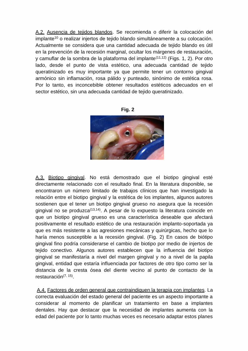
A.2. Ausencia de tejidos blandos. Se recomienda o diferir la colocación del
implante10 o realizar injertos de tejido blando simultáneamente a su colocación.
Actualmente se considera que una cantidad adecuada de tejido blando es útil
en la prevención de la recesión marginal, ocultar los márgenes de restauración,
y camuflar de la sombra de la plataforma del implante(11,12) (Figs. 1, 2). Por otro
lado, desde el punto de vista estético, una adecuada cantidad de tejido
queratinizado es muy importante ya que permite tener un contorno gingival
armónico sin inflamación, rosa pálido y punteado, sinónimo de estética rosa.
Por lo tanto, es inconcebible obtener resultados estéticos adecuados en el
sector estético, sin una adecuada cantidad de tejido queratinizado.
Fig. 2
A.3. Biotipo gingival. No está demostrado que el biotipo gingival esté
directamente relacionado con el resultado final. En la literatura disponible, se
encontraron un número limitado de trabajos clínicos que han investigado la
relación entre el biotipo gingival y la estética de los implantes, algunos autores
sostienen que el tener un biotipo gingival grueso no asegura que la recesión
gingival no se produzca(13,14). A pesar de lo expuesto la literatura coincide en
que un biotipo gingival grueso es una característica deseable que afectará
positivamente el resultado estético de una restauración implanto-soportada ya
que es más resistente a las agresiones mecánicas y quirúrgicas, hecho que lo
haría menos susceptible a la recesión gingival. (Fig. 2) En casos de biótipo
gingival fino podría considerarse el cambio de biotipo por medio de injertos de
tejido conectivo. Algunos autores establecen que la influencia del biotipo
gingival se manifestaría a nivel del margen gingival y no a nivel de la papila
gingival, entidad que estaría influenciada por factores de otro tipo como ser la
distancia de la cresta ósea del diente vecino al punto de contacto de la
restauración(7, 15).
A.4. Factores de orden general que contraindiquen la terapia con implantes. La
correcta evaluación del estado general del paciente es un aspecto importante a
considerar al momento de planificar un tratamiento en base a implantes
dentales. Hay que destacar que la necesidad de implantes aumenta con la
edad del paciente por lo tanto muchas veces es necesario adaptar estos planes
de tratamiento a su estado general. El tratamiento de los pacientes de alto
riesgo, por enfermedades generales crónicas y tabaquismo o medicación que
afecte al tejido óseo, debe realizarse con cautela ya que en ellos los resultados
estéticos son menos predecibles(6).
B. Factores extrínsecos
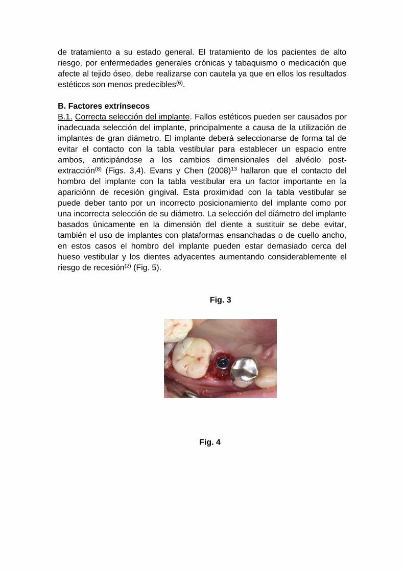
B.1. Correcta selección del implante. Fallos estéticos pueden ser causados por
inadecuada selección del implante, principalmente a causa de la utilización de
implantes de gran diámetro. El implante deberá seleccionarse de forma tal de
evitar el contacto con la tabla vestibular para establecer un espacio entre
ambos, anticipándose a los cambios dimensionales del alvéolo post-
extracción(8) (Figs. 3,4). Evans y Chen (2008)13 hallaron que el contacto del
hombro del implante con la tabla vestibular era un factor importante en la
apariciónn de recesión gingival. Esta proximidad con la tabla vestibular se
puede deber tanto por un incorrecto posicionamiento del implante como por
una incorrecta selección de su diámetro. La selección del diámetro del implante
basados únicamente en la dimensión del diente a sustituir se debe evitar,
también el uso de implantes con plataformas ensanchadas o de cuello ancho,
en estos casos el hombro del implante pueden estar demasiado cerca del
hueso vestibular y los dientes adyacentes aumentando considerablemente el
riesgo de recesión(2) (Fig. 5).
Fig. 3
Fig. 4
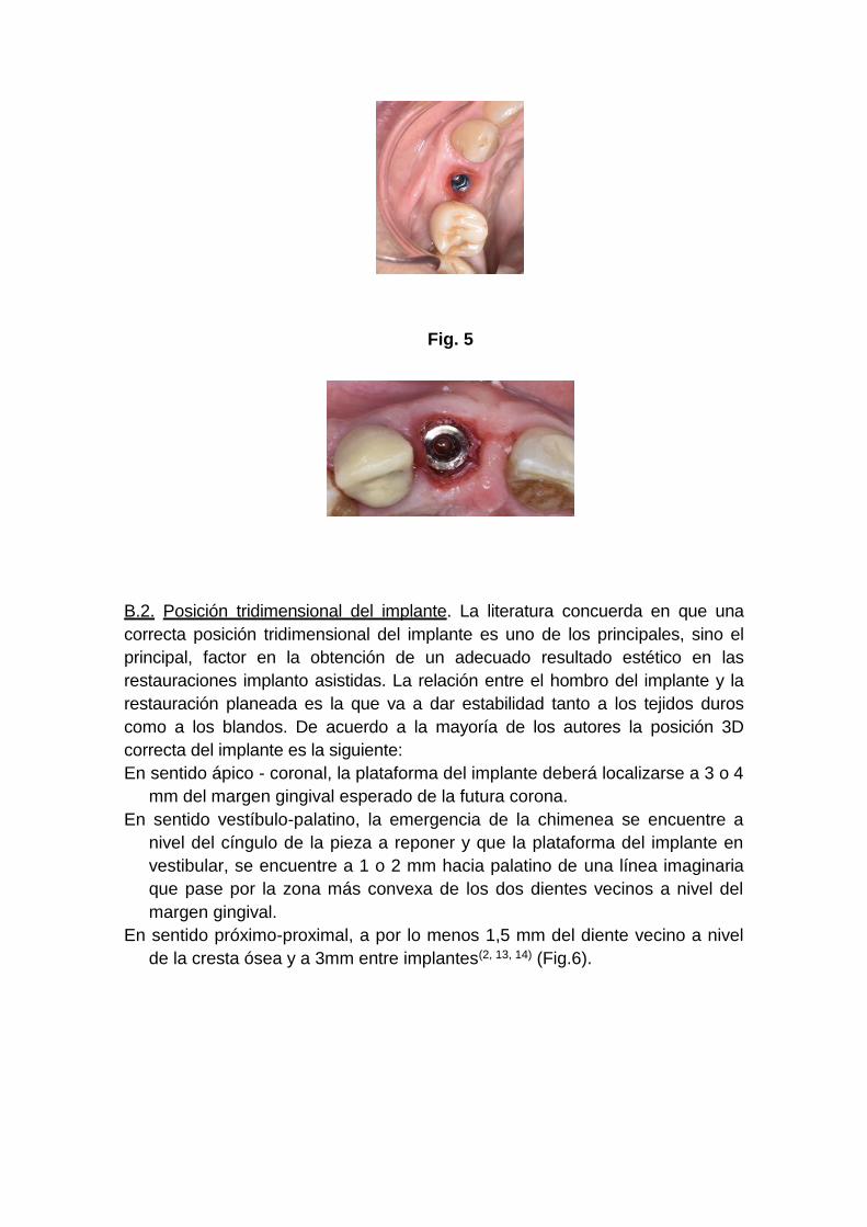
Fig. 5
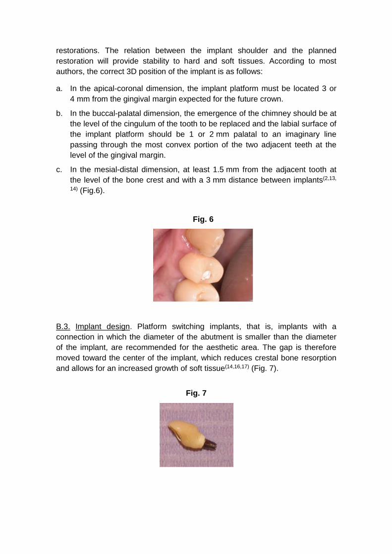
B.2. Posición tridimensional del implante. La literatura concuerda en que una
correcta posición tridimensional del implante es uno de los principales, sino el
principal, factor en la obtención de un adecuado resultado estético en las
restauraciones implanto asistidas. La relación entre el hombro del implante y la
restauración planeada es la que va a dar estabilidad tanto a los tejidos duros
como a los blandos. De acuerdo a la mayoría de los autores la posición 3D
correcta del implante es la siguiente:
En sentido ápico - coronal, la plataforma del implante deberá localizarse a 3 o 4
mm del margen gingival esperado de la futura corona.
En sentido vestíbulo-palatino, la emergencia de la chimenea se encuentre a
nivel del cíngulo de la pieza a reponer y que la plataforma del implante en
vestibular, se encuentre a 1 o 2 mm hacia palatino de una línea imaginaria
que pase por la zona más convexa de los dos dientes vecinos a nivel del
margen gingival.
En sentido próximo-proximal, a por lo menos 1,5 mm del diente vecino a nivel
de la cresta ósea y a 3mm entre implantes(2, 13, 14) (Fig.6).

Fig. 6
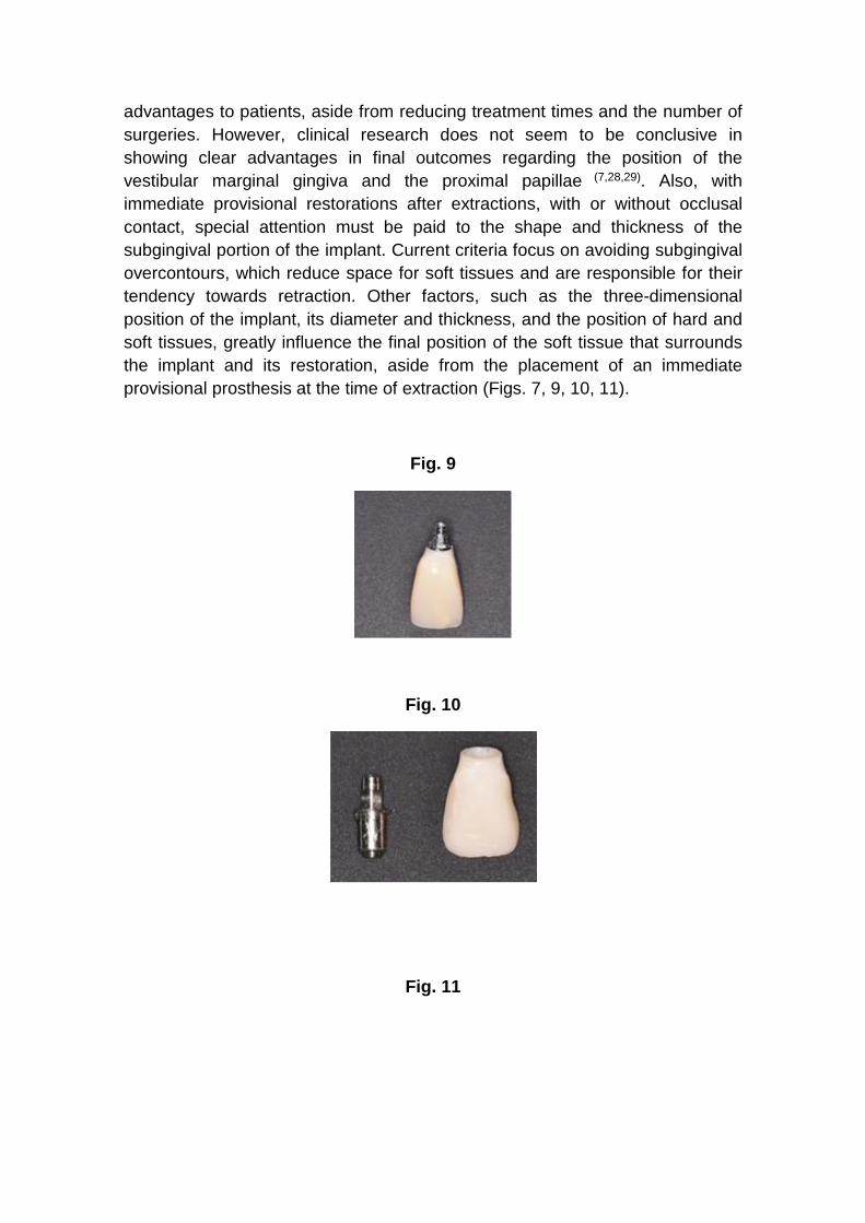
B.3. Diseño del implante. Para la zona estética, se recomienda la selecciónn de
implantes con cambio de plataforma o platform switching es decir la conexión
de un pilar de menor diámetro al del implante. De esta forma se desplaza el
gap hacia el centro del implante reduciendo así la reabsorción del hueso crestal
y permitiendo mayor crecimiento del tejido blando(14, 16, 17) (Fig. 7).
Fig. 7
B.4. Técnica quirúrgica. Para finalizar con la problemática de la recesión
gingival, cabe destacar que si bien existe bibliografía, no es determinante en
cuanto a que las técnicas sin colgajo den mejores resultados que las técnicas
con colgajo(18). En el caso de implantes inmediatos, sí existe evidencia
científica que con colocación sin colgajo y su provisionalización inmediata se
obtiene mejor estabilidad gingival.
Pregunta N°2. ¿Qué importancia tiene el manejo del provisional en la
conformación de los tejidos peri-implantares en la zona estética?
-Anatomía gingival en la dentición natural. Los tejidos blandos que rodean las
coronas de los dientes naturales, presentan una forma festoneada. Dicha forma
de los tejidos blandos, acompaña al diseño también festoneado de las crestas
óseas de los alvéolos dentales, que a su vez copian al festoneado del límite
amelo cementario de las piezas dentarias. Dicha unión amelo cementaria se
encuentra más cerca de apical en el cenit de las caras libres, y unos 2 o 3mm
más hacia incisal en el centro de las caras proximales(19, 20).
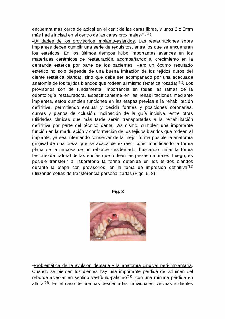
-Utilidades de los provisorios implanto-asistidos. Las restauraciones sobre
implantes deben cumplir una serie de requisitos, entre los que se encuentran
los estéticos. En los últimos tiempos hubo importantes avances en los
materiales cerámicos de restauración, acompañando al crecimiento en la
demanda estética por parte de los pacientes. Pero un óptimo resultado
estético no solo depende de una buena imitación de los tejidos duros del
diente (estética blanca), sino que debe ser acompañado por una adecuada
anatomía de los tejidos blandos que rodean al mismo (estética rosada) (21). Los
provisorios son de fundamental importancia en todas las ramas de la
odontología restauradora. Específicamente en las rehabilitaciones mediante
implantes, estos cumplen funciones en las etapas previas a la rehabilitación
definitiva, permitiendo evaluar y decidir formas y posiciones coronarias,
curvas y planos de oclusión, inclinación de la guía incisiva, entre otras
utilidades clínicas que más tarde serán transportadas a la rehabilitación
definitiva por parte del técnico dental. Asimismo, cumplen una importante
función en la maduración y conformación de los tejidos blandos que rodean al
implante, ya sea intentando conservar de la mejor forma posible la anatomía
gingival de una pieza que se acaba de extraer, como modificando la forma
plana de la mucosa de un reborde desdentado, buscando imitar la forma
festoneada natural de las encías que rodean las piezas naturales. Luego, es
posible transferir al laboratorio la forma obtenida en los tejidos blandos
durante la etapa con provisorios, en la toma de impresión definitiva (22)
utilizando cofias de transferencia personalizadas (Figs. 6, 8).
Fig. 8
-Problemática de la avulsión dentaria y la anatomía gingival peri-implantaría.
Cuando se pierden los dientes hay una importante pérdida de volumen del
reborde alveolar en sentido vestíbulo-palatino(23), con una mínima pérdida en
altura(24). En el caso de brechas desdentadas individuales, vecinas a dientes
naturales con su periodonto sano, la cresta ósea alveolar proximal, se va a
mantener en su lugar gracias a la conservación del periodonto del diente
vecino. En los rebordes desdentados ya remodelados donde faltan dos o más
dientes contiguos, la anatomía ósea tiene una altura de aproximadamente 1mm
más hacia apical de donde estaba la cresta ósea vestibular de las piezas
ausentes y es normalmente plana, ya no festoneada, debido a la pérdida de la
cresta ósea proximal que existía entre los dientes. La mucosa bucal que tapiza
dicha cresta ósea posee también una conformación aplanada. Es así que, en el
caso de brechas extensas cuando recién instalamos un provisorio sobre un
implante dental, encontraremos una anatomía aplanada en los tejidos blandos,
debido a la anatomía también plana de la cresta ósea desdentada. Los tejidos
blandos peri-implantarios tendrán un déficit de altura en la parte
correspondiente a las papilas. Sin embargo y dependiendo de la correcta
ubicación tridimensional del implante(2) tendremos muchas veces un excedente
de tejido blando en la cara vestibular. Debemos tener presente que el tejido
blando es capaz de rellenar un máximo de 5mm en sentido vertical entre dos
dientes naturales, tomando la distancia desde la cresta ósea hasta la base del
punto de contacto(25). Sin embargo, las mediciones clínicas de la altura del
tejido blando entre dos implantes, dan un promedio de 3,4mm(26). A esto se
debe agregar como elemento negativo, la pérdida de altura de la cresta ósea
interdental luego de la extracción de dos dientes vecinos. Es entonces que
entre dos implantes contiguos tendremos un tejido blando aproximadamente
1,5 mm más delgado y una cresta ósea interproximal unos 3 mm más hacia
apical respecto a lo que sucede entre dos dientes naturales con su periodonto
sano.
-Utilidad de los provisionales en los casos de los implantes inmediatos post
extracción. En los últimos tiempos se preconizó mucho la técnica del implante
inmediato post extracción con la esperanza de que el estímulo funcional del
implante evitaría la reabsorción del hueso alveolar. En los primeros años de este
siglo los estudios de Araujo(27), entre otros, dejaron bien en claro que esa
suposición era errónea. De forma similar se sostiene que la provisionalización
inmediata del implante inmediato post extracción va a mantener incambiada la
posición de la encía marginal, dado que el colocar una corona artificial que
sostenga los tejidos blandos va a evitar el colapso de los mismos. Es bien claro
que la provisionalización inmediata ofrece grandes ventajas, sicológicas y
funcionales al paciente, además de la reducción en los tiempos de tratamiento y
el menor número de cirugías. Sin embargo, las investigaciones clínicas no
parecen ser determinantes en mostrar claras ventajas en los resultados finales
referentes a la posición de la encía marginal vestibular y las papilas
proximales(7,28, 29). A su vez en el caso de los provisorios inmediatos post
extracción, con o sin contacto oclusal, se debe tener muy en cuenta la forma y el
grosor de la porción subgingival del mismo. Los criterios actuales están
orientados a evitar los sobrecontornos subgingivales, que reducen el espacio
para los tejidos blandos y provocan la tendencia a la retracción de los mismos.
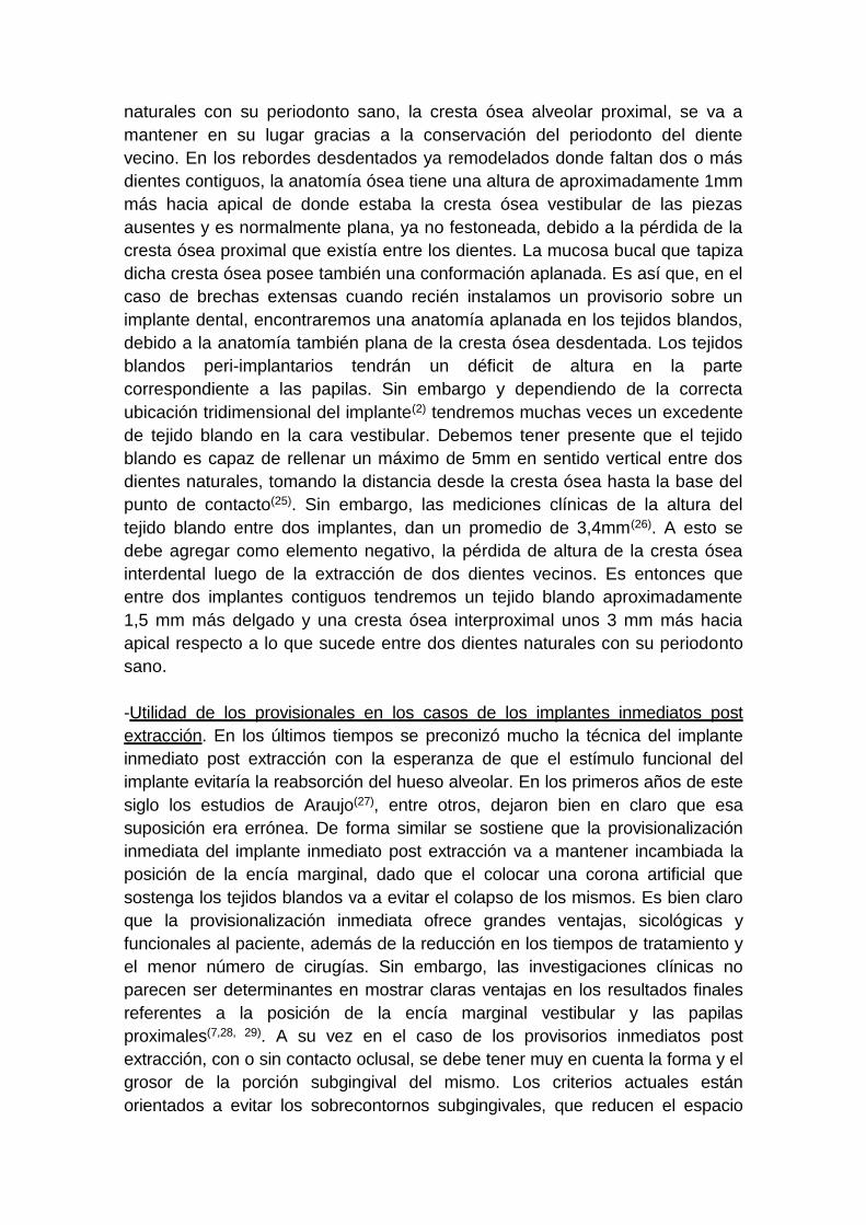
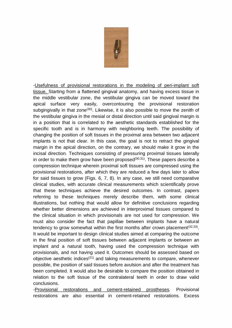
Otros factores, como la posición tridimensional del implante, su diámetro y el
grosor y posición de los tejidos duros y blandos tienen gran influencia en la
posición final de los tejidos blandos que rodean al implante y su restauración
además de la colocación de un provisorio inmediato en el momento de la
extracción (Figs.7, 9, 10, 11).
Fig. 9
Fig. 10
Fig. 11

-Utilidad de los provisorios en el modelado de los tejidos blandos peri-
implantarios. Cuando partimos de una anatomía gingival aplanada, y con un
excedente de tejido en la zona media vestibular, se puede desplazar hacia
apical de una forma muy sencilla dicha encía vestibular, sobre-contorneando
sub-gingivalmente el provisorio en esa zona(30). Del mismo modo también es
posible desplazar hacia mesial o distal el zenit de la encía vestibular hasta
obtener una posición de ese margen gingival que guarde relación con los
cánones estéticos definidos para la pieza en cuestión y su armonía con los
dientes vecinos. No es tan clara la posibilidad de modificar la posición de los
tejidos blandos en la zona proximal, entre dos implantes vecinos. En este caso
no se busca retraer el margen gingival hacia apical, sino por el contrario
deberíamos hacerlo crecer hacia incisal. Se han propuesto técnicas
consistentes en presionar lateralmente los tejidos proximales para lograr el
crecimiento de dichos tejidos(30,31). Estos artículos describen una técnica de
compresión de los tejidos blandos proximales mediante los provisionales,
seguida por un desgaste de los mismos algunos días después para permitir el
crecimiento de dichos tejidos (Figs. 6, 7, 8). De todos modos siguen faltando
estudios clínicos comparativos, con mediciones clínicas certeras que
demuestren científicamente que dichas técnicas logran los resultados
buscados. Por el contrario, los artículos que refieren a estas técnicas se limitan
a describirlas, con alguna ilustración clínica pero nada que permita sacar
conclusiones determinantes respecto a que se logren mejores dimensiones en
los tejidos blandos interproximales comparados con la situación clínica en la
que se trabaje sin comprimir mediante provisorios. Se debe tener también en
cuenta que las papilas entre implantes tienen una tendencia natural a tener
cierto crecimiento en los primeros meses luego de instalada la corona(32, 33).
Sería importante diseñar estudios clínicos tendientes a comparar el resultado
en la posición final de los tejidos blandos entre implantes contiguos o entre
implante y diente natural, habiendo utilizado y no, la técnica de la compresión
mediante provisorios. Se deberían evaluar los resultados basándose en índices
estéticos objetivos(21) y realizando mediciones que comparen, cuando sea
posible, la posición de dichos tejidos antes de la avulsión y luego de terminado
el tratamiento. Del mismo modo sería deseable comparar la posición obtenida
respecto a los tejidos blandos de las piezas contralaterales para sacar
conclusiones valederas en esta materia.
-Provisorios y las prótesis cementadas. También los provisorios son de gran
importancia en los casos de restauraciones cementadas. Últimamente se está
citando como factor de riesgo de enfermedad periimplantaria a los excesos de
cemento subgingival(34). Se ha sugerido que los márgenes no deben localizarse
más profundos que 2mm respecto al borde libre de la encía periimplantaria. Si
la restauración es finalizada luego de un período suficiente de maduración de
los tejidos con provisionales atornillados, podremos dejar los márgenes de la
restauración a escasa distancia del borde libre de la encía, con escaso riesgo
de visibilidad en el futuro. Por los motivos anteriormente citados, es
recomendable el uso de provisorios atornillados, por sobre los cementados.
Cuando se realizan coronas cementadas, una perforación mínima de 1 mm de
diámetro, ubicada en oclusal o palatino, permite evitar el efecto de
confinamiento del material cementante, orientando al mismo a emerger por la
perforación y en consecuencia, no proyectarse más allá del margen de la
corona. Como conclusión, además de las ventajas sicológicas y funcionales
que le aporta al paciente, el tener restauraciones provisionales fijas durante un
período previo a la rehabilitación definitiva, la evaluación estética tanto por
parte del paciente como del profesional, durante períodos prolongados de
tiempo, permite llegar a mejores resultados finales. Por otra parte, trabajando
de esta manera, al momento de las impresiones definitivas, los tejidos blandos
tendrán una forma suficientemente evaluada y estable como para establecer
las posiciones finales de la restauración definitiva, y también será menos
probable que existan cambios no esperados en las etapas posteriores a la
instalación. Más allá de las limitaciones en el resultado final de los tejidos
blandos interimplantes, los provisionales juegan un rol muy importante en toda
la etapa que transcurre desde el fin del período de oseointegración hasta la
instalación de la restauración final.
Pregunta Nº 3. ¿Que se considera un manejo óptimo de los pilares en
relación a su diseño, los materiales y la estética? Diferentes causas fueron
propuestas en la bibliografía para atribuir la pérdida de la cresta ósea peri-
implantaria, entre ellas se encuentran: acumulación de stress debido a las
fuerzas oclusales, falta de espesor del tejido óseo y de la mucosa peri
implantar y recuperación del ancho biológico en forma natural, trauma
quirúrgico y a la conexión implante-pilar entre otras. El mantenimiento de la
cresta ósea es un objetivo de fundamental importancia en la zona estética,
como forma de lograr una arquitectura gingival armónica y evitar así la recesión
gingival. La selección del pilar juega un rol fundamental en ello. De la
bibliografía revisada se establecieron pautas para su correcta selección:
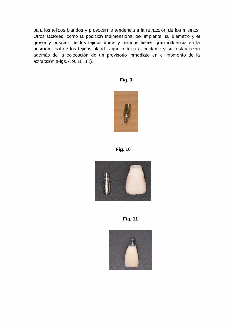
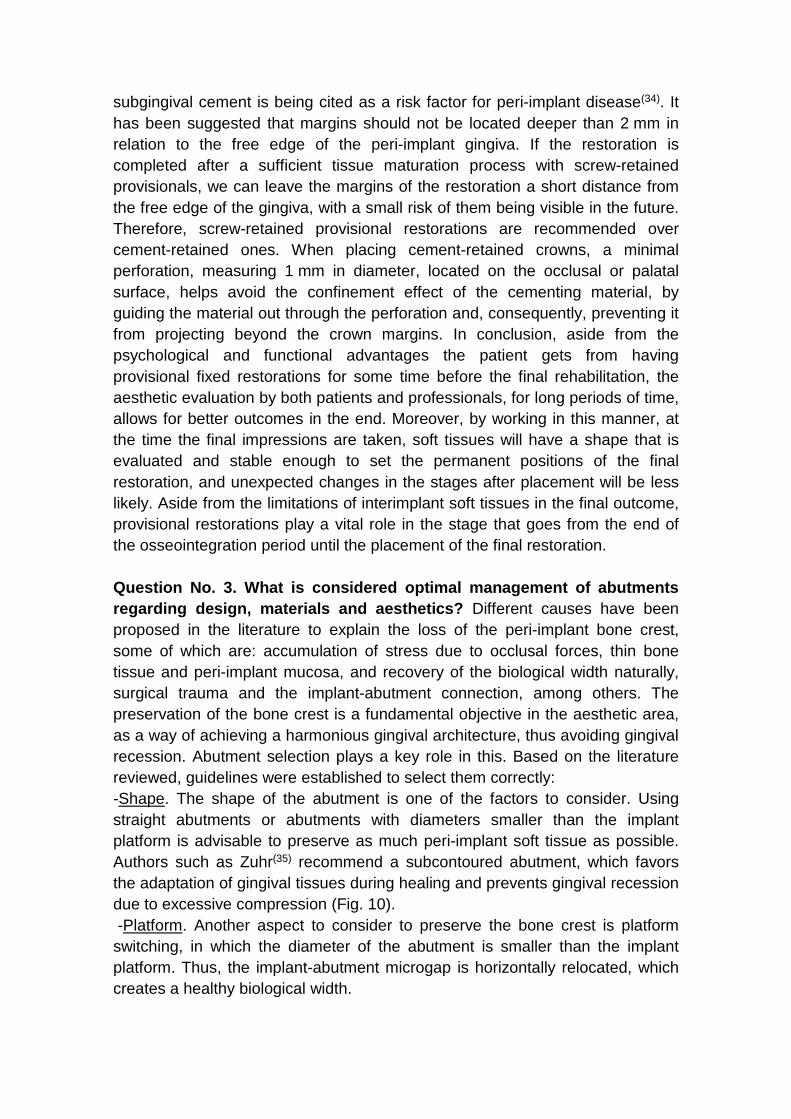
-Forma. La forma del pilar es uno de los factores a considerar. Para conservar
la mayor cantidad de los tejidos blandos peri implantares, es recomendable el
uso de pilares rectos o con diámetros más pequeños que la plataforma del
implante. Autores como Zuhr(35) aconsejan un pilar sub contorneado,que
favorece la adaptación de los tejidos gingivales durante la cicatrización y evita
la recesión gingival por excesiva compresión (Fig. 10).
-Plataforma. Para mantener la cresta ósea, otro concepto a considerar es el de
cambio de plataforma (Platform Switching), en el cual el diámetro del pilar es
menor a la plataforma del implante. De este modo se reubica horizontalmente
el micro-gap implante-pilar, logrando un espacio biológico saludable.
-Tipo de conexión. La bibliografía no es clara en determinar que un tipo sea el
más favorable en el mantenimiento de la cresta ósea y ninguna ha demostrado
tener un micro-gap inactivo ya que en todos los sistemas se verifica la
contaminación microbiana a través del espacio implante-pilar(36, 37).
- Espesor de los tejidos gingivales. Es un factor a considerar en la selección del
material del pilar. Para espesores de encía menores a 2mm la zirconia muestra
un mejor comportamiento óptico que los pilares metálicos(38).
-Bio-compatibilidad de los diferentes materiales. En algunos estudios el uso de
pilares de zirconia pareció favorecer la salud de los tejidos gingivales ya que
existiría una menor adherencia del biofilm que en los pilares de titanio(39). Sin
embargo, otros autores no encontraron diferencias comparando estos
materiales(40). En relación a la adherencia epitelial, un trabajo realizado por
Belser y col.(40) mostró un mejor desempeño en pilares de titanio y de cerámica
en comparación con pilares de aleación de oro y de cerámica fundida sobre
metal. Otros autores mencionan que estos resultados dependen de las
propiedades adhesivas de los materiales estudiados y las diferencias en cuanto
a la resistencia a la corrosión39. De acuerdo a la bibliografía con la que
contamos al día de hoy es difícil concluir sobre las ventajas de un material por
sobre otros, ya que hay variables que no se contemplaron en los distintos
estudios, entre ellas, el grado de micro-rugosidad del pilar o el tratamiento de
limpieza y esterilización del mismo previo a su uso. Se deberían diseñar
estudios que contemplen todas esas variables de modo de poder sacar
conclusiones definivas para seleccionar el material ideal del pilar. Existe
coincidencia en la indicación de pilares de zirconia en el sector estético, sin
embargo diferentes autores han recomendado su uso con cautela dada la poca
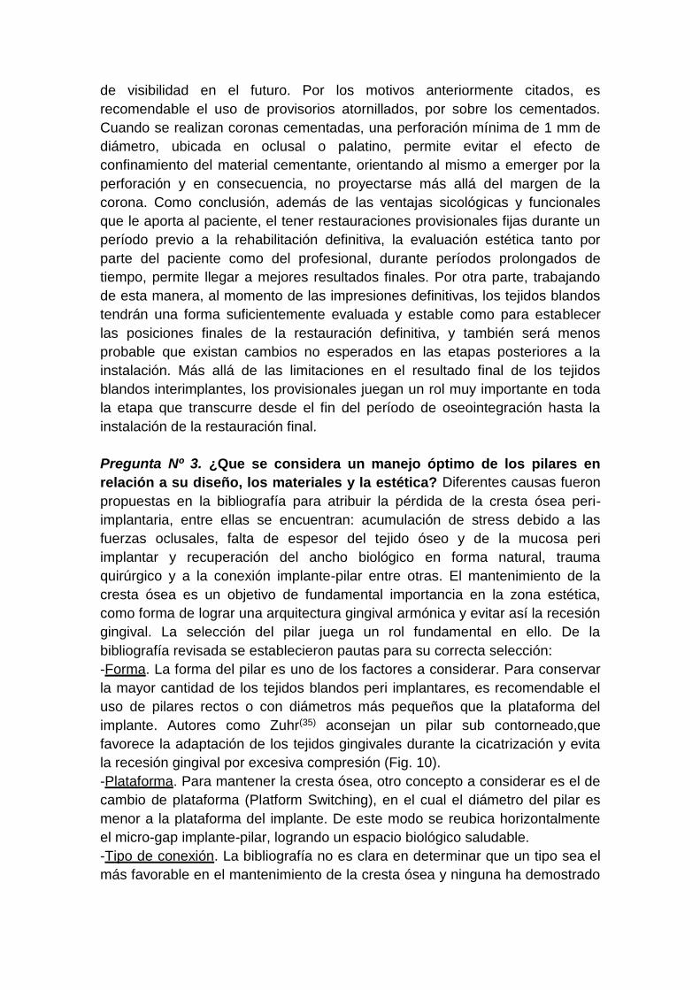
evidencia científica con resultados a largo plazo(41). Los autores del presente
trabajo opinan que existen problemas técnicos que surgen de la utilización de
la zirconia como material para pilares, tales como la imposibilidad de reproducir
algunos sistemas de conexión interna, la fractura del pilar al aplicar torque al
tornillo de retención o el daño al hexágono del implante. Para solucionar estos
problemas es que la industria proporciona pilares de zirconia con base
metálica, pudiendo ésta, ser cementada (Ti Base, Biomet) situación que tiende
a aumentar el volumen del pilar en su emergencia a nivel del implante. Este
hecho, en algunas situaciones clínicas, por ejemplo, en casos de incisivos
laterales, puede ser desfavorable (Figs.11, 12).
Fig. 12
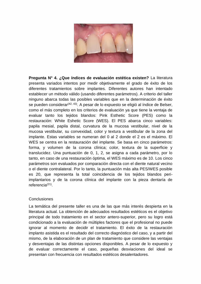
Pregunta N° 4. ¿Que índices de evaluación estética existen? La literatura
presenta variados intentos por medir objetivamente el grado de éxito de los
diferentes tratamientos sobre implantes. Diferentes autores han intentado
establecer un método válido (usando diferentes parámetros). A criterio del taller
ninguno abarca todas las posibles variables que en la determinación de éxito
se pueden considerar(42, 43). A pesar de lo expuesto se eligió al índice de Belser,
como el más completo en los criterios de evaluación ya que tiene la ventaja de
evaluar tanto los tejidos blandos: Pink Esthetic Score (PES) como la
restauración: White Eshetic Score (WES). El PES abarca cinco variables:
papila mesial, papila distal, curvatura de la mucosa vestibular, nivel de la
mucosa vestibular, su convexidad, color y textura a vestibular de la zona del
implante. Estas variables se numeran del 0 al 2 donde el 2 es el máximo. El
WES se centra en la restauración del implante. Se basa en cinco parámetros:
forma, y volumen de la corona clínica; color, textura de la superficie y
translucidez. Una puntuación de 0, 1, 2, se asigna a cada parámetro, por lo
tanto, en caso de una restauración óptima, el WES máximo es de 10. Los cinco
parámetros son evaluados por comparación directa con el diente natural vecino
o el diente contralateral. Por lo tanto, la puntuación más alta PES/WES posible
es 20, que representa la total coincidencia de los tejidos blandos peri-
implantarios y de la corona clínica del implante con la pieza dentaría de
referencia(21).
Conclusiones
La temática del presente taller es una de las que más interés despierta en la
literatura actual. La obtención de adecuados resultados estéticos es el objetivo
principal de todo tratamiento en el sector antero-superior, pero su logro está
condicionado a la evaluación de múltiples factores que el profesional no puede
ignorar al momento de decidir el tratamiento. El éxito de la restauración
implanto asistida es el resultado del correcto diagnóstico del caso, y a partir del
mismo, de la elaboración de un plan de tratamiento que considere las ventajas
y desventajas de las distintas opciones disponibles. A pesar de lo expuesto y
de evaluar correctamente el caso, pequeñas desviaciones del ideal se
presentan con frecuencia con resultados estéticos desalentadores.

Evaluación científica. Los responsables del taller seleccionaron artículos
representativos de la temática a desarrollar, a partir de los cuales formularon
preguntas que abarcaron los contenidos de los mismos, permitiendo de esta
forma que se desarrollara un rico intercambio científico entre los participantes.
La planificación y coordinación del taller cumplió con los tiempos asignados.
Los participantes presentaron gran diferencia de nivel y participación, pocos de
los cursantes conocían la literatura propuesta y su valor en base al tipo de
trabajo (revisión sistemática, meta-análisis, serie de casos clínicos). Del
intercambio surgieron las conclusiones que se publican. Estas conclusiones
son representativas de la bibliografía consultada y del desarrollo científico del
taller.
Referencias
1. Papaspyridakos P, Chen CJ, Singh M, Weber HP and Gallucci GO. Success
criteria in implant dentistry: a systematic review. J Dent Res. 2012; 91: 242-
8.
2. Buser D, Martin W and Belser UC. Optimizing esthetics for implant
restorations in the anterior maxilla: anatomic and surgical considerations. Int
J Oral Maxillofac Implants. 2004; 19 Suppl: 43-61.
3. Hammerle C, S.TChen and Wilson TG. Consensus statements and
recommended clinical procedures regarding the placement of implants in
extraction sockets. Int J Oral Maxillofac Implants. 2004; 19: 26-8.
4. Chen ST and Buser D. Clinical and esthetic outcomes of implants placed in
postextraction sites. Int J Oral Maxillofac Implants. 2009; 24 Suppl: 186-217.
5. Tortamano P, Camargo LO, Bello-Silva MS and Kanashiro LH. Immediate
implant placement and restoration in the esthetic zone: a prospective study
with 18 months of follow-up. Int J Oral Maxillofac Implants. 2010; 25: 345-50.
6. Belser U, Buser D and Higginbottom F. Consensus statements and
recommended clinical procedures regarding esthetics in implant dentistry. Int
J Oral Maxillofac Implants. 2004; 19 Suppl: 73-4.
7. Kan JY, Rungcharassaeng K, Lozada JL and Zimmerman G. Facial gingival
tissue stability following immediate placement and provisionalization of
maxillary anterior single implants: a 2- to 8-year follow-up. Int J Oral
Maxillofac Implants. 2011; 26: 179-87.
8. Levin BP and Wilk BL. Immediate provisionalization of immediate implants in
the esthetic zone: a prospective case series evaluating implant survival,
esthetics, and bone maintenance. Compend Contin Educ Dent. 2013; 34:
352-61.
9. Martins Da Rosa,JC. Implantes con Carga Inmediata en Alveolos
Comprometidos. Edit Santos Brasil, 2012.
10. Buser D, Chen ST, Weber HP and Belser UC. Early implant placement
following single-tooth extraction in the esthetic zone: biologic rationale and
surgical procedures. Int J Periodontics Restorative Dent. 2008; 28: 441-51.
11. Chung DM, Oh TJ, Shotwell JL, Misch CE and Wang HL. Significance of
keratinized mucosa in maintenance of dental implants with different
surfaces. J Periodontol. 2006; 77: 1410-20.
12. Schrott AR, Jimenez M, Hwang JW, Fiorellini J and Weber HP. Five-year
evaluation of the influence of keratinized mucosa on peri-implant soft-tissue
health and stability around implants supporting full-arch mandibular fixed
prostheses. Clin Oral Implants Res. 2009; 20: 1170-7.
13. Evans CD and Chen ST. Esthetic outcomes of immediate implant
placements. Clin Oral Implants Res. 2008; 19: 73-80.
14. Fu JH, Lee A and Wang HL. Influence of tissue biotype on implant esthetics.
Int J Oral Maxillofac Implants. 2011; 26: 499-508.
15. Chow YC, Eber RM, Tsao YP, Shotwell JL and Wang HL. Factors
associated with the appearance of gingival papillae. J Clin Periodontol.
2010; 37: 719-27.
16. Lazzara RJ and Porter SS. Platform switching: a new concept in implant
dentistry for controlling postrestorative crestal bone levels. Int J Periodontics
Restorative Dent. 2006; 26: 9-17.
17. Canullo L, Fedele GR, Iannello G and Jepsen S. Platform switching and
marginal bone-level alterations: the results of a randomized-controlled trial.
Clin Oral Implants Res. 2010; 21: 115-21.
18. Caneva M, Botticelli D, Salata LA, Souza SL, Bressan E and Lang NP. Flap
vs. "flapless" surgical approach at immediate implants: a histomorphometric
study in dogs. Clin Oral Implants Res. 2010; 21: 1314-9.
19. Becker W, Ochsenbein C, Tibbetts L and Becker BE. Alveolar bone
anatomic profiles as measured from dry skulls. Clinical ramifications. J Clin
Periodontol. 1997; 24: 727-31.
20. Gallucci GO, Belser UC, Bernard JP and Magne P. Modeling and
characterization of the CEJ for optimization of esthetic implant design. Int J
Periodontics Restorative Dent. 2004; 24: 19-29.
21. Belser UC, Grutter L, Vailati F, Bornstein MM, Weber HP and Buser D.
Outcome evaluation of early placed maxillary anterior single-tooth implants
using objective esthetic criteria: a cross-sectional, retrospective study in 45
patients with a 2- to 4-year follow-up using pink and white esthetic scores. J
Periodontol. 2009; 80: 140-51.
22. Elian N, Tabourian G, Jalbout ZN, et al. Accurate transfer of peri-implant
soft tissue emergence profile from the provisional crown to the final
prosthesis using an emergence profile cast. J Esthet Restor Dent. 2007; 19:
306-14; discussion 15.
23. Botticelli D, Berglundh T and Lindhe J. Hard-tissue alterations following
immediate implant placement in extraction sites. J Clin Periodontol. 2004;
31: 820-8.
24. Schropp L, Wenzel A, Kostopoulos L and Karring T. Bone healing and soft
tissue contour changes following single-tooth extraction: a clinical and
radiographic 12-month prospective study. Int J Periodontics Restorative
Dent. 2003; 23: 313-23.
25. Tarnow D, Magner A and Fletcher P. The effect of the distance from the
contact point to the crest of bone on the presence or absence of the
interproximal dental papilla. J Periodontol. 1992; 63: 995-6.
26. Tarnow D, Elian N, Fletcher P, et al. Vertical distance from the crest of bone
to the height of the interproximal papilla between adjacent implants. J
Periodontol. 2003; 74: 1785-8.
27. Araujo MG, Sukekava F, Wennstrom JL and Lindhe J. Ridge alterations
following implant placement in fresh extraction sockets: an experimental
study in the dog. J Clin Periodontol. 2005; 32: 645-52.
28. Tsuda H, Rungcharassaeng K, Kan JY, Roe P, Lozada JL and Zimmerman
G. Peri-implant tissue response following connective tissue and bone
grafting in conjunction with immediate single-tooth replacement in the
esthetic zone: a case series. Int J Oral Maxillofac Implants. 2011; 26: 427-
36.
29. Cabello G, Rioboo M and Fabrega JG. Immediate placement and
restoration of implants in the aesthetic zone with a trimodal approach: soft
tissue alterations and its relation to gingival biotype. Clin Oral Implants Res.
2013; 24: 1094-100.
30. Su H, Gonzalez-Martin O, Weisgold A and Lee E. Considerations of implant
abutment and crown contour: critical contour and subcritical contour. Int J
Periodontics Restorative Dent. 2010; 30: 335-43.
31. Wittneben JG, Buser D, Belser UC and Bragger U. Peri-implant soft tissue
conditioning with provisional restorations in the esthetic zone: the dynamic
compression technique. Int J Periodontics Restorative Dent. 2013; 33: 447-
55.
32. Jemt T. Restoring the gingival contour by means of provisional resin crowns
after single-implant treatment. Int J Periodontics Restorative Dent. 1999; 19:
20-9.
33. Priest G. Predictability of soft tissue form around single-tooth implant
restorations. Int J Periodontics Restorative Dent. 2003; 23: 19-27.
34. Wilson TG, Jr. The positive relationship between excess cement and peri-
implant disease: a prospective clinical endoscopic study. J Periodontol.
2009; 80: 1388-92.
35. OttoZuhr GS. Maintenance of the Original Emergence Profilefor Natural
Esthetics with Implant-SupportedRestorations. Implant Prosthodontics.
2002.
36. Dibart S, Warbington M, Su MF and Skobe Z. In vitro evaluation of the
implant-abutment bacterial seal: the locking taper system. Int J Oral
Maxillofac Implants. 2005; 20: 732-7.
37. Schwarz F, Hegewald A and Becker J. Impact of implant-abutment
connection and positioning of the machined collar/microgap on crestal bone
level changes: a systematic review. Clin Oral Implants Res. 2014; 25: 417-
25.
38. Iñaki Gamborena MBB. The Gray ZoneAround Dental Implants:Keys to
Esthetic Success. The American Journal of Esthetic Dentistry. 2011; 1: 26-
46.
39. Nakamura K, Kanno T, Milleding P and Ortengren U. Zirconia as a dental
implant abutment material: a systematic review. Int J Prosthodont. 2010; 23:
299-309.
40. Belser UC, Schmid B, Higginbottom F and Buser D. Outcome analysis of
implant restorations located in the anterior maxilla: a review of the recent
literature. Int J Oral Maxillofac Implants. 2004; 19 Suppl: 30-42.
41. Guess PC, Att W and Strub JR. Zirconia in fixed implant prosthodontics.
Clin Implant Dent Relat Res. 2012; 14: 633-45.
42. Furhauser R, Florescu D, Benesch T, Haas R, Mailath G and Watzek G.
Evaluation of soft tissue around single-tooth implant crowns: the pink
esthetic score. Clin Oral Implants Res. 2005; 16: 639-44.
43. Annibali S, Bignozzi I, La Monaca G and Cristalli MP. Usefulness of the
aesthetic result as a success criterion for implant therapy: a review. Clin Implant
Dent Relat Res. 2012; 14: 3-40.
Workshop 4 - Challenges of implant-supported rehabilitation in
the aesthetic area
INTRODUCTION
The outcomes of the practice of replacing missing dental organs with implants
have reached such a degree of universalization and certainty(1) that it has
become an everyday tool in the clinical practice. Nevertheless, the better
outcomes brought about greater demands from patients and also from
professionals, who strive to reach dental aesthetics and, especially, the gingival
aesthetics of natural teeth.
Achieving pleasing gingival aesthetics in teeth restored with implants has
proven the main challenge to address in this branch of the profession. The
literature review on this subject matter to be discussed in a workshop
environment, aiming towards a consensus, was designed to make a
contribution in this area.
Workshop methodology. The workshop was structured in the following
stages:
- Literature review. The people leading the seminar accessed the Timbó,
Pubmed, Medline and Lilacs portals, reviewed the literature of the last ten
years, and selected 23 studies, all of them conducted on humans, considered
to be representative of the topic under discussion.
- Guiding questions. Four guiding questions representing the subject matter
of the seminar and the literature selected were formulated. The questions
were:
1. What are the causes of gingival recession?
2. How important is the management of the provisional restoration in
shaping peri-implant tissues in the aesthetic area?
3. What is considered optimal management of abutments regarding design,
materials and aesthetics?
4. What aesthetic evaluation indices are there?
- Discussion with workshop participants. The selected literature and the
guiding questions for discussion were emailed to participants for them to read
and evaluate, and they were invited to two prior meetings to begin the
scientific exchange.
- Scientific review. It was decided that a scientific reviewer should be present
at the workshop without participating in the discussion. He would evaluate the
quality of the suggested literature, the representativeness of the guiding
questions, the scientific level reached during the discussion on the day of the
seminar, and the connection between the conclusions of the seminar and the
guiding questions and bibliography.
Workshop participants. The following professionals attended the workshop:
Drs. Javier Trinidad, Enrique Elhordoy, Mariana Seoane, Natalia Panissa,
Viviana Rocha, Sergio Montenegro, Carla Laurino, David Durán, Fernando
Indart, and Susana Borrás.
Workshop.
Question No. 1. What are the causes of gingival recession linked to dental
implants? In the workshop it was determined that gingival recession is
associated to the following kinds of factors: A) Intrinsic (related to the patient)
and B) Extrinsic (related to technical aspects), and that they are probably
closely related to each other.
A. Intrinsic factors
A.1. Complete or partial absence of the vestibular table at the time of implant
placement. The evidence from the literature consistently indicates that the risk
of gingival recession in these cases is very high. Placing implants in sites with
vestibular bone defects frequently leads to soft-tissue recession, with the
potential risk of altering the harmony of the gingival margin(2,3). In this situation,
it is recommended to delay implant placement. In a literature review, Chen
(2009)(4) concluded that regeneration procedures are effective in reconstructing
defects in the vestibular table, in type 1 (immediate placement) and type 2
(early placement) implant placement situations. Despite this, less vestibular
bone loss was observed with type 1, which has consequences in the aesthetic
area. On the other hand, if the vestibular table is intact, and provided there is no
acute disease, the implant could be placed immediately(5,6). There is no
scientific evidence that early placement guarantees a better outcome than
immediate placement when correctly indicated, whether with or without
provisionalization(5,7,8). In order to reduce the treatment time, in 2008 Da
Rosa(9)proposed, for cases with partial or total loss of the vestibular table, the
Immediate Dentoalveolar Restoration (IDR) technique. The purpose of this
technique is to repair the defect in the socket with a corticomedullary bone graft
from the maxillary tuberosity and, simultaneously, to place the implant and
perform immediate non-occlusal loading.
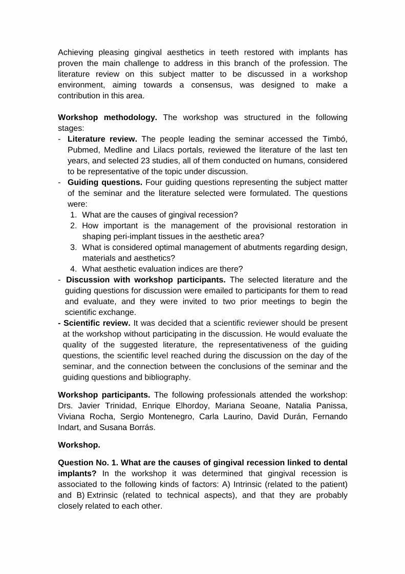
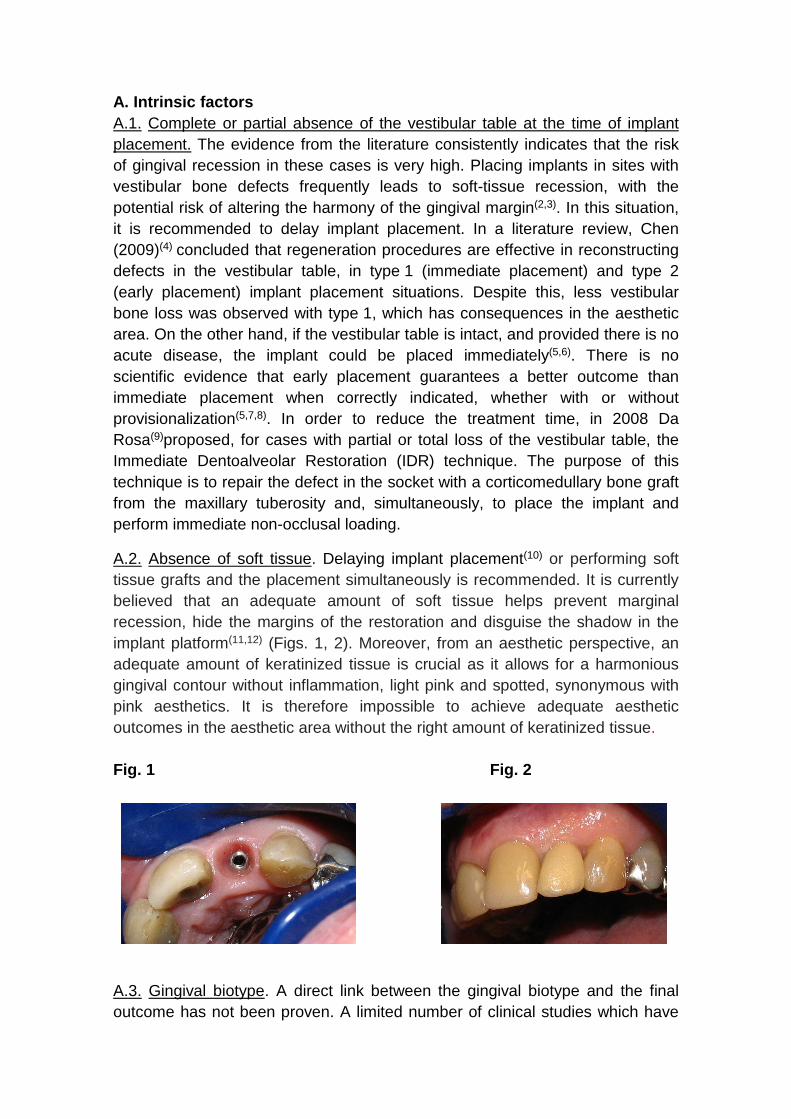
A.2. Absence of soft tissue. Delaying implant placement(10) or performing soft
tissue grafts and the placement simultaneously is recommended. It is currently
believed that an adequate amount of soft tissue helps prevent marginal
recession, hide the margins of the restoration and disguise the shadow in the
implant platform(11,12) (Figs. 1, 2). Moreover, from an aesthetic perspective, an
adequate amount of keratinized tissue is crucial as it allows for a harmonious
gingival contour without inflammation, light pink and spotted, synonymous with
pink aesthetics. It is therefore impossible to achieve adequate aesthetic
outcomes in the aesthetic area without the right amount of keratinized tissue.
Fig. 1 Fig. 2
A.3. Gingival biotype. A direct link between the gingival biotype and the final
outcome has not been proven. A limited number of clinical studies which have
researched the link between the gingival biotype and the aesthetics of implants
were found in the available literature, with some authors claiming that a thick
gingival biotype does not guarantee that there will not be any gingival
recession(13,14). Despite this, the literature agrees that a thick gingival biotype is
a desirable characteristic which will positively impact the aesthetic outcome of
an implant-supported restoration since it is more resistant to mechanical and
surgical insult, making it less susceptible to gingival recession. (Fig. 2).
Changing the biotype with connective tissue grafts could be considered for thin
gingival biotypes. Some authors claim that the influence of the gingival biotype
would manifest in the gingival margin, and not in the gingival papillae, an entity
that could be influenced by other kinds of factors, such as the distance from the
bone crest of the adjacent tooth to the contact point of the restoration(7,15).
A.4. General factors which may be contraindications for implant therapy. A
suitable assessment of the general condition of the patient is something
important to be considered when planning a dental implant-based treatment. It
is worth noting that the need for implants increases with the age of the patient,
therefore, these treatment plans very often need to be tailored to their general
condition. Patients who are deemed high risk due to chronic general diseases,
and use of tobacco or a medication which affects bone tissue, should be treated
with caution, since aesthetic outcomes are less predictable for them(6).
B. Extrinsic factors
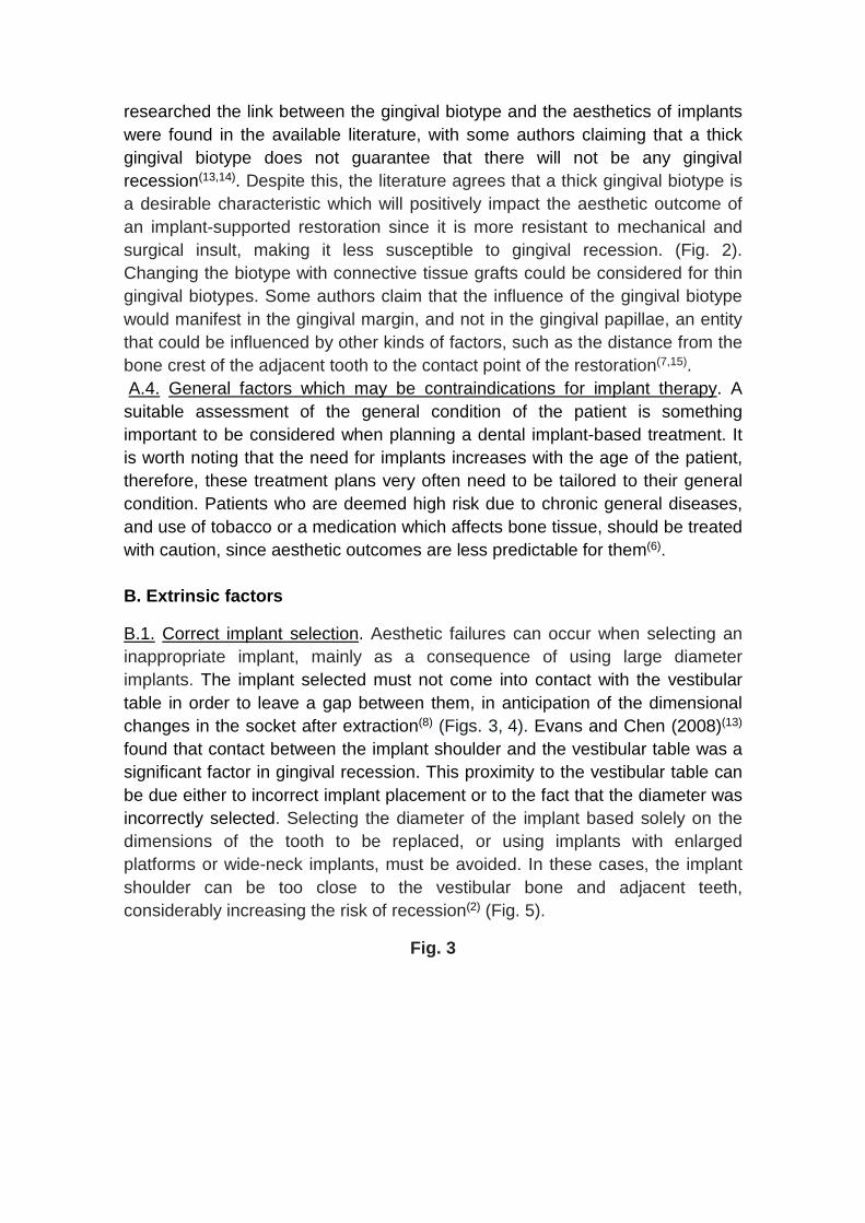
B.1. Correct implant selection. Aesthetic failures can occur when selecting an
inappropriate implant, mainly as a consequence of using large diameter
implants. The implant selected must not come into contact with the vestibular
table in order to leave a gap between them, in anticipation of the dimensional
changes in the socket after extraction(8) (Figs. 3, 4). Evans and Chen (2008)(13)
found that contact between the implant shoulder and the vestibular table was a
significant factor in gingival recession. This proximity to the vestibular table can
be due either to incorrect implant placement or to the fact that the diameter was
incorrectly selected. Selecting the diameter of the implant based solely on the
dimensions of the tooth to be replaced, or using implants with enlarged
platforms or wide-neck implants, must be avoided. In these cases, the implant
shoulder can be too close to the vestibular bone and adjacent teeth,
considerably increasing the risk of recession(2) (Fig. 5).
Fig. 3
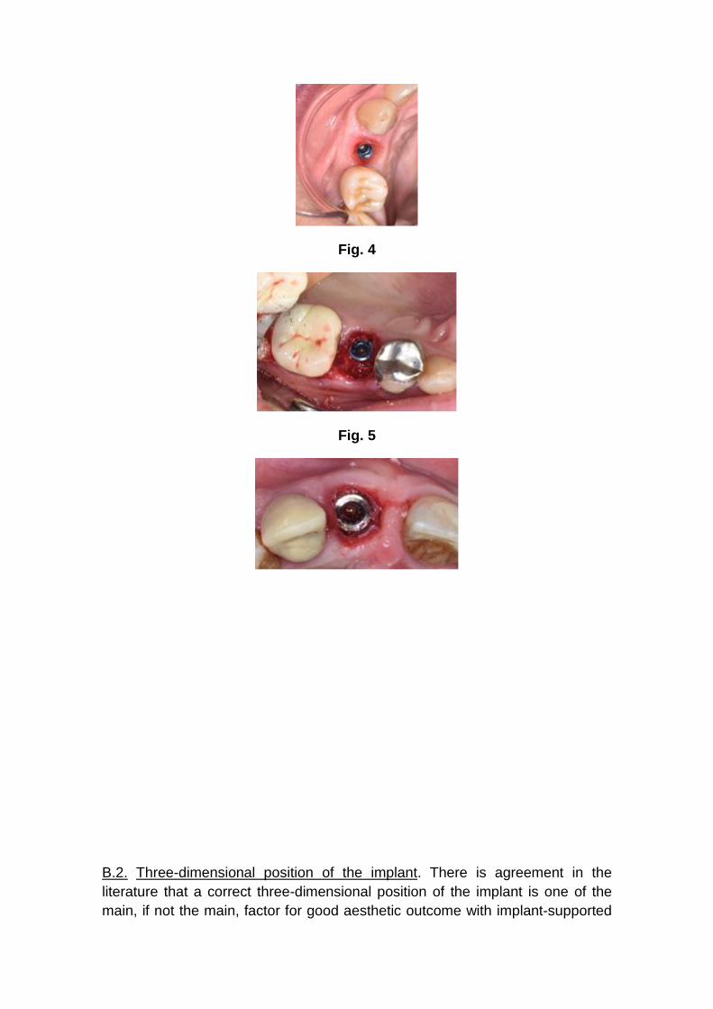
Fig. 4
Fig. 5
B.2. Three-dimensional position of the implant. There is agreement in the
literature that a correct three-dimensional position of the implant is one of the
main, if not the main, factor for good aesthetic outcome with implant-supported
restorations. The relation between the implant shoulder and the planned
restoration will provide stability to hard and soft tissues. According to most
authors, the correct 3D position of the implant is as follows:
a. In the apical-coronal dimension, the implant platform must be located 3 or
4 mm from the gingival margin expected for the future crown.
b. In the buccal-palatal dimension, the emergence of the chimney should be at
the level of the cingulum of the tooth to be replaced and the labial surface of
the implant platform should be 1 or 2 mm palatal to an imaginary line
passing through the most convex portion of the two adjacent teeth at the
level of the gingival margin.
c. In the mesial-distal dimension, at least 1.5 mm from the adjacent tooth at
the level of the bone crest and with a 3 mm distance between implants(2,13,
14) (Fig.6).
Fig. 6
B.3. Implant design. Platform switching implants, that is, implants with a
connection in which the diameter of the abutment is smaller than the diameter
of the implant, are recommended for the aesthetic area. The gap is therefore
moved toward the center of the implant, which reduces crestal bone resorption
and allows for an increased growth of soft tissue(14,16,17) (Fig. 7).
Fig. 7

B.4. Surgical technique. To finish with gingival recession, it is worth noting that
although there is literature on it, it is not conclusive as to whether flapless
techniques have better outcomes than flap techniques(18). As for immediate
implants, there is scientific evidence that flapless placement and immediate
provisionalization result in better gingival stability.
Question No. 2. How important is the management of the provisional
restoration in shaping peri-implant tissues in the aesthetic area?
-Gingival anatomy in natural dentition. The soft tissue that surrounds the crowns
of natural teeth has a scalloped shape. This shape follows the design of the
bone crests of the dental sockets –which are also scalloped– and these, in turn,
mirror the scallop of the cementoenamel junction of the teeth. This junction is
closer to the apical portion in the zenith of the free surfaces, and about 2 or
3 mm more toward incisal in the center of the proximal faces(19, 20).
-Usefulness of implant-supported provisional restorations. Restorations on
implants must meet a series of requirements, some of which are aesthetic.
Significant progress has been made in ceramic restoration materials in recent
times, along with the growth in the aesthetic demands of patients. But an
optimal aesthetic outcome does not depend solely on correctly replicating the
hard tissues of the tooth (white aesthetics); it must be accompanied by the right
anatomy of the surrounding soft tissue (pink aesthetics)(21). Provisional
restorations are fundamental in all areas of restorative dentistry. Specifically, in
implant rehabilitations, they serve different purposes in the stages prior to the
final rehabilitation, making it possible to assess and decide on crown shapes
and positions, curvatures and occlusal planes, incisal guide angle, among other
clinical benefits which the dental technician will then transfer to the final
restoration. They also play an important part in the maturation and shaping of
the soft tissues which surround the implant, both by attempting to maintain in
the best way possible the gingival anatomy of a tooth which was just removed,
and altering the flat shape of the mucosa of an edentulous ridge, trying to
replicate the naturally scalloped shape of the gums surrounding natural teeth.
Afterwards, it is possible to transfer to the laboratory the shape obtained in the
soft tissues during the phase with provisional restorations, when the final
impression the dental technician(22) is taken using personalized transfer copings
(Figs. 6, 8).
Fig. 8

-The problems of dental avulsion and the peri-implant anatomy of gums. When
teeth are missing, there is major loss of volume of the alveolar ridge in the
buccal-palatal dimension(23), with minimal height bone loss(24). In the case of
individual edentulous gaps, which are adjacent to natural teeth with a healthy
periodontium, the gap will remain in its place thanks to the conservation of the
periodontium in the adjacent tooth. In edentulous ridges that have already been
remodeled, where two or more adjacent teeth are missing, the height of the
bone anatomy is approximately 1 mm more apical to where the vestibular bone
crest of the missing teeth was, and is usually flat, not scalloped, due to the loss
of the proximal bone crest that was between the teeth. The oral mucosa that
lines said bone crest also has a flat shape. This is why in large gaps, after
placing a provisional prosthesis on a dental implant, we find that the anatomy of
soft tissues is flat because the anatomy of the edentulous bone crest is also flat.
There will be a height deficit in peri-implant soft tissues in the part
corresponding to the papillae. However, and depending on whether the three-
dimensional location of the implant is correct(2), we will often have excess soft
tissue in the vestibular surface. We must remember that the soft tissue can fill a
maximum of 5 mm vertically between two natural teeth, measuring the distance
from the bone crest to the base of the contact point(25). Nevertheless, average
clinical measurements of soft tissue height between two implants were
3.4 mm(26). Another negative aspect to consider is the loss of height of the
interdental bone crest after the two adjacent teeth have been removed.
Therefore, soft tissue between two adjacent implants will be approximately
1.5 mm thinner, and the interproximal bone crest will be 3 mm more apical in
relation to what happens between two natural teeth with a healthy periodontium.
-Usefulness of provisional restorations in post-extraction immediate implants.
Immediate placement techniques, performed in the hope that the functional
stimulation provided by the implant would prevent alveolar bone resorption,
have been widely recommended in recent years. In the first years of this
century, the research of Araujo, among others(27), made it very clear that this
assumption was wrong. Similarly, it is claimed that the immediate
provisionalization of the implant placed right after extraction will maintain the
position of the marginal gingiva unchanged, since placing an artificial crown that
supports the soft tissues will prevent them from collapsing. It is very clear that
immediate provisionalization provides great psychological and functional
advantages to patients, aside from reducing treatment times and the number of
surgeries. However, clinical research does not seem to be conclusive in
showing clear advantages in final outcomes regarding the position of the
vestibular marginal gingiva and the proximal papillae (7,28,29). Also, with
immediate provisional restorations after extractions, with or without occlusal
contact, special attention must be paid to the shape and thickness of the
subgingival portion of the implant. Current criteria focus on avoiding subgingival
overcontours, which reduce space for soft tissues and are responsible for their
tendency towards retraction. Other factors, such as the three-dimensional
position of the implant, its diameter and thickness, and the position of hard and
soft tissues, greatly influence the final position of the soft tissue that surrounds
the implant and its restoration, aside from the placement of an immediate
provisional prosthesis at the time of extraction (Figs. 7, 9, 10, 11).
Fig. 9
Fig. 10
Fig. 11
-Usefulness of provisional restorations in the modeling of peri-implant soft
tissue. Starting from a flattened gingival anatomy, and having excess tissue in
the middle vestibular zone, the vestibular gingiva can be moved toward the
apical surface very easily, overcontouring the provisional restoration
subgingivally in that zone(30). Likewise, it is also possible to move the zenith of
the vestibular gingiva in the mesial or distal direction until said gingival margin is
in a position that is correlated to the aesthetic standards established for the
specific tooth and is in harmony with neighboring teeth. The possibility of
changing the position of soft tissues in the proximal area between two adjacent
implants is not that clear. In this case, the goal is not to retract the gingival
margin in the apical direction, on the contrary, we should make it grow in the
incisal direction. Techniques consisting of pressuring proximal tissues laterally
in order to make them grow have been proposed(30,31). These papers describe a
compression technique wherein proximal soft tissues are compressed using the
provisional restorations, after which they are reduced a few days later to allow
for said tissues to grow (Figs. 6, 7, 8). In any case, we still need comparative
clinical studies, with accurate clinical measurements which scientifically prove
that these techniques achieve the desired outcomes. In contrast, papers
referring to these techniques merely describe them, with some clinical
illustrations, but nothing that would allow for definitive conclusions regarding
whether better dimensions are achieved in interproximal tissues compared to
the clinical situation in which provisionals are not used for compression. We
must also consider the fact that papillae between implants have a natural
tendency to grow somewhat within the first months after crown placement(32,33).
It would be important to design clinical studies aimed at comparing the outcome
in the final position of soft tissues between adjacent implants or between an
implant and a natural tooth, having used the compression technique with
provisionals, and not having used it. Outcomes should be assessed based on
objective aesthetic indices(21) and taking measurements to compare, whenever
possible, the position of said tissues before avulsion and after the treatment has
been completed. It would also be desirable to compare the position obtained in
relation to the soft tissue of the contralateral teeth in order to draw valid
conclusions.
-Provisional restorations and cement-retained prostheses. Provisional
restorations are also essential in cement-retained restorations. Excess
subgingival cement is being cited as a risk factor for peri-implant disease(34). It
has been suggested that margins should not be located deeper than 2 mm in
relation to the free edge of the peri-implant gingiva. If the restoration is
completed after a sufficient tissue maturation process with screw-retained
provisionals, we can leave the margins of the restoration a short distance from
the free edge of the gingiva, with a small risk of them being visible in the future.
Therefore, screw-retained provisional restorations are recommended over
cement-retained ones. When placing cement-retained crowns, a minimal
perforation, measuring 1 mm in diameter, located on the occlusal or palatal
surface, helps avoid the confinement effect of the cementing material, by
guiding the material out through the perforation and, consequently, preventing it
from projecting beyond the crown margins. In conclusion, aside from the
psychological and functional advantages the patient gets from having
provisional fixed restorations for some time before the final rehabilitation, the
aesthetic evaluation by both patients and professionals, for long periods of time,
allows for better outcomes in the end. Moreover, by working in this manner, at
the time the final impressions are taken, soft tissues will have a shape that is
evaluated and stable enough to set the permanent positions of the final
restoration, and unexpected changes in the stages after placement will be less
likely. Aside from the limitations of interimplant soft tissues in the final outcome,
provisional restorations play a vital role in the stage that goes from the end of
the osseointegration period until the placement of the final restoration.
Question No. 3. What is considered optimal management of abutments
regarding design, materials and aesthetics? Different causes have been
proposed in the literature to explain the loss of the peri-implant bone crest,
some of which are: accumulation of stress due to occlusal forces, thin bone
tissue and peri-implant mucosa, and recovery of the biological width naturally,
surgical trauma and the implant-abutment connection, among others. The
preservation of the bone crest is a fundamental objective in the aesthetic area,
as a way of achieving a harmonious gingival architecture, thus avoiding gingival
recession. Abutment selection plays a key role in this. Based on the literature
reviewed, guidelines were established to select them correctly:
-Shape. The shape of the abutment is one of the factors to consider. Using
straight abutments or abutments with diameters smaller than the implant
platform is advisable to preserve as much peri-implant soft tissue as possible.
Authors such as Zuhr(35) recommend a subcontoured abutment, which favors
the adaptation of gingival tissues during healing and prevents gingival recession
due to excessive compression (Fig. 10).
-Platform. Another aspect to consider to preserve the bone crest is platform
switching, in which the diameter of the abutment is smaller than the implant
platform. Thus, the implant-abutment microgap is horizontally relocated, which
creates a healthy biological width.
-Type of connection. The literature is not clear in determining whether one type
is more favorable to preserve the bone crest, and none has been shown to have
an inactive microgap since microbial contamination through the implant-
abutment space is found in all systems(36,37).
- Thickness of the gingival tissues. It is a significant factor in the selection of the
abutment material. For gingival thicknesses smaller than 2 mm, zirconia
displays better optical properties than metallic abutments(38).
-Biocompatibility of the different materials. In some studies, zirconia abutments
seemed to favor the health of gingival tissues given that there might be less
biofilm adherence than with titanium abutments(39). Other authors, however,
found no differences when comparing these materials(40). Regarding epithelial
adhesion, in a study conducted by Belser et al.(40) titanium and ceramic
abutments showed a better performance compared to gold alloy and ceramic
fused to metal abutments. Other authors mention that these outcomes depend
on the adhesion properties of the materials studied and their differences in
terms of resistance to corrosion(39). According to the literature available today, it
is difficult to reach a conclusion on the advantages of one material over others,
since there are variables that were not considered in the different studies,
including the degree of micro-roughness of the abutment, or the cleaning and
sterilization procedure it requires before use. Studies that include all those
variables are necessary to reach final conclusions for selecting the ideal
material for abutments. There is agreement in the aesthetic sector in indicating
zirconia abutments. However, different authors have recommended caution
when using them due to the small scientific evidence available of long-term
outcomes(41). The authors of this work think that there are technical problems as
a result of using zirconia as the material for abutments, such as the impossibility
of replicating some internal connection systems, the fracture of the abutment
when torque is applied to the retention screw, or the damage to the hexagon of
the implant. To solve these problems, the industry offers zirconia abutments
with metal bases that can be cement-retained (Ti Base, Biomet), which tends to
increase the volume of the abutments in the emergence at the implant level.
This can be a disadvantage in some clinical situations, for example, for lateral
incisors (Figs. 11, 12).
Fig. 12
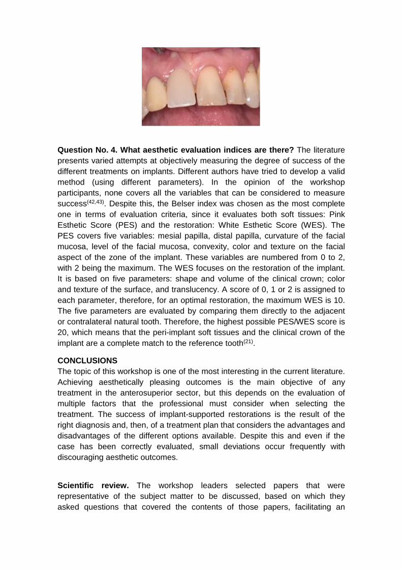
Question No. 4. What aesthetic evaluation indices are there? The literature
presents varied attempts at objectively measuring the degree of success of the
different treatments on implants. Different authors have tried to develop a valid
method (using different parameters). In the opinion of the workshop
participants, none covers all the variables that can be considered to measure
success(42,43). Despite this, the Belser index was chosen as the most complete
one in terms of evaluation criteria, since it evaluates both soft tissues: Pink
Esthetic Score (PES) and the restoration: White Esthetic Score (WES). The
PES covers five variables: mesial papilla, distal papilla, curvature of the facial
mucosa, level of the facial mucosa, convexity, color and texture on the facial
aspect of the zone of the implant. These variables are numbered from 0 to 2,
with 2 being the maximum. The WES focuses on the restoration of the implant.
It is based on five parameters: shape and volume of the clinical crown; color
and texture of the surface, and translucency. A score of 0, 1 or 2 is assigned to
each parameter, therefore, for an optimal restoration, the maximum WES is 10.
The five parameters are evaluated by comparing them directly to the adjacent
or contralateral natural tooth. Therefore, the highest possible PES/WES score is
20, which means that the peri-implant soft tissues and the clinical crown of the
implant are a complete match to the reference tooth(21).
CONCLUSIONS
The topic of this workshop is one of the most interesting in the current literature.
Achieving aesthetically pleasing outcomes is the main objective of any
treatment in the anterosuperior sector, but this depends on the evaluation of
multiple factors that the professional must consider when selecting the
treatment. The success of implant-supported restorations is the result of the
right diagnosis and, then, of a treatment plan that considers the advantages and
disadvantages of the different options available. Despite this and even if the
case has been correctly evaluated, small deviations occur frequently with
discouraging aesthetic outcomes.
Scientific review. The workshop leaders selected papers that were
representative of the subject matter to be discussed, based on which they
asked questions that covered the contents of those papers, facilitating an

enriching scientific exchange among participants. The workshop followed the
planning and coordination schedule. The members of the workshop showed
varying degrees of level and participation. Few participants knew the literature
suggested and the value of publications based on the type of study (systematic
review, meta-analysis, a series of clinical cases). The discussion resulted in the
conclusions presented in this paper. These conclusions are representative of
the literature reviewed and the scientific development of the workshop.