Práctica # 3: Platelmintos · sistema excretor está formado por protonefridios y el aparato...
13
Práctica # 3: Platelmintos INTRODUCCIÓN: Los Platelmintos son un filo de animales bilaterales protostomados acelomados. Los individuos se caracterizan por poseer un cuerpo aplanado dorsoventralmente y de estructura acelomada, es decir, el espacio corporal donde se encuentran los órganos y la pared del cuerpo está lleno de un tejido llamado parénquima. El aparato digestivo, cuando existe, es ciego (o sea, con una sola apertura: la boca). El sistema excretor está formado por protonefridios y el aparato reproductor es hermafrodita. Cuatro Clases integran el grupo de Platelmintos, una de ellas comprende los gusanos planos de vida libre y las otras tres a los gusanos planos parásitos. Clase TURBELLARIA: Gusanos planos de vida libre. Predominantemente acuáticos. La boca conduce a una faringe y luego a un sistema digestivo cerrado. La epidermis celular es generalmente ciliada. Clase MONOGENEA: gusanos planos monogenéticos: el ciclo de vida involucra un huésped. El cuerpo está cubierto por un tegumento. Presentan un prohaptor (organo de fijación anterior) como ventosa, granulaciones o glándulas pegajosas y un opisthaptor posterior con ganchos esclerotizados. Son ectoparásitos (generalmente en peces) y algunos son endoparásitos de vertebrados ectotérmicos (ej. Reptiles). Clase TREMATODA: gusanos planos digenéticos; su ciclo de vida incluye dos o más huéspedes. La mayoría son endoparásitos. Poseen una o más ventosas. Clase CESTODA: Exclusivamente endoparásitos. Cuerpo cubierto por tegumento. En la mayoría el cuerpo consiste de un éscolex anterior seguido de estróbilos compuesto de segmentos o proglótidas. No presentan tracto digestivo. Tradicionalmente se consideró que Platelmintos era un grupo monofilético y hermano de todos los demás Bilateria. La visión actual es que el redefinido grupo Platelmintos se encuentra contenido en un grupo mayor, Platyzoa, un grupo de protostomados cercanamente relacionados con Lophotrochozoa. MATERIAL: Microscopio estereoscópico Microscopio compuesto 3 cajas de Petri Gotero Pipeta Pinzas de disección Portaobjetos y cubreobjetos Martillo Botella con agua de mar METODOLOGIA: Observación de Clase Trematoda. Subclase Digenea. Diseccionar caracol con martillo. Observar la parte posterior del caracol sin concha debajo del estereoscopio y buscar la presencia de trematodos. En caso de encontrar trematodos, observar los diferentes estadios y método de movimiento de las cercarías. Utilizar la guía taxonómica para determinar la especie de trematodo. Investigue: ¿Cuáles son los principales parásitos de la División Platelmintos en la región? ¿Qué problemas representan estos parásitos en el estado de Baja California (factores económicos y sociales, factores ecológicos, etc)?
Transcript of Práctica # 3: Platelmintos · sistema excretor está formado por protonefridios y el aparato...
Práctica # 3: Platelmintos INTRODUCCIÓN: Los Platelmintos son un filo de animales bilaterales protostomados acelomados. Los individuos se caracterizan por poseer un cuerpo aplanado dorsoventralmente y de estructura acelomada, es decir, el espacio corporal donde se encuentran los órganos y la pared del cuerpo está lleno de un tejido llamado parénquima. El aparato digestivo, cuando existe, es ciego (o sea, con una sola apertura: la boca). El sistema excretor está formado por protonefridios y el aparato reproductor es hermafrodita. Cuatro Clases integran el grupo de Platelmintos, una de ellas comprende los gusanos planos de vida libre y las otras tres a los gusanos planos parásitos. Clase TURBELLARIA: Gusanos planos de vida libre. Predominantemente acuáticos. La boca conduce a una faringe y luego a un sistema digestivo cerrado. La epidermis celular es generalmente ciliada. Clase MONOGENEA: gusanos planos monogenéticos: el ciclo de vida involucra un huésped. El cuerpo está cubierto por un tegumento. Presentan un prohaptor (organo de fijación anterior) como ventosa, granulaciones o glándulas pegajosas y un opisthaptor posterior con ganchos esclerotizados. Son ectoparásitos (generalmente en peces) y algunos son endoparásitos de vertebrados ectotérmicos (ej. Reptiles). Clase TREMATODA: gusanos planos digenéticos; su ciclo de vida incluye dos o más huéspedes. La mayoría son endoparásitos. Poseen una o más ventosas. Clase CESTODA: Exclusivamente endoparásitos. Cuerpo cubierto por tegumento. En la mayoría el cuerpo consiste de un éscolex anterior seguido de estróbilos compuesto de segmentos o proglótidas. No presentan tracto digestivo. Tradicionalmente se consideró que Platelmintos era un grupo monofilético y hermano de todos los demás Bilateria. La visión actual es que el redefinido grupo Platelmintos se encuentra contenido en un grupo mayor, Platyzoa, un grupo de protostomados cercanamente relacionados con Lophotrochozoa. MATERIAL: Microscopio estereoscópico Microscopio compuesto 3 cajas de Petri Gotero Pipeta Pinzas de disección Portaobjetos y cubreobjetos Martillo Botella con agua de mar METODOLOGIA: Observación de Clase Trematoda. Subclase Digenea. Diseccionar caracol con martillo. Observar la parte posterior del caracol sin concha debajo del estereoscopio y buscar la presencia de trematodos. En caso de encontrar trematodos, observar los diferentes estadios y método de movimiento de las cercarías. Utilizar la guía taxonómica para determinar la especie de trematodo. Investigue: ¿Cuáles son los principales parásitos de la División Platelmintos en la región? ¿Qué problemas representan estos parásitos en el estado de Baja California (factores económicos y sociales, factores ecológicos, etc)?
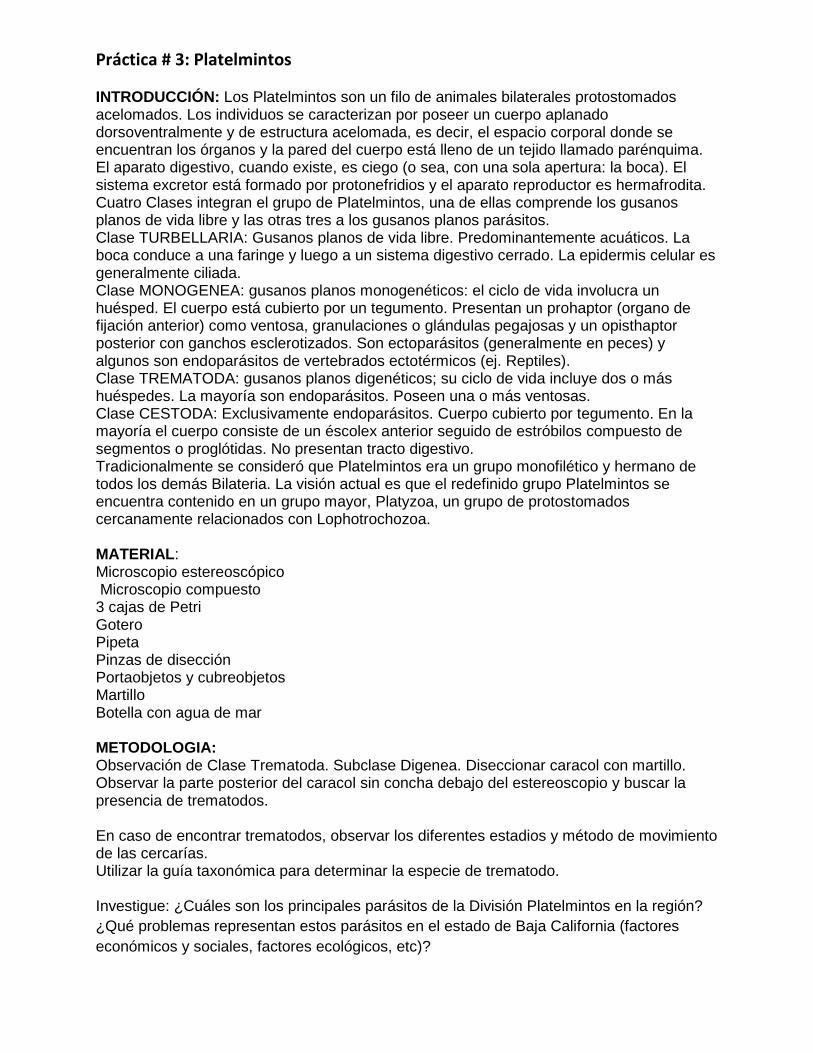
Key to larval trematode infections in Cerithidea californica & other Cerithidea spp.
Based on Martin (1972) & revised by Todd C. Huspeni & Ryan F. Hechinger
1. a. Tail forked on cercaria .................. 2
b. Tail non-forked on cercaria .................. 4
2. a. Cercaria with eyespots .................. AUST
Austrobilharzia sp. Cercaria develop in opaque bright white sporocysts which occupy the gonad and
(commonly) the green vesicular tissue posterior to kidney. Frequently occurs in double infections. In
cercaria, when oral sucker is pushed out, it appears that there are some spines or bristles around it.
Swims in an "S" motion rapidly then stops and contracts head and tail then resumes swimming.
Cercariae found primarily in the water column rather than on the bottom of the dish. Prevalence
generally < 1%. May be Austrobilharzia penneri which uses Cerithidea scalariformis as intermediate
host. See Martin 1972 for more details on infection experiments.
Austrobilharzia sp. & Acanthoparyphium spinulosum double infection in C. californica
Austrobilharzia sp. sporocysts (white) Acanthoparyphium rediae (orange)

Austrobilharzia sp. sporocysts (white) Acanthoparyphium rediae (orange)
Cercaria of Austrobilharzia sp. in C. californica
b. Cercaria without eyespots .................. 3

3. a. Cercariae are released from stringy vermicelli-like whitish sporocysts located in gonad & digestive
gland; flame cells in cercaria in groups of twos;
.................. SMCY
Small cyathocotylid - often found in double infections with heterophyids. Prevalence varies from rare
(< 1%) to over 40% at some sites. Cercariae encyst in the muscles of boney fishes. Cercaria is smaller
than 3b.
Pic of SMCY infection Pic of SMCY cercaria
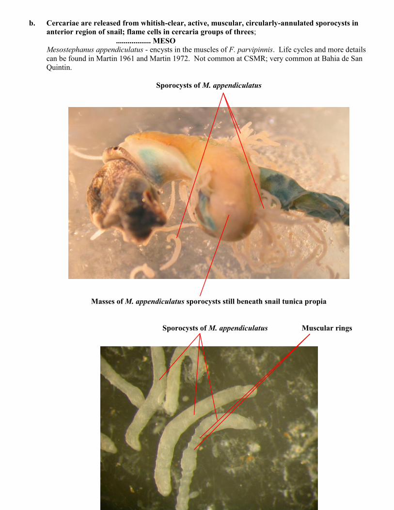
b. Cercariae are released from whitish-clear, active, muscular, circularly-annulated sporocysts in
anterior region of snail; flame cells in cercaria groups of threes;
.................. MESO Mesostephanus appendiculatus - encysts in the muscles of F. parvipinnis. Life cycles and more details
can be found in Martin 1961 and Martin 1972. Not common at CSMR; very common at Bahia de San
Quintin.
Sporocysts of M. appendiculatus
Masses of M. appendiculatus sporocysts still beneath snail tunica propia
Sporocysts of M. appendiculatus Muscular rings
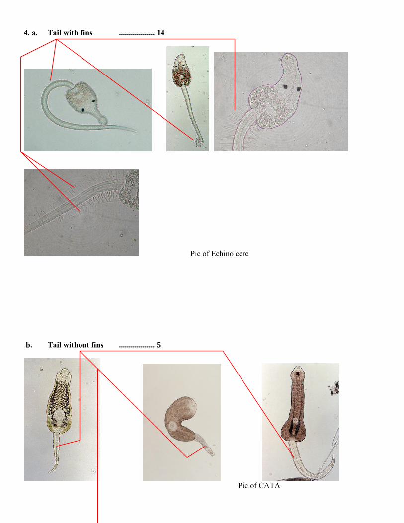
4. a. Tail with fins .................. 14
Pic of Echino cerc
b. Tail without fins .................. 5
Pic of CATA

5. a. With eyespots .................. 6
Pic of CATA
b. Without eyespots .................. 8
6. a. Eyespotted cercariae released from orange to yellow rediae in “mantle” region of snail; cercariae
with a pouch at each posterior-lateral margin of the body
.................. CATA Catatropis johnstoni - Encysts on almost any object within minutes of contact. Creeps along bottom of
dish before encysting. See Martin 1956 and Martin 1972 for the life cycle and more details.
Pic of CATA cerc. Pic of CATA infection
b. Cercariae not released from yellowish orange rediae in “mantle” of snail; cercariae without a
pouch at each posterior-lateral margin of the body
.................. 7

7. a. Large cercariae have a black median pigment spot between the eyespots which can be seen easily
through the body wall of the large (X mm X Ymm) whitish-gray rediae located in the snail gonad;
Tails of cercariae form a “figure 8” when swimming; Cercariae constantly swim and will readily
encyst on almost any object
.................. HIMA
Himasthla rhigedana - See Adams and Martin 1963 and Martin 1972 for the life cycle and more details.
Digestive gland Rediae of Himasthla rhigedana
Kidney
Green vesicular tissue
H. rhigedana cercaria
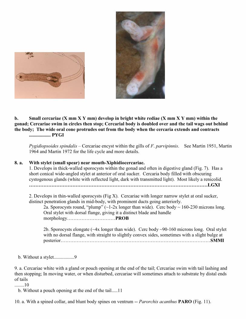
b. Small cercariae (X mm X Y mm) develop in bright white rediae (X mm X Y mm) within the
gonad; Cercariae swim in circles then stop; Cercarial body is doubled over and the tail wags out behind
the body; The wide oral cone protrudes out from the body when the cercaria extends and contracts
.................. PYGI
Pygidiopsoides spindalis – Cercariae encyst within the gills of F. parvipinnis. See Martin 1951, Martin
1964 and Martin 1972 for the life cycle and more details.
8. a. With stylet (small spear) near mouth-Xiphidiocercariae.
1. Develops in thick-walled sporocysts within the gonad and often in digestive gland (Fig. 7). Has a
short conical wide-angled stylet at anterior of oral sucker. Cercaria body filled with obscuring
cystogenous glands (white with reflected light, dark with transmitted light). Most likely a renicolid.
…………………………………………………………………………………………………LGXI
2. Develops in thin-walled sporocysts (Fig X). Cercariae with longer narrow stylet at oral sucker,
distinct penetration glands in mid-body, with prominent ducts going anteriorly.
2a. Sporocysts round, “plump” (~1-2x longer than wide). Cerc body ~ 160-230 microns long.
Oral stylet with dorsal flange, giving it a distinct blade and handle
morphology…………………………PROB
2b. Sporocysts elongate (~4x longer than wide). Cerc body ~90-160 microns long. Oral stylet
with no dorsal flange, with straight to slightly convex sides, sometimes with a slight bulge at
posterior…………………………………………………………………………………SMMI
b. Without a stylet.................9
9. a. Cercariae white with a gland or pouch opening at the end of the tail; Cercariae swim with tail lashing and
then stopping; In moving water, or when disturbed, cercariae will sometimes attach to substrate by distal ends
of tails
........10
b. Without a pouch opening at the end of the tail.....11
10. a. With a spined collar, and blunt body spines on ventrum -- Parorchis acanthus PARO (Fig. 11).
Develops in very large white rediae within the gonad of the snail. Few cercariae per redia. Swims as
body lashes back and forth fully extended in an "S" motion, similar to a nematode thrashing. Cercariae
encyst on almost any object. Use green filter on the light source of the microscope to better view spines
around the collar of the oral sucker. Cercaria has cystogenous glands dorsal to the acetabulum, making
an opaque “acetabular-window”. See Cable 1932, Cable and Martin 1935, and Martin 1972 for the life
cycle and more details. Also see Huspeni 2000 for information on multiple species of Parorchis in
North American Cerithidea spp.
b. Without a spined collar -- Cloacitrema michiganensis (CLOA)
Develops in white rediae within the gonad of the snail. Rediae and cercariae are smaller than P.
acanthus. Cloacitrema michiganensis cercariae do not have any body spines, nor spines around the oral
sucker. Cercaria appears clear or “see-through” at the region of the acetabulum (“acetabular-
window”). Cercariae encyst on almost any object.
11. a. With a spined collar...............12
b. Without a spined collar............13
12. a. Encysts in radular muscle of snail – Acanthoparyphium
spinulosum (Fig. 10). Develops in orange rediae within the gonad of the snail. Cercariae encyst in the
radular muscle of the snail and in clam feet. Swims much like HIMB but stops intermittently. Cercariae
and rediae are smaller than HIMB. Rediae appear orange under the dissecting microscope. Identified
by the absence of a dorso-ventral tail fin. See Martin and Adams 1960 and Martin 1972 for the life
cycle and more details.
b. Encysts in feces of the snail -- Acanthoparyphium sp. (Fig.
12). Develops in rediae within the gonad of the snail. Cercariae encyst in the snail feces and in certain
annelids. See Martin and Steele and Martin 1972 for the life cycle and more details. Huspeni doubts the
existence of this. Ryan and Josh are currently figuring out what the deal is with this one. Martin and his
student never published on this observation.
13. a. Can form clusters by adhesions of proximal portions of
tails, only after having voluntarily shed from the snail -- Renicola buchanani (Fig. 13). Develops in
large sporocysts within the mantle cavity of the snail. Sporocysts are bright orange. Cercariae encyst
within the liver of F. parvipinnis and Gillichthys mirabilis. See Martin 1971 and Martin 1972 for the
life cycle and more details.
b. Does not form clusters -- Renicola cerithidicola (Fig. 14).
Develops in orange sporocysts within the mantle wall of the snail. Cercariae are very small and they do
not have a stylet as some renicolids do. Sporocysts are “stickier” than sporocysts of R. buchanani, and
will usually stick to one another when snail is cracked and sporocysts are released. Cercariae encyst
within the gills of F. parvipinnis. See Martin 1971 and Martin 1972 for the life cycle and more details.
14. a. With dorso-ventral tail fin only............15
b. With dorso-ventral and lateral tail fins....16
15. a. With eye-spots -- Phocitremoides ovale (Fig. 15). Develops
in rediae within the gonad of the snail. Cercariae easily confused with Euhaplorchis californiensis
unless viewed under compound microscope, as swimming behaviors, rediae and cercarial sizes are very
similar. The collar that is found at the junction of the head and tail regions is not as prominent as the
figure shows. The oral sucker is very prominent, as is the V-shaped excretory bladder at the posterior
part of the head region. Cercariae encyst under the scales of F. parvipinnis and Atherinops californica.
See Martin 1950c and Martin 1972 for the life cycle and more details.
b. Without eyespots – Himasthla sp. B. (Fig. 17). Develops in
large rediae within the gonad of the snail. Rediae appear orange to whitish yellow under the dissecting
microscope, and there are few cercariae per redia. Cercariae swim continuously upon emerging from
rediae. Ventral sucker very prominent in cercariae. Identified easily on dissecting microscope by
presence of a large dorso-ventral tail fin. This fin is best observed on recently emerged cercariae that
have not yet begun to swim. Cercariae swim in tight circles with the body doubled over and the tail
lashing out behind. Martin (1972) attributed this cercaria to the genus Echinoparyphium, a freshwater
genus. Subsequent examination by Huspeni (2000) indicates this cercaria belongs in the genus
Himasthla. Metacercariae of this trematode have been found in naturally infected Acteocina inculta and
<10mm C. californica (Huspeni, per obs). Metacercariae of this trematode are also found in the heart
region in >10mm Cerithidea californica, but the metacercariae are nearly always dead.
16. a. Flame cells in groups of twos; penetration gland ducts begin in posterior of body: – Euhaplorchis
californiensis (Fig. 16). Develops in rediae within the gonad of the snail. Cercariae are identified by
looking for the lateral fins that are on the anterior part of the tail. Swims and stops within the water
column and also on the bottom of the dish. Flame cells are best seen near the eye spot between the spot
and the outer margin of the body, after the slide has dried out considerably and the cercaria is nearly
dead. T. Stevens has confirmed that the most common species found in CSMR is this one. Cercariae
encyst on the brain of F. parvipinnis, forming a layer of metacercariae that is one or two deep. This
layer can be easily peeled off the brain matter. See Martin 1950, Martin 1950a, Bamberg and Martin
1951 and Martin 1972 for the life cycle and more details.
b. Flame cells in groups of threes; penetration gland ducts anterior to excretory bladder;lateral fins wider than
EUHA– Stictodora (Parastictodora) hancocki (Fig. 18). Develops in rediae within the gonad of the snail.
Cercariae encyst in various tissues of F. parvipinnis and Gillichthys mirabilis. Rarely recorded from CSMR.
See Martin 1950b, Martin 1950 and Martin 1972 for the life cycle and more details.

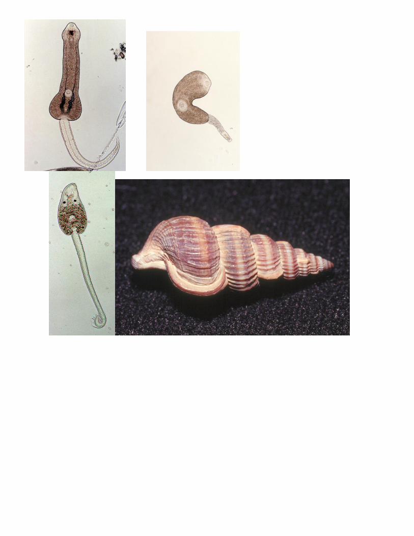
Some typical cercariae from infections in Cerithidea
spp.: