Low-Dose Radiation Exposure with 56MnO2 Powder Changes ...
10
International Journal of Molecular Sciences Article Low-Dose Radiation Exposure with 56 MnO 2 Powder Changes Gene Expressions in the Testes and the Prostate in Rats Nariaki Fujimoto 1, * , Gaukhar Amantayeva 2 , Nailya Chaizhunussova 2 , Dariya Shabdarbayeva 2 , Zhaslan Abishev 2 , Bakhyt Ruslanova 2 , Yersin Zhunussov 2 , Almas Azhimkhanov 3 , Kassym Zhumadilov 4 , Aleksey Petukhov 5 , Valeriy Stepanenko 5 and Masaharu Hoshi 6 1 Research Institute for Radiation Biology and Medicine, Hiroshima University, Hiroshima 7340037, Japan 2 Semey Medical University, Semey 071400, Kazakhstan; [email protected] (G.A.); [email protected] (N.C.); [email protected] (D.S.); [email protected] (Z.A.); [email protected] (B.R.); [email protected] (Y.Z.) 3 National Nuclear Center of the Republic of Kazakhstan, Kurchatov 071100, Kazakhstan; [email protected] 4 L.N. Gumilyov Eurasian National University, Nur-Sultan 010000, Kazakhstan; [email protected] 5 A. Tsyb Medical Radiological Research Center-National Medical Research Center of Radiology, Ministry of Health of Russian Federation, 249031 Obninsk, Russia; [email protected] (A.P.); [email protected] (V.S.) 6 The Center for Peace, Hiroshima University, Hiroshima 7300053, Japan; [email protected] * Correspondence: [email protected]; Tel./Fax: +81-(82)-257-5820 Received: 18 June 2020; Accepted: 13 July 2020; Published: 15 July 2020 Abstract: To investigate the biological effects of internal exposure of radioactive 56 MnO 2 powder, the major radioisotope dust in the soil after atomic bomb explosions, on male reproductive function, the gene expression of the testes and the prostate was examined. Ten-week-old male Wistar rats were exposed to three doses of radioactive 56 MnO 2 powder (41–100 mGy in whole body doses), stable MnO 2 powder, or external 60 Co γ-rays (2 Gy). Animals were necropsied on Days 3 and 61 postexposure. The mRNA expressions of testicular marker protein genes and prostatic secretory protein genes were quantified by Q-RT-PCR. On Day 3 postexposure, the testicular gene expressions of steroidogenesis-related enzymes, Cyp17a1 and Hsd3b1, decreased in 56 MnO 2 -exposed groups. Germ cell-specific Spag4 and Zpbp mRNA levels were also reduced. On postexposure Day 61, the Cyp11a1 gene expression became significantly reduced in the testes in the group exposed to the highest dose of 56 MnO 2 , while another steroidogenesis-related StAR gene mRNA level reduced in the 60 Co γ-rays group. There were no differences in Spag4 and Zpbp mRNA levels among groups on Day 61. No histopathological changes were observed in the testes in any group following exposure. Expression in the prostatic protein genes, including CRP1, KS3, and PSP94, significantly decreased in 56 MnO 2 -exposed groups as well as in the 60 Co γ-rays group on Day 61 postexposure. These data suggest that the internal exposure to 56 MnO 2 powder, at doses of less than 100 mGy, affected the gene expressions in the testis and the prostate, while 2 Gy of external γ-irradiation was less effective. Keywords: residual radiation; internal radiation exposure; radiation injury; testis; prostatic function; rat 1. Introduction Notwithstanding the effects of the initial radiation from the atomic bombings in Hiroshima and Nagasaki, there have been concerns regarding the potentially significant influence of the residual Int. J. Mol. Sci. 2020, 21, 4989; doi:10.3390/ijms21144989 www.mdpi.com/journal/ijms
Transcript of Low-Dose Radiation Exposure with 56MnO2 Powder Changes ...
International Journal of
Molecular Sciences
Article
Low-Dose Radiation Exposure with 56MnO2 PowderChanges Gene Expressions in the Testes and theProstate in Rats
Nariaki Fujimoto 1,* , Gaukhar Amantayeva 2, Nailya Chaizhunussova 2,Dariya Shabdarbayeva 2, Zhaslan Abishev 2, Bakhyt Ruslanova 2, Yersin Zhunussov 2,Almas Azhimkhanov 3, Kassym Zhumadilov 4, Aleksey Petukhov 5, Valeriy Stepanenko 5
and Masaharu Hoshi 6
1 Research Institute for Radiation Biology and Medicine, Hiroshima University, Hiroshima 7340037, Japan2 Semey Medical University, Semey 071400, Kazakhstan; [email protected] (G.A.); [email protected] (N.C.);
[email protected] (D.S.); [email protected] (Z.A.); [email protected] (B.R.);[email protected] (Y.Z.)
3 National Nuclear Center of the Republic of Kazakhstan, Kurchatov 071100, Kazakhstan; [email protected] L.N. Gumilyov Eurasian National University, Nur-Sultan 010000, Kazakhstan; [email protected] A. Tsyb Medical Radiological Research Center-National Medical Research Center of Radiology, Ministry of
Health of Russian Federation, 249031 Obninsk, Russia; [email protected] (A.P.);[email protected] (V.S.)
6 The Center for Peace, Hiroshima University, Hiroshima 7300053, Japan; [email protected]* Correspondence: [email protected]; Tel./Fax: +81-(82)-257-5820
Received: 18 June 2020; Accepted: 13 July 2020; Published: 15 July 2020�����������������
Abstract: To investigate the biological effects of internal exposure of radioactive 56MnO2 powder,the major radioisotope dust in the soil after atomic bomb explosions, on male reproductive function,the gene expression of the testes and the prostate was examined. Ten-week-old male Wistar ratswere exposed to three doses of radioactive 56MnO2 powder (41–100 mGy in whole body doses),stable MnO2 powder, or external 60Co γ-rays (2 Gy). Animals were necropsied on Days 3 and 61postexposure. The mRNA expressions of testicular marker protein genes and prostatic secretoryprotein genes were quantified by Q-RT-PCR. On Day 3 postexposure, the testicular gene expressionsof steroidogenesis-related enzymes, Cyp17a1 and Hsd3b1, decreased in 56MnO2-exposed groups.Germ cell-specific Spag4 and Zpbp mRNA levels were also reduced. On postexposure Day 61,the Cyp11a1 gene expression became significantly reduced in the testes in the group exposed to thehighest dose of 56MnO2, while another steroidogenesis-related StAR gene mRNA level reduced inthe 60Co γ-rays group. There were no differences in Spag4 and Zpbp mRNA levels among groups onDay 61. No histopathological changes were observed in the testes in any group following exposure.Expression in the prostatic protein genes, including CRP1, KS3, and PSP94, significantly decreased in56MnO2-exposed groups as well as in the 60Co γ-rays group on Day 61 postexposure. These datasuggest that the internal exposure to 56MnO2 powder, at doses of less than 100 mGy, affected the geneexpressions in the testis and the prostate, while 2 Gy of external γ-irradiation was less effective.
Keywords: residual radiation; internal radiation exposure; radiation injury; testis; prostaticfunction; rat
1. Introduction
Notwithstanding the effects of the initial radiation from the atomic bombings in Hiroshima andNagasaki, there have been concerns regarding the potentially significant influence of the residual
Int. J. Mol. Sci. 2020, 21, 4989; doi:10.3390/ijms21144989 www.mdpi.com/journal/ijms
Int. J. Mol. Sci. 2020, 21, 4989 2 of 10
radioactive dust on the health of those exposed. The people who moved to these cities a short timeafter detonation were only exposed to residual radiation, likely via inhaling radioactive dust, and werereported to suffer from acute radiation syndromes [1]. A primary source of residual radiation was56Mn, a radioisotope produced in the soil by the neutron beam from an atomic bomb explosion [2].We investigated the biological effects of neutron-activated 56MnO2 powder in Wistar rats to gain abetter understanding of the significance of residual radiation [3–5]. Our dosimetry data demonstratethat the highest absorbed doses from internal radiation were found in the gastrointestinal tract, skin,and lungs, while the highest whole-body dose was 100 mGy. Interestingly, at these low radiation doses,internal exposure to 56MnO2 significantly increased the serum alanine aminotransferase (ALT) level [3].Histopathological changes were also noted in the small intestine and lungs [4].
The male reproductive system is a sensitive target for radiation exposure [6–8]. The Chernobylaccident victims exposed to radioactive particles from the reactor (1–5.5 Gy) suffered from theimpairment of exocrine and endocrine testicular function [9]. The effects of residual radioactive dustafter an atomic bombing on the male function have been considered but are not yet understoodwell [10]. Therefore, we investigated the effects of 56MnO2 particles on the male function in our model.The rat spermatogenesis is disturbed in the testes by radiation exposure, although the sensitivities varydepending on the strain of rats, with lower sensitivities in SD and Wistar rats and higher sensitivities inBrown–Norway and Lewis rats [6,11]. As the majority of studies, including ours, use SD and Wistar rats,when examining the toxicological effect of chemicals or radiation, morphological changes in the testesare not a sensitive endpoint unless doses over 5 Gy of external radiation are applied. In our previousstudies of rats exposed to 56MnO2, testicular histology was examined on postexposure Days 3, 14, and60, without showing any significant changes as well [4]. However, recent studies demonstrated thatchanges in testicular gene expressions are useful markers to identify the functional changes in the testesdue to ionizing radiation or chemicals [12–14]. In the present study, mRNA levels were determined bythe quantitative RT-PCR to assess the effects of low-dose radiation exposure with 56MnO2 on the testes.In addition, mRNA expressions of prostatic secretory protein genes were measured in the prostate tofurther analyze the effects of 56MnO2 on male reproductive function. Animals were examined onlyon postexposure Days 3 and 60 in the present study to increase the number of rats in each group forquantitative analysis since the capacity of the 56MnO2 exposure system was limited.
2. Results
2.1. Estimated Doses of Internal Irradiation
The estimated accumulated doses of internal irradiation from 56MnO2 in each organ were describedpreviously [3]. The whole-body doses of internal irradiation were 41 ± 8, 91 ± 3, and 100 ± 10 mGy inMn56×1, Mn56×2 and Mn56×3 groups, respectively. The higher absorbed doses were found in thecolon (90 ± 61, 520 ± 110, and 760 ± 170 mGy) and skin (71 ± 23, 110 ± 2.3, and 140 ± 170 mGy) forthe Mn56×1, Mn56×2, and Mn56×3 groups, respectively. The calculated absorbed doses for the testesand the prostate were less than 0.3 mGy (Mn56×1 group), 0.6 mGy (Mn56×2 group), and 1.0 mGy(Mn56×3 group).
2.2. Body and Testes Weight and Serum Testosterone Level
Body weight and relative testes weight on Days 3 and 61 after exposure are summarized in Table 1.There were no significant differences in the testes weight on either day. Serum testosterone levelsdecreased significantly in the Mn56×2 and Co-60 groups on Day 61 postexposure.
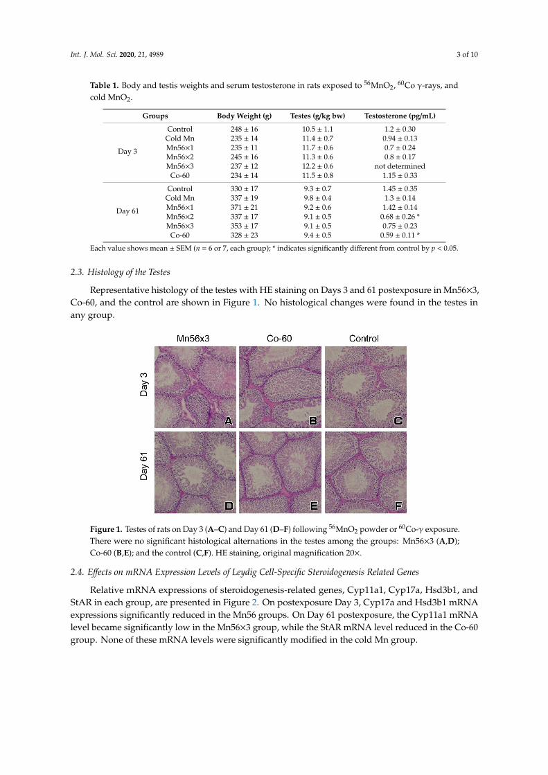
Int. J. Mol. Sci. 2020, 21, 4989 3 of 10
Table 1. Body and testis weights and serum testosterone in rats exposed to 56MnO2, 60Co γ-rays, andcold MnO2.
Groups Body Weight (g) Testes (g/kg bw) Testosterone (pg/mL)
Day 3
Control 248 ± 16 10.5 ± 1.1 1.2 ± 0.30Cold Mn 235 ± 14 11.4 ± 0.7 0.94 ± 0.13Mn56×1 235 ± 11 11.7 ± 0.6 0.7 ± 0.24Mn56×2 245 ± 16 11.3 ± 0.6 0.8 ± 0.17Mn56×3 237 ± 12 12.2 ± 0.6 not determined
Co-60 234 ± 14 11.5 ± 0.8 1.15 ± 0.33
Day 61
Control 330 ± 17 9.3 ± 0.7 1.45 ± 0.35Cold Mn 337 ± 19 9.8 ± 0.4 1.3 ± 0.14Mn56×1 371 ± 21 9.2 ± 0.6 1.42 ± 0.14Mn56×2 337 ± 17 9.1 ± 0.5 0.68 ± 0.26 *Mn56×3 353 ± 17 9.1 ± 0.5 0.75 ± 0.23
Co-60 328 ± 23 9.4 ± 0.5 0.59 ± 0.11 *
Each value shows mean ± SEM (n = 6 or 7, each group); * indicates significantly different from control by p < 0.05.
2.3. Histology of the Testes
Representative histology of the testes with HE staining on Days 3 and 61 postexposure in Mn56×3,Co-60, and the control are shown in Figure 1. No histological changes were found in the testes inany group.
Int. J. Mol. Sci. 2020, 21, x 3 of 10
Table 1. Body and testis weights and serum testosterone in rats exposed to 56MnO2, 60Co γ-rays, and cold MnO2.
Groups Body Weight (g) Testes (g/kg bw) Testosterone (pg/mL)
Day 3
Control 248 ± 16 10.5 ± 1.1 1.2 ± 0.30 Cold Mn 235 ± 14 11.4 ± 0.7 0.94 ± 0.13 Mn56×1 235 ± 11 11.7 ± 0.6 0.7 ± 0.24 Mn56×2 245 ± 16 11.3 ± 0.6 0.8 ± 0.17 Mn56×3 237 ± 12 12.2 ± 0.6 not determined Co-60 234 ± 14 11.5 ± 0.8 1.15 ± 0.33
Day 61
Control 330 ± 17 9.3 ± 0.7 1.45 ± 0.35 Cold Mn 337 ± 19 9.8 ± 0.4 1.3 ± 0.14 Mn56×1 371 ± 21 9.2 ± 0.6 1.42 ± 0.14 Mn56×2 337 ± 17 9.1 ± 0.5 0.68 ± 0.26 * Mn56×3 353 ± 17 9.1 ± 0.5 0.75 ± 0.23 Co-60 328 ± 23 9.4 ± 0.5 0.59 ± 0.11 *
Each value shows mean ± SEM (n = 6 or 7, each group); * indicates significantly different from control by p < 0.05.
2.3. Histology of the Testes
Representative histology of the testes with HE staining on Days 3 and 61 postexposure in Mn56×3, Co-60, and the control are shown in Figure 1. No histological changes were found in the testes in any group.
Figure 1. Testes of rats on Day 3 (A–C) and Day 61 (D–F) following 56MnO2 powder or 60Co-γ exposure. There were no significant histological alternations in the testes among the groups: Mn56×3 (A,D); Co-60 (B,E); and the control (C,F). HE staining, original magnification 20×.
2.4. Effects on mRNA Expression Levels of Leydig Cell-Specific Steroidogenesis Related Genes
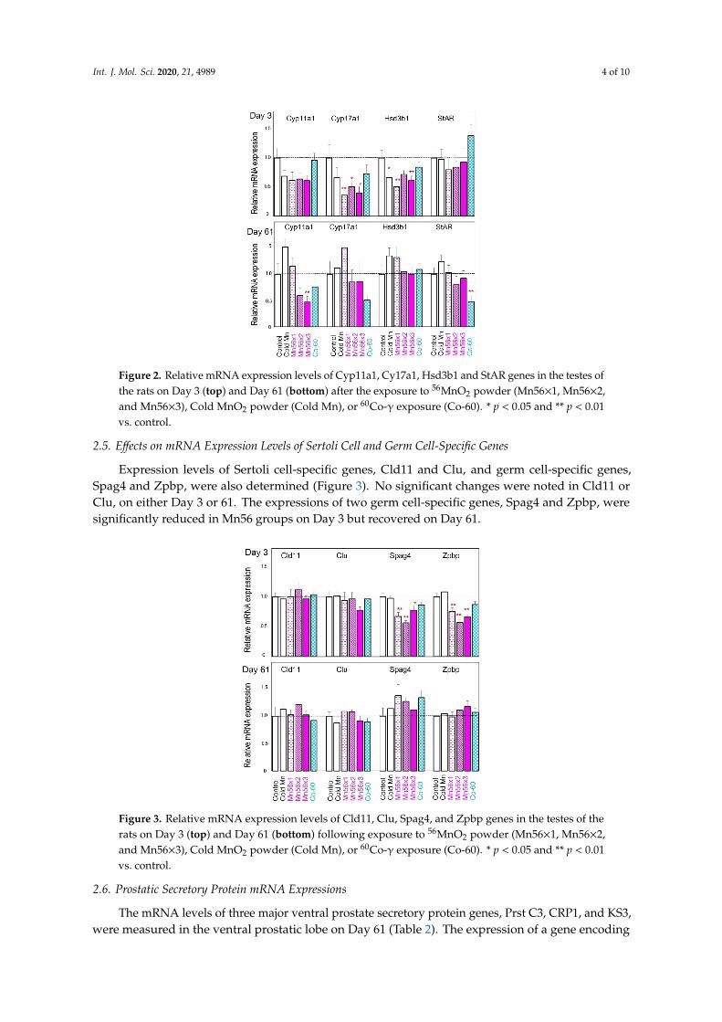
Relative mRNA expressions of steroidogenesis-related genes, Cyp11a1, Cyp17a, Hsd3b1, and StAR in each group, are presented in Figure 2. On postexposure Day 3, Cyp17a and Hsd3b1 mRNA expressions significantly reduced in the Mn56 groups. On Day 61 postexposure, the Cyp11a1 mRNA level became significantly low in the Mn56×3 group, while the StAR mRNA level reduced in the Co-60 group. None of these mRNA levels were significantly modified in the cold Mn group.
Figure 1. Testes of rats on Day 3 (A–C) and Day 61 (D–F) following 56MnO2 powder or 60Co-γ exposure.There were no significant histological alternations in the testes among the groups: Mn56×3 (A,D);Co-60 (B,E); and the control (C,F). HE staining, original magnification 20×.
2.4. Effects on mRNA Expression Levels of Leydig Cell-Specific Steroidogenesis Related Genes
Relative mRNA expressions of steroidogenesis-related genes, Cyp11a1, Cyp17a, Hsd3b1, andStAR in each group, are presented in Figure 2. On postexposure Day 3, Cyp17a and Hsd3b1 mRNAexpressions significantly reduced in the Mn56 groups. On Day 61 postexposure, the Cyp11a1 mRNAlevel became significantly low in the Mn56×3 group, while the StAR mRNA level reduced in the Co-60group. None of these mRNA levels were significantly modified in the cold Mn group.
Int. J. Mol. Sci. 2020, 21, 4989 4 of 10Int. J. Mol. Sci. 2020, 21, x 4 of 10
Figure 2. Relative mRNA expression levels of Cyp11a1, Cy17a1, Hsd3b1 and StAR genes in the testes of the rats on Day 3 (top) and Day 61 (bottom) after the exposure to 56MnO2 powder (Mn56×1, Mn56×2, and Mn56×3), Cold MnO2 powder (Cold Mn), or 60Co-γ exposure (Co-60). * p < 0.05 and ** p < 0.01 vs. control.
2.5. Effects on mRNA Expression Levels of Sertoli Cell and Germ Cell-Specific Genes
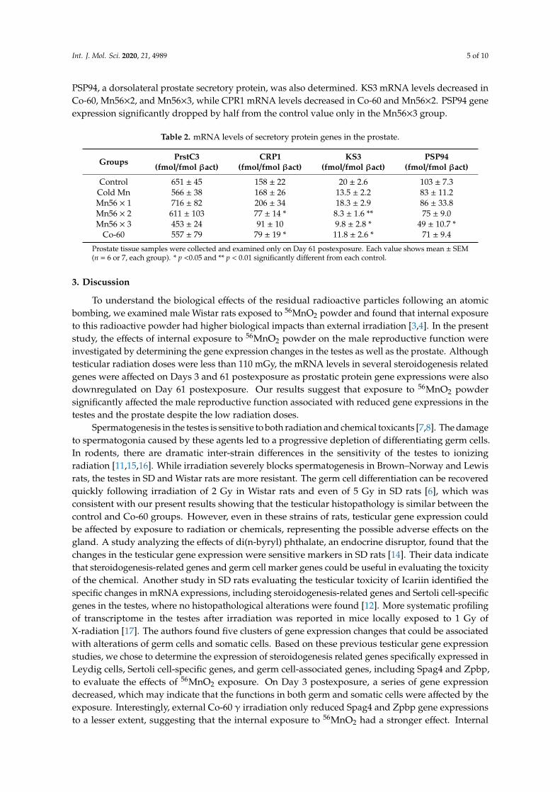
Expression levels of Sertoli cell-specific genes, Cld11 and Clu, and germ cell-specific genes, Spag4 and Zpbp, were also determined (Figure 3). No significant changes were noted in Cld11 or Clu, on either Day 3 or 61. The expressions of two germ cell-specific genes, Spag4 and Zpbp, were significantly reduced in Mn56 groups on Day 3 but recovered on Day 61.
Figure 3. Relative mRNA expression levels of Cld11, Clu, Spag4, and Zpbp genes in the testes of the rats on Day 3 (top) and Day 61 (bottom) following exposure to 56MnO2 powder (Mn56×1, Mn56×2, and Mn56×3), Cold MnO2 powder (Cold Mn), or 60Co-γ exposure (Co-60). * p < 0.05 and ** p < 0.01 vs. control.
Figure 2. Relative mRNA expression levels of Cyp11a1, Cy17a1, Hsd3b1 and StAR genes in the testes ofthe rats on Day 3 (top) and Day 61 (bottom) after the exposure to 56MnO2 powder (Mn56×1, Mn56×2,and Mn56×3), Cold MnO2 powder (Cold Mn), or 60Co-γ exposure (Co-60). * p < 0.05 and ** p < 0.01vs. control.
2.5. Effects on mRNA Expression Levels of Sertoli Cell and Germ Cell-Specific Genes
Expression levels of Sertoli cell-specific genes, Cld11 and Clu, and germ cell-specific genes,Spag4 and Zpbp, were also determined (Figure 3). No significant changes were noted in Cld11 orClu, on either Day 3 or 61. The expressions of two germ cell-specific genes, Spag4 and Zpbp, weresignificantly reduced in Mn56 groups on Day 3 but recovered on Day 61.
Int. J. Mol. Sci. 2020, 21, x 4 of 10
Figure 2. Relative mRNA expression levels of Cyp11a1, Cy17a1, Hsd3b1 and StAR genes in the testes of the rats on Day 3 (top) and Day 61 (bottom) after the exposure to 56MnO2 powder (Mn56×1, Mn56×2, and Mn56×3), Cold MnO2 powder (Cold Mn), or 60Co-γ exposure (Co-60). * p < 0.05 and ** p < 0.01 vs. control.
2.5. Effects on mRNA Expression Levels of Sertoli Cell and Germ Cell-Specific Genes
Expression levels of Sertoli cell-specific genes, Cld11 and Clu, and germ cell-specific genes, Spag4 and Zpbp, were also determined (Figure 3). No significant changes were noted in Cld11 or Clu, on either Day 3 or 61. The expressions of two germ cell-specific genes, Spag4 and Zpbp, were significantly reduced in Mn56 groups on Day 3 but recovered on Day 61.
Figure 3. Relative mRNA expression levels of Cld11, Clu, Spag4, and Zpbp genes in the testes of the rats on Day 3 (top) and Day 61 (bottom) following exposure to 56MnO2 powder (Mn56×1, Mn56×2, and Mn56×3), Cold MnO2 powder (Cold Mn), or 60Co-γ exposure (Co-60). * p < 0.05 and ** p < 0.01 vs. control.
Figure 3. Relative mRNA expression levels of Cld11, Clu, Spag4, and Zpbp genes in the testes of therats on Day 3 (top) and Day 61 (bottom) following exposure to 56MnO2 powder (Mn56×1, Mn56×2,and Mn56×3), Cold MnO2 powder (Cold Mn), or 60Co-γ exposure (Co-60). * p < 0.05 and ** p < 0.01vs. control.
2.6. Prostatic Secretory Protein mRNA Expressions
The mRNA levels of three major ventral prostate secretory protein genes, Prst C3, CRP1, and KS3,were measured in the ventral prostatic lobe on Day 61 (Table 2). The expression of a gene encoding
Int. J. Mol. Sci. 2020, 21, 4989 5 of 10
PSP94, a dorsolateral prostate secretory protein, was also determined. KS3 mRNA levels decreased inCo-60, Mn56×2, and Mn56×3, while CPR1 mRNA levels decreased in Co-60 and Mn56×2. PSP94 geneexpression significantly dropped by half from the control value only in the Mn56×3 group.
Table 2. mRNA levels of secretory protein genes in the prostate.
Groups PrstC3(fmol/fmol βact)
CRP1(fmol/fmol βact)
KS3(fmol/fmol βact)
PSP94(fmol/fmol βact)
Control 651 ± 45 158 ± 22 20 ± 2.6 103 ± 7.3Cold Mn 566 ± 38 168 ± 26 13.5 ± 2.2 83 ± 11.2Mn56 × 1 716 ± 82 206 ± 34 18.3 ± 2.9 86 ± 33.8Mn56 × 2 611 ± 103 77 ± 14 * 8.3 ± 1.6 ** 75 ± 9.0Mn56 × 3 453 ± 24 91 ± 10 9.8 ± 2.8 * 49 ± 10.7 *
Co-60 557 ± 79 79 ± 19 * 11.8 ± 2.6 * 71 ± 9.4
Prostate tissue samples were collected and examined only on Day 61 postexposure. Each value shows mean ± SEM(n = 6 or 7, each group). * p <0.05 and ** p < 0.01 significantly different from each control.
3. Discussion
To understand the biological effects of the residual radioactive particles following an atomicbombing, we examined male Wistar rats exposed to 56MnO2 powder and found that internal exposureto this radioactive powder had higher biological impacts than external irradiation [3,4]. In the presentstudy, the effects of internal exposure to 56MnO2 powder on the male reproductive function wereinvestigated by determining the gene expression changes in the testes as well as the prostate. Althoughtesticular radiation doses were less than 110 mGy, the mRNA levels in several steroidogenesis relatedgenes were affected on Days 3 and 61 postexposure as prostatic protein gene expressions were alsodownregulated on Day 61 postexposure. Our results suggest that exposure to 56MnO2 powdersignificantly affected the male reproductive function associated with reduced gene expressions in thetestes and the prostate despite the low radiation doses.
Spermatogenesis in the testes is sensitive to both radiation and chemical toxicants [7,8]. The damageto spermatogonia caused by these agents led to a progressive depletion of differentiating germ cells.In rodents, there are dramatic inter-strain differences in the sensitivity of the testes to ionizingradiation [11,15,16]. While irradiation severely blocks spermatogenesis in Brown–Norway and Lewisrats, the testes in SD and Wistar rats are more resistant. The germ cell differentiation can be recoveredquickly following irradiation of 2 Gy in Wistar rats and even of 5 Gy in SD rats [6], which wasconsistent with our present results showing that the testicular histopathology is similar between thecontrol and Co-60 groups. However, even in these strains of rats, testicular gene expression couldbe affected by exposure to radiation or chemicals, representing the possible adverse effects on thegland. A study analyzing the effects of di(n-byryl) phthalate, an endocrine disruptor, found that thechanges in the testicular gene expression were sensitive markers in SD rats [14]. Their data indicatethat steroidogenesis-related genes and germ cell marker genes could be useful in evaluating the toxicityof the chemical. Another study in SD rats evaluating the testicular toxicity of Icariin identified thespecific changes in mRNA expressions, including steroidogenesis-related genes and Sertoli cell-specificgenes in the testes, where no histopathological alterations were found [12]. More systematic profilingof transcriptome in the testes after irradiation was reported in mice locally exposed to 1 Gy ofX-radiation [17]. The authors found five clusters of gene expression changes that could be associatedwith alterations of germ cells and somatic cells. Based on these previous testicular gene expressionstudies, we chose to determine the expression of steroidogenesis related genes specifically expressed inLeydig cells, Sertoli cell-specific genes, and germ cell-associated genes, including Spag4 and Zpbp,to evaluate the effects of 56MnO2 exposure. On Day 3 postexposure, a series of gene expressiondecreased, which may indicate that the functions in both germ and somatic cells were affected by theexposure. Interestingly, external Co-60 γ irradiation only reduced Spag4 and Zpbp gene expressionsto a lesser extent, suggesting that the internal exposure to 56MnO2 had a stronger effect. Internal
Int. J. Mol. Sci. 2020, 21, 4989 6 of 10
exposure to 56MnO2 and external exposure to 60Co-γ rays had a differential impact on long-term geneexpressions in the testes. On Day 61 postexposure, the Cyp11a1 mRNA level was significantly lowerin the Mn56×3 group, while StAR gene expression became significantly reduced in the Co-60 group.A recent study examining the testicular gene expression in Wistar rats also found a similar effect:a marked decrease in StAR gene expression after gamma irradiation [13]. It is interesting that bothinternal 56MnO2 exposure and external γ-ray exposure affect the steroidogenesis gene expressions,likely resulting in reduced testosterone production. The functional changes in Leydig cells by ionizingradiation are not likely related to the histological changes in the testes [18,19]. Similarly, the alterationin steroidogenesis related gene expressions regulating the Leydig cell function may not lead to anymorphological changes, as shown in this study. Previous investigations suggested that Sertoli cells aremore resistant to radiation than Leydig cells in rats [18,19], which correlates with our gene expressionresults, showing that Cld11 and Clu gene mRNA levels did no changes after exposure, save for a slightreduction in Clu mRNA level in Mn56×3 on Day 3.
To further evaluate the effect of 56MnO2 on male reproductive function, we examined prostaticactivities. The rodent prostate consists of morphologically-separated ventral, dorsolateral, andanterior (coagulating gland) portions, each of which secretes different proteins representing prostaticdevelopment and activity [20]. We determined mRNA levels of ventral prostatic secreted proteins,prstC3, CRP1, and KS3, and a dorsolateral prostatic protein, PSP94. These mRNA expressionssignificantly decreased in the Mn56×3 group in Day 61 postexposure, suggesting that the 56MnO2
exposure posed the prostatic function, probably via the reduction in serum testosterone levels.The gene expressions significantly changed in the testes and the prostate in Mn56 groups, despite
their low internal radiation doses (less than 1 mGy). In this respect, it should be noted that estimationsof internal irradiation of each organ were based upon the measured radioactivity of the organs.However, in the case of the testes, which were anatomically located outside of the body not beingretracted inside, as the animals were asleep during the exposure period, they were additionallysubjected to irradiation from adjacent skin directly in contact with 56MnO2 powder. Consideringthe maximal energy of 2.85 MeV of β rays from 56Mn with a range of approximately 4 mm in thetissue, the additional testicular dose should have been 110 mGy in the Mn56×3 group according tothe calculation method we previously reported [21]. However, this was not the case in the prostate,an internal organ. The reduction in mRNA levels of prostatic proteins was likely the result of decreasedserum testosterone levels.
The body distribution of certain radionuclides following internal exposure depends on theirspecific chemical nature [22,23] When rats are exposed to stable and insoluble molecules such as MnO2,these molecules are delivered to the gastrointestinal tract, skin, and lungs. It is believed that irradiationfrom radioactive particles may be less hazardous in terms of carcinogenesis than the same activityuniformly distributed [24]. However, radioactive particles could induce stronger biological impacts in“target organs”, as we previously found histological changes in the lung and the small intestine in ratsinternally exposed to 56MnO2 powder [24]. The present study again indicated high biological impactson certain target organs by the radioactive particles. The effects on the testicular gene expression by56MnO2 were evidently higher than the effects caused by 2 Gy of external γ irradiation.
Concerns of the effects of ionizing radiation on male reproductive function have been raised asradiation exposure becomes more common in both diagnostic and therapeutic procedures [25]. Humantestes are known to be more radiosensitive than those of rodents. Human spermatogenesis could bedisturbed following external irradiation as low as 0.3 Gy [26]. Doses greater than 1 Gy significantlyreduced the number of spermatogonia and spermatocytes, which require 9–19 months to recover.In cases of victims of the Chernobyl accident, one study examined 12 men receiving doses varyingfrom 1 to 5.5 Gy. It reported that the radiation exposure was associated with impairment of exocrineand endocrine testicular function [9]. Another study investigated men who cleaned the region aroundthe reactor and received doses of 0.16 ± 0.08 Gy; it did not find any disruption in the endocrine statusand spermatogenesis. However, this study was conducted 7–9 years following the exposure [27].
Int. J. Mol. Sci. 2020, 21, 4989 7 of 10
People in northeastern Kazakhstan adjacent to the Semipalatinsk nuclear test site had been exposed toradioactive fallout after nuclear tests conducted by the former Soviet Union [28]. One study examinedthe sex ratio in the offspring of these people as an indicator of the radiation effect on reproductive healthand did not find any significant changes [10]. However, this result does not exclude the possibility ofadverse effects caused by radiation fallout on the reproductive function in the exposed population,since sex-linked lethal mutations in germ cells, which require high doses of radiation exposure, havebeen postulated to cause changes in the sex ratio of the offspring [29]. The present study demonstratedthat rats exposed to 56MnO2 powder decreased the gene expressions related to male reproductivefunction, including steroidogenesis related genes in Leydig cells and prostatic secreted protein genes.Further studies are recommended to determine whether these changes lead to any adverse effect onmale reproductive health.
4. Materials and Methods
4.1. Animals
The animal experiment design was described previously [3]. Ten-week-old male Wistar rats werepurchased from Kazakh Scientific Center of Quarantine and Zoonotic Diseases, Almaty, Kazakhstan.Animals were housed in plastic cages (two rats/cage) and maintained with free access to basal dietand tap water. The room was maintained at 19–22 ◦C with a relative humidity of 30–70% and a12-h light cycle. The animal facility was of the conventional type, although the purchased rats werespecific-pathogen free animals. Rats were randomly divided into 6 groups, adjusting the averagebody weights to be similar among groups: Mn56×1 group (n = 17), Mn56×2 group (n = 17), Mn56×3group (n = 17), Co-60 group (n = 14), cold Mn group (n = 14), and control group (n = 14). Mn56×1,Mn56×2, and Mn56×3 groups were exposed to 3 different activities of 56MnO2 powder (100 mg) of2.7 × 108, 5.5 × 10⁸, and 8 × 10⁸ Bq, respectively. The cold Mn group was exposed to nonradioactiveMnO2 powder (100 mg). Three rats from each Mn56 group were necropsied to assess absorbeddose 0.5 h following exposure. The Co-60 group received 2 Gy of external 60Co γ-ray whole-bodyirradiation. From each group, 7 rats were necropsied on postexposure Days 3 and 61 between 11:00and 17:00. The rats were placed into a box containing isoflurane anesthetic gas (Fujifilm Wako PureChemical Co., Tokyo, Japan), and then euthanized by removing and collecting whole blood from anabdominal artery. The testes, the ventral prostate, the dorsolateral prostate, and the seminal vesicleswere dissected. Half of the testes were stored in RNA Save solution (Biological Industries Ltd., Beit Alfa,Israel) for RNA extraction, and the other half was fixed in 10% formalin and embedded in paraffin.Sections of 4 µm thickness were prepared and stained with hematoxylin and eosin. Prostate tissuesamples were collected only on Day 61 and stored in RNA Save solution. Ethics Committee Approval(document #3-30.11.2018) was received from the Animal Experiment Ethics Committee of SemeyMedical University, Semey Kazakhstan.
4.2. Irradiation and Dosimetry
Details of irradiation using 56MnO2 powder and the internal dose estimation have been describedpreviously [5]. Briefly, MnO2 powder (Rare Metallic Co., Tokyo, Japan, particle diameters ranging from1–19 µm) was radio-activated by neutron beam in the Baikal-1 nuclear reactor at the National NuclearCenter, Kurchatov, Kazakhstan. Thermal neutron fluencies of 4 × 10¹⁴, 8 × 10¹⁴, and 1.2 × 10¹⁵ n/cm2
were applied to each 100 mg of MnO2 powder to produce 56MnO2 with activities of 2.7 × 10⁸, 5.5 × 10⁸,and 8 × 10⁸ Bq/100 mg (for Mn56×1, Mn56×2, and Mn56×3 groups, respectively). This 56MnO2 powderwas then air-pressure sprayed into sealed exposure boxes containing 8 or 9 rats per box. Following1 h of exposure, the animals were moved to new cages. Thirty minutes later, three rats per groupwere euthanized. A section of each organ was dissected, and the radioactivity was measured with aγ-spectrometer. Absorbed fractions of energy from β and γ-irradiation of 56Mn in each organ, as wellas that for the whole body, were calculated using the Monte Carlo code (version MCNP-4C) and the
Int. J. Mol. Sci. 2020, 21, 4989 8 of 10
mathematical phantom of the rat. Absorbed doses of internal radiation were estimated on the basis ofmeasured radioactivity of 56Mn in each organ using calculated values of absorbed fractions of energyin these organs. All groups of animals were brought to the 56Mn exposure facility. A 60Co radiotherapymachine, Teragam K-2 unit (UJP Praha, Praha-Zbraslav, Czech Republic), was used for whole-bodyγ-ray irradiation of 2 Gy (1.0 Gy/min).
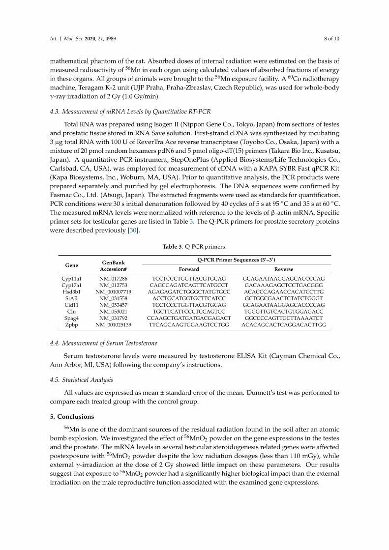
4.3. Measurement of mRNA Levels by Quantitative RT-PCR
Total RNA was prepared using Isogen II (Nippon Gene Co., Tokyo, Japan) from sections of testesand prostatic tissue stored in RNA Save solution. First-strand cDNA was synthesized by incubating3 µg total RNA with 100 U of ReverTra Ace reverse transcriptase (Toyobo Co., Osaka, Japan) with amixture of 20 pmol random hexamers pdN6 and 5 pmol oligo-dT(15) primers (Takara Bio Inc., Kusatsu,Japan). A quantitative PCR instrument, StepOnePlus (Applied Biosystems/Life Technologies Co.,Carlsbad, CA, USA), was employed for measurement of cDNA with a KAPA SYBR Fast qPCR Kit(Kapa Biosystems, Inc., Woburn, MA, USA). Prior to quantitative analysis, the PCR products wereprepared separately and purified by gel electrophoresis. The DNA sequences were confirmed byFasmac Co., Ltd. (Atsugi, Japan). The extracted fragments were used as standards for quantification.PCR conditions were 30 s initial denaturation followed by 40 cycles of 5 s at 95 ◦C and 35 s at 60 ◦C.The measured mRNA levels were normalized with reference to the levels of β-actin mRNA. Specificprimer sets for testicular genes are listed in Table 3. The Q-PCR primers for prostate secretory proteinswere described previously [30].
Table 3. Q-PCR primers.
Gene GenBankAccession#
Q-PCR Primer Sequences (5′–3′)
Forward Reverse
Cyp11a1 NM_017286 TCCTCCCTGGTTACGTGCAG GCAGAATAAGGAGCACCCCAGCyp17a1 NM_012753 CAGCCAGATCAGTTCATGCCT GACAAAGAGCTCCTGACGGGHsd3b1 NM_001007719 AGAGAGATCTGGGCTATGTGCC ACACCCAGAACCACATCCTTG
StAR NM_031558 ACCTGCATGGTGCTTCATCC GCTGGCGAACTCTATCTGGGTCld11 NM_053457 TCCTCCCTGGTTACGTGCAG GCAGAATAAGGAGCACCCCAG
Clu NM_053021 TGCTTCATTCCCTCCAGTCC TGGGTTGTCACTGTGGAGACCSpag4 NM_031792 CCAAGCTGATGATGACGAGACT GGCCCCAGTTGCTTAAAATCTZpbp NM_001025139 TTCAGCAAGTGGAAGTCCTGG ACACAGCACTCAGGACACTTGG
4.4. Measurement of Serum Testosterone
Serum testosterone levels were measured by testosterone ELISA Kit (Cayman Chemical Co.,Ann Arbor, MI, USA) following the company’s instructions.
4.5. Statistical Analysis
All values are expressed as mean ± standard error of the mean. Dunnett’s test was performed tocompare each treated group with the control group.
5. Conclusions
56Mn is one of the dominant sources of the residual radiation found in the soil after an atomicbomb explosion. We investigated the effect of 56MnO2 powder on the gene expressions in the testesand the prostate. The mRNA levels in several testicular steroidogenesis related genes were affectedpostexposure with 56MnO2 powder despite the low radiation dosages (less than 110 mGy), whileexternal γ-irradiation at the dose of 2 Gy showed little impact on these parameters. Our resultssuggest that exposure to 56MnO2 powder had a significantly higher biological impact than the externalirradiation on the male reproductive function associated with the examined gene expressions.
Int. J. Mol. Sci. 2020, 21, 4989 9 of 10
Author Contributions: Conceptualization, M.H., and V.S.; Methodology, N.F., and V.S.; Investigation and analysis,N.F, G.A., Z.A., B.R., A.A., K.Z., A.P., and V.S.; Writing—original draft preparation, N.F.; Supervision, N.C., andD.S.; and Funding acquisition, N.C., Y.Z., M.H., and N.F. All authors have read the manuscript and agree tobe published.
Funding: This work was supported by Semey Medical University, Kazakhstan, JSPS Open Partnership JointResearch Projects Grants (H30-31), and JSPS Kakenhi Grants (19H01149 and 19KK0266).
Acknowledgments: The authors thank Vyacheslav Gnyrya and Alexander Kolbayenkov at National NuclearCenter of the Republic of Kazakhstan in Kurchatov for their excellent technical help exposing animals to 56MnO2powder and Dastan Erzhanov and Oryngazy Aliyev at Semey Medical University for their expert technicalassistance at the necropsy of animals.
Conflicts of Interest: All authors have read the journal’s authorship statement and policy on disclosure of potentialconflicts of interest and have disclosed no conflict of interest.
References
1. Imanaka, T.; Endo, S.; Tanaka, K.; Shizuma, K. Gamma-ray exposure from neutron-induced radionuclides insoil in Hiroshima and Nagasaki based on DS02 calculations. Radiat. Environ. Biophys. 2008, 47, 331–336.[CrossRef] [PubMed]
2. Tanaka, K.; Endo, S.; Imanaka, T.; Shizuma, K.; Hasai, H.; Hoshi, M. Skin dose from neutron-activatedsoil for early entrants following the A-bomb detonation in Hiroshima: Contribution from β and γ rays.Radiat. Environ. Biophys. 2008, 47, 323–330. [CrossRef] [PubMed]
3. Fujimoto, N.; Baurzhan, A.; Chaizhunusova, N.; Amantayeva, G.; Kairkhanova, Y.; Shabdarbaeva, D.;Zhunussov, Y.; Zhumadilov, K.; Stepanenko, V.; Gnyrya, V.; et al. Effects of Internal Exposure to 56MnO2
Powder on Blood Parameters in Rats. Eurasian J. Med. 2020, 52, 52–56. [CrossRef]4. Shichijo, K.; Fujimoto, N.; Uzbekov, D.; Kairkhanova, Y.; Saimova, A.; Chaizhunusova, N.; Sayakenov, N.;
Shabdarbaeva, D.; Aukenov, N.; Azimkhanov, A.; et al. Internal exposure to neutron-activated 56Mn dioxidepowder in Wistar rats—Part 2: Pathological effects. Radiat. Environ. Biophys. 2017, 56, 55–61. [CrossRef][PubMed]
5. Stepanenko, V.; Rakhypbekov, T.; Otani, K.; Endo, S.; Satoh, K.; Kawano, N.; Shichijo, K.; Nakashima, M.;Takatsuji, T.; Sakaguchi, A.; et al. Internal exposure to neutron-activated 56Mn dioxide powder in Wistarrats: Part 1: Dosimetry. Radiat. Environ. Biophys. 2017, 56, 47–54. [CrossRef] [PubMed]
6. Delic, J.I.; Schlappack, O.K.; Harwood, J.R.; Stanley, J.A. Comparative effects of X irradiation on the testes ofadult Sprague-Dawley and Wistar rats. Radiat. Res. 1987, 112, 99–104. [CrossRef]
7. Gunn, S.A.; Gould, T.C.; Anderson, W.A. The effect of x-irradiation on the morphology and function of therat testis. Am. J. Pathol. 1960, 37, 203–213. [PubMed]
8. Huckins, C. Behavior of Stem Cell Spermatogonia in the Adult Rat Irradiated Testis. Biol. Reprod. 1978, 19,747–760. [CrossRef]
9. Birioukov, A.; Meurer, M.; Peter, R.U.; Braun-Falco, O.; Plewig, G. Male reproductive system in patientsexposed to ionizing irradiation in the chernobyl accident. Syst. Biol. Reprod. Med. 1993, 30, 99–104. [CrossRef]
10. Mudie, N.Y.; Gusev, B.I.; Pivina, L.M.; Schoemaker, M.J.; Rijinkova, O.N.; Apsalikov, K.N.; Swerdlow, A.J.Sex Ratio in the Offspring of Parents with Chronic Radiation Exposure from Nuclear Testing in Kazakhstan.Radiat. Res. 2007, 168, 600–607. [CrossRef]
11. Abuelhija, M.; Weng, C.C.; Shetty, G.; Meistrich, M.L. Differences in radiation sensitivity of recovery ofspermatogenesis between rat strains. Toxicol. Sci. 2012, 126, 545–553. [CrossRef]
12. Chen, M.; Hao, J.; Yang, Q.; Li, G. Effects of icariin on reproductive functions in male rats. Molecules 2014, 19,9502–9514. [CrossRef] [PubMed]
13. Ibrahim, A.A.; Karam, H.M.; Shaaban, E.A.; Safar, M.M.; El-Yamany, M.F. MitoQ ameliorates testiculardamage induced by gamma irradiation in rats: Modulation of mitochondrial apoptosis and steroidogenesis.Life Sci. 2019, 232, 116655. [CrossRef] [PubMed]
14. Ryu, J.Y.; Lee, B.M.; Kacew, S.; Kim, H.S. Identification of differentially expressed genes in the testis ofSprague-Dawley rats treated with di(n-butyl) phthalate. Toxicology 2007, 234, 103–112. [CrossRef] [PubMed]
15. Malashenko, A.M.; Beskova, T.B.; Pomerantseva, M.D.; Ramaiya, L.K. Comparison of Three Inbred MouseStrains 1053. Russ. J. Genet. 2003, 39, 1052–1055. [CrossRef]
Int. J. Mol. Sci. 2020, 21, 4989 10 of 10
16. Meistrich, M.L.; Finch, M.; Lu, C.C.; de Ruiter-Bootsma, A.L.; de Rooij, D.G.; Davids, J.A.G. Strain Differencesin the Response of Mouse Testicular Stem Cells to Fractionated Radiation. Radiat. Res. 1984, 97, 478.[CrossRef]
17. Belling, K.C.; Tanaka, M.; Dalgaard, M.D.; Nielsen, J.E.; Nielsen, H.B.; Brunak, S.; Almstrup, K.; Leffers, H.Transcriptome profiling of mice testes following low dose irradiation. Reprod. Biol. Endocrin. 2013, 11, 1–13.[CrossRef]
18. Delic, J.I.; Hendry, J.H.; Morris, I.D.; Shalet, S.M. Dose and time related responses of the irradiated prepubertalrat testis. I. Leydig cell function. Int. J. Androl. 1985, 8, 459–471. [CrossRef] [PubMed]
19. Pinon-Lataillade, G.; Viguier-Martinez, M.C.; Touzalin, A.M.; Maas, J.; Jégou, B. Effect of an acute exposureof rat testes to gamma rays on germ cells and on Sertoli and Leydig cell functions. Reprod. Nutr. Dev. 1991,31, 617–629. [CrossRef]
20. Cunha, G.R.; Donjacour, A.A.; Cooke, P.S.; Mee, S.; Bigsby, R.M.; Higgins, S.J.; Sugimura, Y. The endocrinologyand developmental biology of the prostate. Endocr. Rev. 1987, 8, 338–362. [CrossRef]
21. Stepanenko, V.F.; Yaskova, E.K.; Petriev, V.M.; Skvortsov, V.G.; Kolyzhenkov, T.V.; Petukhov, A.D.; Dubov, D.V.The calculation of internal irradiation of nano-, micro- and macro-biostructures by electrons, beta particlesand quantum radiation of different energy for the development and research of new radiopharmaceuticalsin nuclear medicine. Radiat. Risk 2015, 24, 35–60.
22. Harrison, J.D.; Stather, J.W. The assessment of doses and effects from intakes of radioactive particles. J. Anat.1996, 189, 521–530. [PubMed]
23. Lang, S.; Raunemaa, T. Behavior of Neutron-Activated Uranium Dioxide Dust Particles in the GastrointestinalTract of the Rat. Radiat. Res. 1991, 126, 273. [CrossRef] [PubMed]
24. Limpanussorn, J.; Simon, L.; Dayan, A.D. Transepithelial transport of large particles in rat: A new model forthe quantitative study of particle uptake. J. Pharm. Pharmacol. 1998, 50, 753–760. [CrossRef]
25. Ahmad, G.; Agarwal, A. Ionizing Radiation and Male Fertility. In Male Infertility: A Clinical Approach;Gunasekaran, K., Pandiyan, N., Eds.; Springer: New Delhi, India, 2017; pp. 185–195. [CrossRef]
26. Ogilvy-Stuart, A.L.; Shalet, S.M. Effect of radiation on the human reproductive system. Environ. HealthPerspect. 1993, 101, 109–116. [CrossRef] [PubMed]
27. Goncharov, N.P.; Katsiya, G.V.; Kolesnikova, G.S.; Dobracheva, G.A.D.; Todua, T.N.; Vax, V.V.; Giwercman, A.;Waites, G.M.H. Endocrine and reproductive health status of men who had experienced short-term radiationexposure at Chernobyl. Int. J. Androl. 1998, 21, 271–276. [CrossRef]
28. Grosche, B. Semipalatinsk test site: Introduction. Radiat. Environ. Biophys. 2002, 41, 53–55. [CrossRef]29. Schull, W.J.; Neel, J.V. Radiation and the sex ratio in man. Science 1958, 128, 343–348. [CrossRef]30. Fujimoto, N.; Suzuki, T.; Ohta, S.; Kitamura, S. Identification of rat prostatic secreted proteins using mass
spectrometric analysis and androgen-dependent mRNA expression. J. Androl. 2009, 30, 669–678. [CrossRef]
© 2020 by the authors. Licensee MDPI, Basel, Switzerland. This article is an open accessarticle distributed under the terms and conditions of the Creative Commons Attribution(CC BY) license (http://creativecommons.org/licenses/by/4.0/).


![Servicios Científicos Scientific Facilities Servicios... · [130] Protección Radiológica | Radiation Safety [131] Proteómica y Genómica | ... generation of composite overview](https://static.fdocumento.com/doc/165x107/5ba4054d09d3f238618c6e0b/servicios-cienticos-scientic-servicios-130-proteccion-radiologica.jpg)