1, Oscar Campuzano , Paola Berne Josep Brugada , Pedro ... · 34 Georgia Sarquella-Brugada, Oscar...
31
In: Comas and Syncope: Causes, Prevention and Treatment ISBN 978-1-62100-603-9 Editors: Eduardo Silva and Gabriel Cruz. ©2012 Nova Science Publishers, Inc. Chapter 2 SUDDEN CARDIAC DEATH IN PEDIATRICS: GENETIC BASIS AND CLINICAL TREATMENT Georgia Sarquella-Brugada 1 , Oscar Campuzano 2 , Paola Berne 3 , Josep Brugada 3 , Pedro Brugada 4 and Ramon Brugada 2 1 Unit of Arrhythmias, Hospital Sant Joan de Deu, University of Barcelona, (Spain) 2 Cardiovascular Genetics Center, University of Girona–IdIBGi, Girona (Spain) 3 Unit of Arrhythmias, Hospital Clinic de Barcelona,University of Barcelona, (Spain) 4 Heart Rhythm Management Center, Cardiovascular Division,UZ Brussel-Vrije Universiteit Brussel, Brussels, (Belgium) ABSTRACT In last 15 years, cardiology has experienced an amazing progress in genetics and molecular biology. These advances help to develop new methods of prevention, diagnosis and clinical treatment of cardiac pathologies. The majority of these diseases have an incomplete penetrance; therefore patients may be unaware of their illness. Sudden cardiac death (SCD) is a rare but socially devastating event. In several cases, physical activity can be the trigger for SCD as first symptom. Most common causes of SCD are congenital electrical disorders and structural heart diseases although a significant number of SCD remain unexplained. In the pediatric population, in up to 50% of SCD cases, sudden death (SD) is the first and only clinical manifestation of an inherited cardiac disease that had remained undetected by conventional clinical investigations. Concretely, sudden infant death syndrome (SIDS) accounts for 10% of infant mortality in the first year of life. Most of the SCD in pediatric population are due to channelopathies and cardiomyopathies. This review focuses on recent advances in genetic channelopathies and cardiomyopathies in pediatric population and their clinical treatment. Corresponding Author: Oscar Campuzano Larrea, PhD Cardiovascular Genetics Center Parc Cientific i Tecnologic, Pic Peguera 11 Girona 17003, Spain, Email: [email protected] No part of this digital document may be reproduced, stored in a retrieval system or transmitted commercially in any form or by any means. The publisher has taken reasonable care in the preparation of this digital document, but makes no expressed or implied warranty of any kind and assumes no responsibility for any errors or omissions. No liability is assumed for incidental or consequential damages in connection with or arising out of information contained herein. This digital document is sold with the clear understanding that the publisher is not engaged in rendering legal, medical or any other professional services.
Transcript of 1, Oscar Campuzano , Paola Berne Josep Brugada , Pedro ... · 34 Georgia Sarquella-Brugada, Oscar...
In: Comas and Syncope: Causes, Prevention and Treatment ISBN 978-1-62100-603-9
Editors: Eduardo Silva and Gabriel Cruz. ©2012 Nova Science Publishers, Inc.
Chapter 2
SUDDEN CARDIAC DEATH IN PEDIATRICS:
GENETIC BASIS AND CLINICAL TREATMENT
Georgia Sarquella-Brugada1, Oscar Campuzano
2, Paola Berne3,
Josep Brugada3, Pedro Brugada
4 and Ramon Brugada
2
1Unit of Arrhythmias, Hospital Sant Joan de Deu, University of Barcelona, (Spain)
2Cardiovascular Genetics Center, University of Girona–IdIBGi, Girona (Spain)
3Unit of Arrhythmias, Hospital Clinic de Barcelona,University of Barcelona, (Spain)
4Heart Rhythm Management Center, Cardiovascular Division,UZ Brussel-Vrije
Universiteit Brussel, Brussels, (Belgium)
ABSTRACT
In last 15 years, cardiology has experienced an amazing progress in genetics and
molecular biology. These advances help to develop new methods of prevention, diagnosis
and clinical treatment of cardiac pathologies. The majority of these diseases have an
incomplete penetrance; therefore patients may be unaware of their illness. Sudden cardiac
death (SCD) is a rare but socially devastating event. In several cases, physical activity
can be the trigger for SCD as first symptom. Most common causes of SCD are congenital
electrical disorders and structural heart diseases although a significant number of SCD
remain unexplained. In the pediatric population, in up to 50% of SCD cases, sudden
death (SD) is the first and only clinical manifestation of an inherited cardiac disease that
had remained undetected by conventional clinical investigations. Concretely, sudden
infant death syndrome (SIDS) accounts for 10% of infant mortality in the first year of
life. Most of the SCD in pediatric population are due to channelopathies and
cardiomyopathies. This review focuses on recent advances in genetic channelopathies
and cardiomyopathies in pediatric population and their clinical treatment.
Corresponding Author: Oscar Campuzano Larrea, PhD Cardiovascular Genetics Center Parc Cientific i
Tecnologic, Pic Peguera 11 Girona 17003, Spain, Email: [email protected]
No part of this digital document may be reproduced, stored in a retrieval system or transmitted commercially in any form or by any means. The publisher has taken reasonable care in the preparation of this digital document, but makes no expressed or implied warranty of any kind and assumes no responsibility for any errors or omissions. No liability is assumed for incidental or consequential damages in connection with or arising out of information contained herein. This digital document is sold with the clear understanding that the publisher is not engaged in rendering legal, medical or any other professional services.
Georgia Sarquella-Brugada, Oscar Campuzano, Paola Berne et al. 34
INTRODUCTION
Sudden death syndrome (SDS) in children is defined as the natural and unexpected event
that occurs in an apparently healthy infant or young child or whose disease was not severe
enough to predict a fatal outcome, and in which a thorough postmortem examination fails to
demonstrate an adequate cause of death [1]. Despite these attempts, the definitions and
protocols used for diagnosing SDS still remains a diagnosis of exclusion [2].
SDS affects pediatric population (fetus, newborns, children and adolescents), with an
individual risk of 1-3 per 100,000 person-year [3-5]. It accounts for 10% of infant mortality in
the first year of life [1, 2, 6-9]. The number of quality-adjusted years of life lost is
considerable, as most of these victims would have been expected to have a normal life
expectancy with a minimum of symptoms with the optimal treatment.
SDS is a disorder with many mechanisms resulting in or predisposing to its development,
including infections and several genetic abnormalities [10]. In the last 20 years, the advanced
in genetics of SD has been tremendous and it has been identified hundreds of mutations in
several genes encoding cardiac proteins. The majority of SCD in children and young people
are due to hereditary and congenital cardiac channelopathies, cardiomyopathies, coronary
artery anomalies and/or aortic root dissection [11, 12].
There are various elements necessary to achieve coordinated cardiac activity responsible
for transmitting electrical and mechanical impulse through the cardiac myocytes, including
ionic currents, ion channels and structural proteins. The complexity of this process remains
the major limitation for understanding arrhythmogenesis [13]. Functional analysis of ion
channels has enabled a better understanding of basic arrhythmogenic mechanisms but it was
not until the development of genetics and the discovery of disease causing mutations family
when it has been extrapolated from basic science to clinical practice [14].
CHANNELOPATHIES
Channelopathies are heart diseases induced by mutations in genes encoding cardiac ion
channels. It is not associated with structural cardiac abnormalities and its first manifestation
may be SCD. Moreover, some of these diseases are not accompanied by changes in the
electrocardiogram (ECG), which makes it more difficult diagnosis. Given that these diseases
are determined by a genetic defect, it is hoped that genetic testing can contribute substantially
to the diagnosis, prevention and treatment. In 1976 was published the first association
between SIDS and a cardiac disorder, the long QT syndrome (LQTS) [15, 16]. Since this
association, genetic and/or clinical correlations between channelopathies and SIDS have been
found in several studies [17]. To date, up to 35% of cases of SCD in young people may be
caused by a genetic mutation in ion channels [18].
Long QT Syndrome
LQTS is a clinically and genetically heterogeneous cardiac channelopathy with a
prevalence of 1 in 2500 individuals. It is characterized for a prolongation of the QT interval
Sudden Cardiac Death in Pediatrics: Genetic Basis and Clinical Treatment 35
(QTc>480ms). The clinical presentation can be variable, ranging from asymptomatic patients
to episodes of syncope and SCD due to ventricular tachyarrhythmias (torsade de pointes) in
the setting of a structurally normal heart in otherwise healthy individual [19, 20]. Due to the
ability to identify the individuals at risk through ECG analysis, massive population screening
by ECG has been performed in certain regions, with success of lowering rates of SD among
infants and athletes [21-23]. LQTS is a major cause of SCD among young people [17], and
recent progress in molecular biology has clarified that LQTS partly contributes to SIDS [24,
25].
LQTS can be congenital or acquired: the latter being a consequence of QT prolonging
drugs and less frequently heart block and myocardial infarction (MI) [26]. The congenital
form is associated with mutations in ion channels and/or associated proteins. Inheritance of
LQTS can follow an autosomal dominant (Romano-Ward syndrome) or recessive (Jervell and
Lange-Nielsen syndrome) transmission pattern. To date, more than 600 mutations and splice-
site altering mutations have been identified in 14 LQTS-susceptibility or LQTS overlap-
susceptibility genes (Table1) [27].
Approximately a 75% of clinically definite LQTS are caused by mutations in 3 genes:
kcnq1 (LQT1), kcnh2 (LQT2), and scn5a (LQT3). The remaining 25% have been identified in
a variety of ion channels or channel-interacting proteins [28]. The most common form, LQTS
type 1, caused by mutations in kcnq1 is responsible for 40-50% of the cases of prolonged QT
interval [29, 30]. It was also described the gene kcnh2 (HERG, human-ether-a-go-go-related)
that codifies the -subunit of Ikr. The mutations in this gene suppose a 35%-45% of cases
(LQTS type 2) [31, 32]. The -subunit is codified by kcne2 (MiRP1 protein); the mutations in
kcne2 gene induce LQTS type 6, a rare type (<1%) that also induce a loss of function. Other
gene is kcne1, responsible for 2%-5% of cases (LQTS type 5), which can alter both iks and
ikr [33]. The kcnj2 gene is also implicated in LQTS. It codifies Kir2.1 protein; the mutations
are associated to loss of function (LQTS type 7 or Tawil-Anderson syndrome). The incidence
is very low and infrequently is associated to SCD [34]. Recently, is has been associated kcnj5
gene (Kir3.4 –also named GIRK4-) to LQTS (LQTS type 13). The mutation induces a loss of
function [35]. Mutations in the LQTS give rise especially to a gain of function in the sodium
current (inappropriate prolonged entry of Na+ into the myocyte), by mutations in scn5a
(LQTS type 3), the third gene most prevalent in LQTS [36, 37]. Mutations in this gene induce
gain of function. The LQTS type 10 is caused by mutations in scn4b that codifies for -
subunit of sodium cannel (NaVβ4). The -subunit plays a key role both in the kinetic
regulation and in the a-subunit expression of the sodium channel [38]. In 2008, mutations in
snta-1 gene were associated to LQT (type12) thought a gain of function of the fast sodium
channel [39, 40]. It encodes alpha1-syntrophin protein. The LQTS type 9 is caused by
mutations in Cav3 gene [41]. Mutations in this gene induce gain of function of sodium
channels, similar as LQTS type3. Calcium channels are also associated to LQTS. Type 8
LQTS (Timothy syndrome) has been described with a mutation in the cacna1c gene that
encodes the pore (Cav1.2) of the L-type cardiac calcium channel. This type of LQTS is
uncommon, but it has the highest associated mortality. The mutation induces an enhanced
function with Ica abnormality, and loss of the channel dependent voltage, leading to a
prolongation of the action potential [42, 43].
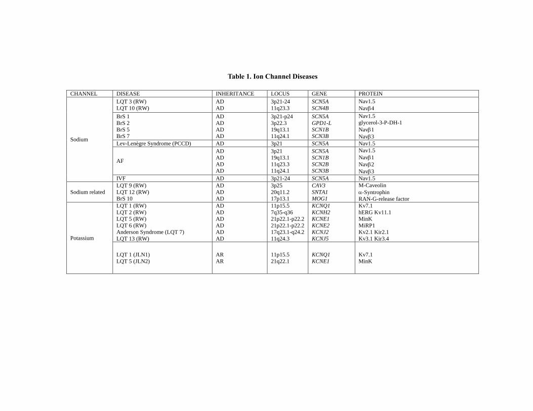
Table 1. Ion Channel Diseases
CHANNEL DISEASE INHERITANCE LOCUS GENE PROTEIN
Sodium
LQT 3 (RW)
LQT 10 (RW)
AD
AD
3p21-24
11q23.3
SCN5A
SCN4B
Nav1.5
Nav4
BrS 1
BrS 2
BrS 5
BrS 7
AD
AD
AD
AD
3p21-p24
3p22.3
19q13.1
11q24.1
SCN5A
GPD1-L
SCN1B
SCN3B
Nav1.5
glycerol-3-P-DH-1
Nav1
Nav3
Lev-Lenègre Syndrome (PCCD) AD 3p21 SCN5A Nav1.5
AF
AD
AD
AD
AD
3p21
19q13.1
11q23.3
11q24.1
SCN5A
SCN1B
SCN2B
SCN3B
Nav1.5
Nav1
Nav2
Nav3
IVF AD 3p21-24 SCN5A Nav1.5
Sodium related
LQT 9 (RW)
LQT 12 (RW)
BrS 10
AD
AD
AD
3p25
20q11.2
17p13.1
CAV3
SNTA1
MOG1
M-Caveolin
-Syntrophin
RAN-G-release factor
Potassium
LQT 1 (RW)
LQT 2 (RW)
LQT 5 (RW)
LQT 6 (RW)
Anderson Syndrome (LQT 7)
LQT 13 (RW)
AD
AD
AD
AD
AD
AD
11p15.5
7q35-q36
21p22.1-p22.2
21p22.1-p22.2
17q23.1-q24.2
11q24.3
KCNQ1
KCNH2
KCNE1
KCNE2
KCNJ2
KCNJ5
Kv7.1
hERG Kv11.1
MinK
MiRP1
Kv2.1 Kir2.1
Kv3.1 Kir3.4
LQT 1 (JLN1)
LQT 5 (JLN2)
AR
AR
11p15.5
21q22.1
KCNQ1
KCNE1
Kv7.1
MinK
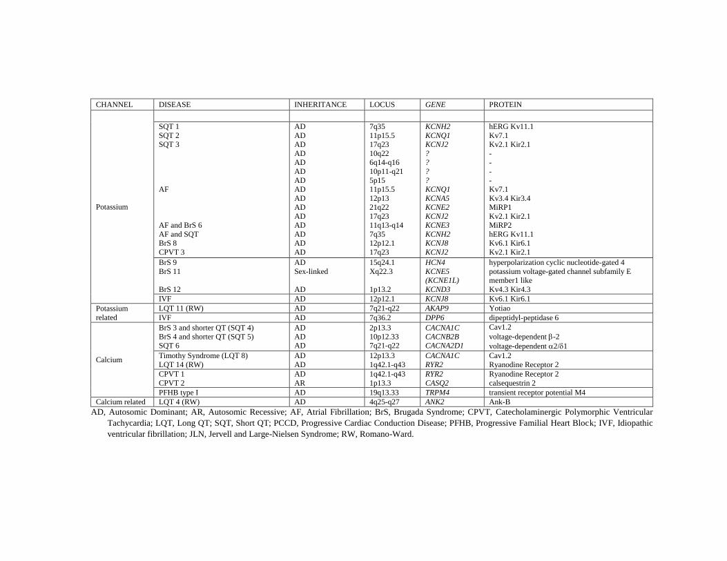
CHANNEL DISEASE INHERITANCE LOCUS GENE PROTEIN
Potassium
SQT 1
SQT 2
SQT 3
AF
AF and BrS 6
AF and SQT
BrS 8
CPVT 3
AD
AD
AD
AD
AD
AD
AD
AD
AD
AD
AD
AD
AD
AD
AD
7q35
11p15.5
17q23
10q22
6q14-q16
10p11-q21
5p15
11p15.5
12p13
21q22
17q23
11q13-q14
7q35
12p12.1
17q23
KCNH2
KCNQ1
KCNJ2
?
?
?
?
KCNQ1
KCNA5
KCNE2
KCNJ2
KCNE3
KCNH2
KCNJ8
KCNJ2
hERG Kv11.1
Kv7.1
Kv2.1 Kir2.1
-
-
-
-
Kv7.1
Kv3.4 Kir3.4
MiRP1
Kv2.1 Kir2.1
MiRP2
hERG Kv11.1
Kv6.1 Kir6.1
Kv2.1 Kir2.1
BrS 9
BrS 11
BrS 12
AD
Sex-linked
AD
15q24.1
Xq22.3
1p13.2
HCN4
KCNE5
(KCNE1L)
KCND3
hyperpolarization cyclic nucleotide-gated 4
potassium voltage-gated channel subfamily E
member1 like
Kv4.3 Kir4.3
IVF AD 12p12.1 KCNJ8 Kv6.1 Kir6.1
Potassium
related
LQT 11 (RW) AD 7q21-q22 AKAP9 Yotiao
IVF AD 7q36.2 DPP6 dipeptidyl-peptidase 6
Calcium
BrS 3 and shorter QT (SQT 4)
BrS 4 and shorter QT (SQT 5)
SQT 6
AD
AD
AD
2p13.3
10p12.33
7q21-q22
CACNA1C
CACNB2B
CACNA2D1
Cav1.2
voltage-dependent -2
voltage-dependent 2/1
Timothy Syndrome (LQT 8)
LQT 14 (RW)
AD
AD
12p13.3
1q42.1-q43
CACNA1C
RYR2
Cav1.2
Ryanodine Receptor 2
CPVT 1
CPVT 2
AD
AR
1q42.1-q43
1p13.3
RYR2
CASQ2
Ryanodine Receptor 2
calsequestrin 2
PFHB type I AD 19q13.33 TRPM4 transient receptor potential M4
Calcium related LQT 4 (RW) AD 4q25-q27 ANK2 Ank-B
AD, Autosomic Dominant; AR, Autosomic Recessive; AF, Atrial Fibrillation; BrS, Brugada Syndrome; CPVT, Catecholaminergic Polymorphic Ventricular
Tachycardia; LQT, Long QT; SQT, Short QT; PCCD, Progressive Cardiac Conduction Disease; PFHB, Progressive Familial Heart Block; IVF, Idiopathic
ventricular fibrillation; JLN, Jervell and Large-Nielsen Syndrome; RW, Romano-Ward.

Georgia Sarquella-Brugada, Oscar Campuzano, Paola Berne et al. 38
This gives rise to an ECG with an extremely long QT interval. Recently, a long QT
interval was also associated with a patient that showed a mutation in the cardiac ryanodine
receptor gene ryr2 [44]; however, further studies were required in order to clarify this case
report. The type 11 LQT is a disorder of the QT interval caused by a mutation (S1570L) in
akap9 gene, which encodes the protein kinase-A anchor protein-9 (chromosome 7q21-q22).
The severity of this type of QT may vary; the most common symptoms are angina, partial or
total loss of consciousness and in some cases the SCD [45]. There are other genes as ank2
(chromosome 4, 4q25-27), which is involved in type 4 LQT syndrome. Although not specific
to a channel is included in the group of channelopathies. This gene encodes the protein
ankyrin-B which is to adapt different structures in the cell membrane as the Na/K ATPase,
Na/Ca and inositol triphosphate receptor. A decrease in the role of ankyrin-B alters calcium
homeostasis prolonging repolarization and fatal ventricular arrhythmias generated [46-48].
The syntrophins are cytoplasmic proteins that are part of the protein complex associated with
dystrophin.
How to Follow a Child with Long QT Syndrome?
In all patients, adults [49] and children [50], beta-blocker administration at high doses is
highly recommended. The dose is adjusted according to the medical tolerance to these drugs
[51]. However, controversy exists regarding the efficacy of cardioselective beta-blockers such
as atenolol [52-56].
Despite beta-blockade, some patients may remain at risk as they persist symptomatic.
Different strategies have been proposed: despite defibrillator implantation, some may
consider left cardiac sympathetic neural denervation [57]. Defibrillator implantation is
mandatory for those patients having had an aborted SD and for those at risk of fatal
arrhythmias [58]. Avoidance of QT prolonging drugs is crucial for these patients
(www.torsades.org). Water activities should be contraindicated in all types of LQTS, as high
intensity exercise [59].
Study of the Relatives of a Patient with LQT
In patients with LQT is important to have a good family history, and do a baseline ECG
to all family members (parents, children, siblings).
Data on the clinical presentation and genotype-phenotype correlation of patients with
congenital LQTS diagnosed at perinatal through infantile period are limited [60-62]. A
nationwide survey was conducted to characterize how LQTS detected during those periods is
different from that in childhood or adolescence. Patients with LQTS who showed life-
threatening arrhythmias at perinatal periods were mostly those with LQT2, LQT3, or no
known mutations. Independent of the genotype, aggressive intervention resulted in effective
suppression of arrhythmias [63].
A prolonged QT interval corrected for heart rate (QTc) is a major risk factor in patients
with LQTS. However, heart rate-related risk in this genetic disorder differs among genotypes.
Therefore, risk stratification for life-threatening cardiac events in LQTS patients can be
improved by incorporating genotype-specific QT correction for heart rate [64].
It is advisable that the genetic study of these patients. In the case of a known mutation,
family members should be studied to rule out that they are carriers [59].

Sudden Cardiac Death in Pediatrics: Genetic Basis and Clinical Treatment 39
What Should We Do with Asymptomatic Gene Carriers?
The gene carrier should avoid competitive sports and should be treated with beta blockers
(level of evidence IIa) because use of beta-blockers decreases the risk of SCD [51] although
do not provide full protection.
Short QT Syndrome
SQTS—first described in 2000 [65] — is a highly malignant condition characterized by a
short QT interval (<330 ms), with a high sharp T wave and a short interval between the peak
and the end of the T wave, leading some clinical manifestations from lack of symptoms to
recurrent syncope, and high risk of SCD [66-68] because of ventricular arrhythmias, but atrial
fibrillation (AF) is also commonly seen in patients with SQTS [69, 70]. Clinical
manifestations may appear as early as childhood, and so it is considered a possible cause of
SD in nursing infants [17, 71, 72].
The genetic origin of this condition has been reported recently, with an autosomal
dominant pattern of transmission and a high penetrance (Table1). The mutations that induce
this syndrome are located on 6 genes, of which 3 (kcnq1, kcnj2, and kcnh2) encode potassium
channels, with enhanced function and, therefore, shortened repolarization [71]. The origin of
this entity has been recently described, with autosomal dominant inheritance pattern and high
penetrance. The burnout syndrome type 1 has been associated with two mutations in kcnh2
(hERG protein) that induce a rapid activation of potassium currents, with a gain of function of
IKr and a shortening of ventricular action potentials. Generally, cardiac events are associated
with adrenergic in situations such as noise or exercise, but also occurred at rest [73]. The
burnout syndrome has been associated with AF in some families [74]. The burnout syndrome
type 2 has been associated with two mutations in the gene kcnq1 (KvLQT1 protein) [30]
which leads to a gain of potassium channel function, leading to a shortening of action
potential with AF. There is a particular entity, by affecting the same gene that is expressed in
uterus in the form of bradycardia in the neonatal period was diagnosed as AF and SQTS [75].
The burnout syndrome type 3 has been associated with a mutation in the gene kcnj2 (Kir2.1
protein) located on chromosome 17, involving a speeding of phase 3 of action potential,
resulting in a gain of function [76]. The association of BrS pattern with a shorter QT interval
has been associated with mutations in the gene cacna1c that cause a change in the unit
calcium channel L-type inducing a loss of channel function related to the association to BrS
and QT interval shorter than normal, with autosomal dominant inheritance pattern. With the
same phenotype and pattern of inheritance, cacnb2b mutations causes a change in the unit-
type calcium channels, L, giving the association BrS and shortened QT interval [77].
Recently, it has been published a relation between cacna2d1 and SQTS [78]. However, more
studies must be performed to establish a clear clinical-genetic association.
How to Follow a Child with Short QT Syndrome?
Being a rare entity, there are no clinical guidelines for the monitoring of these patients.
Quinidine has been tested as a treatment to try to prolong the QT. In cases associated with
AF, has raised the use of antiplatelet drugs to prevent embolic complications. The medication
does not reverse or control AF.
Georgia Sarquella-Brugada, Oscar Campuzano, Paola Berne et al. 40
In cases where the ventricular response is too slow, we recommend the implantation of a
pacemaker. Given the high risk of malignant arrhythmias, may consider the possible
inducibility of arrhythmias and defibrillator implantation, although the young age of these
patients have created much controversy plating in the management. As SQTS is a highly
malignant disease, the genetic study of family members is necessary, and baseline ECG
should be performed in all patients [79].
Brugada Syndrome
Brugada syndrome (BrS) [80], is characterized by an ECG pattern consisting of coved-
type ST-segment elevation in atypical right-bundle branch block in leads V1 to V3 (often
referred to as type-1 Brugada ECG pattern) without structural heart disease. BrS is also
characterized by an increased risk for SCD resulting from episodes of polymorphic
ventricular tachyarrhythmias with an incidence of about 26-38/100.000 person-year.
Although the average age of onset of events is about 40 years, SD can affect people of all
ages, especially men (75%). Of the patients, between 20% -50% had family history of SD
[81].
The penetrance and expressivity of the disorder are highly variable; BrS is generally
considered a disorder involving young male adults, with an arrhythmogenic symptom first
occurring at age of 40 years, and SCD typically occurring during sleep, particularly in Asia,
where it manifests as sudden unexpected nocturnal death syndrome (SUNDS), the most
common cause of natural death in young Asians [82]. The ECG pattern can be present at
baseline or it may be intermittent. In the latter it and can be unmasked during a drug challenge
with a sodium channel blocker (ajmaline or flecainide in children). The description of acute
inducers of the ECG pattern is of paramount importance, as some individuals may be at risk
during anesthesia, when they take some oral medications (antidepressants or antiarrhythmics)
or, of special importance in children, during a febrile episode. Patients with BrS are at risk for
life threatening tachyarrhythmias. After surviving a cardiac arrest or the occurrence of
syncope, the only treatment having any proven effect on the prevention of SD is the
implantable cardioverter-defibrillator (ICD) [81].
To date, 12 genes have been associated to BrS (Table1). Approximately 20% to 30% of
patients with BrS have a mutation in the scn5a gene, classified as BrS type 1. The scn5a gene
(alpha subunit of the cardiac sodium channel) is responsible for the phase 0 of the cardiac
action potential, a key player in the cardiac electrical activity. Mutations in scn5a result in
“loss of function” of the sodium channel [83]. Another sodium channel that has been reported
to induce BrS is gpd1-l gene. It has been shown that mutation of the gpd1-l gene reduces the
surface membrane expression and reduces the inward sodium current. In addition, gpd1-l has
been shown to be the cause of some of the SCD in nursing infants [84, 85]. Additionally, it
has been published mutations in scn1b (sodium channel beta-1 subunit) and scn3b (sodium
channel beta-3 subunit) also associated to BrS [86, 87]. In the heart, β1-subunit modifies
Nav1.5 increasing INa. The mutation described in scn3b alters Nav1.5 trafficking, decreasing
INa. Other gen associated to BrS is kcne3 which codify MiRP2 protein (β-subunit that
regulates the potassium channel Ito). The KCNE peptides modulate some potassium currents
in the heart. The kcne3 gene encodes the regulatory subunit of the potassium channel Ito. The
relationship between mutations in this gene and the BrS were detected in a Danish family
Sudden Cardiac Death in Pediatrics: Genetic Basis and Clinical Treatment 41
[88]. Mutation in the cacna1c gene is responsible for a defective a unit of the type-L calcium
channels. This induces a loss of channel function, linked to the combination of BrS with
shorter QT interval. Transmission follows an autosomal dominant pattern. With the same
phenotype, mutation of the cacnb2b gene leads to a defect in the L-type calcium channel,
giving rise to a combination of BrS and shorter QT interval [77]. In 2009 was associated BrS
to hcn4 gene [89], that codifies for HCN4 channel or If channel. The HCN4 channel controls
the heart rate, and its mutations also predispose to inherited sick sinus syndrome and LQTS
associated with bradycardia. Another gene described associated to BrS is kcnj8, also
previously related to early repolarization syndrome (ERS) [90]. The report implicate kcnj8 as
a novel J-wave syndrome susceptibility gene and a marker gain of function in the cardiac
K(ATP) Kir6.1 channel [91]. Recently, Kattygnarath et al, [92] published a study supporting
that mog1 gene can impair the trafficking of Nav1.5 to the membrane, leading to INa
reduction and clinical manifestation of BrS. Also in 2011, Giudicessi et al. provide the first
molecular and functional evidence implicating novel kcnd3 gain-of-function mutations
(Kir4.3 protein) in the pathogenesis and phenotypic expression of BrS, with the potential for a
lethal arrhythmia being precipitated by a genetically enhanced Ito current gradient within the
right ventricle where kcnd3 gene expression is the highest [92]. Finally, and also in 2011, it is
published a study recommending kcne5 gene screening for BrS or IVF affected patients.
Therefore, kcne5 gene modulates Ito and its novel variants appeared to cause IVF especially
BrS in male patients through gain-of-function effects on Ito [93].
How to Follow a Patient with Brugada Syndrome?
Type 1 Brugada ECG pattern is rare in children. Genetic testing will help to diagnose
these kids. When genetic testing is not available, children being at risk of BrS (brother/sister
or son/daughter of a parent affected with BrS), should be followed as if they had the
syndrome, thus, avoidance of Brugada inducing drugs is mandatory
(www.brugadadrugs.com).
As fever is one of the promoters of the Brugada pattern, fever control is highly
recommended.
We perform flecainide test around the age of 8 years. If it is positive, we perform an
electrophysiological study to assess the inducibility of arrhythmias and possible ICD
implantation. We are aware of the concerns of implanting a defibrillator in a child.
In patients with BrS and presence of syncope, aborted SD, nocturnal agonal breathing or
seizures, is indicated for ICD implantation. If they also show arrhythmias, treatment is
indicated with Quinidine.
Competitive exercise is contraindicated competition, but low-intensity sports are allowed
[94-96].
Particularly challenging is treatment of electrical storm in BrS patients, which has been
anecdotally treated with isoproterenol, disopyramide [97], orciprenaline [98] and quinine
[99].
Special should be considered that BrS can associate AV conduction disturbances and
supraventricular tachycardia, so we question the presence of palpitations and eventually treat
these arrhythmias with ablation.
Georgia Sarquella-Brugada, Oscar Campuzano, Paola Berne et al. 42
Study of the Relatives of a Patient with Brugada Syndrome
In patients with BrS, as in other channelopathies, good family history is crucial. All first
grade relatives should be screened by a basal ECG. When in doubt, flecainide test was
performed.
In the case of a known mutation, family members should be studied to rule out that they
are carriers despite the negativity of the basic ECG and flecainide test.
Lev-Lenègre Syndrome
Lev-Lenègre syndrome is a rare entity characterized by disruption of the conduction
system, in which a block gradually develops, resulting in ventricular arrhythmias or asystolia
[100, 101]. The amount of sodium and the speed with which it enters the cell determine the
velocity of conduction of the electric impulse through the sodium-dependent cells (muscle
cells of the ventricle and atrium and cells of the His-Purkinje system). If a mutation leads to a
reduction in the quantity of sodium that enters the cell, the velocity of conduction of the
impulse is reduced resulting in a loss of function in phase 0 of the action potential (channel
opening). This is the case in the Lev-Lenègre syndrome. In 1995, chromosomal abnormalities
(19q13.2-13.3) associated with bundle branch block were reported [102]. In 1999 the first
mutation was described, located on the scn5a gene [103, 104]. Additionally, it has been
described mutations in nkx2.5 gene (5q35) that codify the transcription factor NKX2.5 (also
named CSX) [105]. The conduction block is due to a congenital heart defect. Recently, it has
also been described a mutation in trpm4 gene (19q) [106]. The trpm4 gene is a causative gene
in isolated cardiac conduction disease with mutations resulting in a gain of function and
TRPM4 channel being highly expressed in cardiac Purkinje fibers (Table1).
Catecholaminergic Polymorphic Ventricular Tachycardia
In 1975 was first reported Catecholaminergic Polymorphic Ventricular Tachycardia
(CPVT) [107]. CPVT is a familial arrhythmogenic disorder characterized by a 2-way
polymorphic ventricular tachycardia characterized by severe arrhythmias in young patients
with apparently normal hearts [108]. It is triggered exclusively by adrenergic stimulus
(vigorous exercise, fear), and has a high mortality (30% by the age of 30 years) [109, 110].
When doubt exists, it is recommended to perform and exercise ECG and Holter monitoring in
order to rule out the bidirectional tachycardia. The CPVT is associated with a normal ECG at
rest (occasionally with bradycardia, and U waves). It was thought that the event happened in
childhood (before age 10) but recent studies show that the first manifestation may occur from
infancy to age 40 [111].
Three genetic variants have been identified (Table1), an autosomal dominant one caused
by mutation in the gene encoding the ryanodine receptor ryr2 and a recessive one, caused by
mutation in the calciquestrina isoform gene (casq2) [112, 113]. Both genes are implicated in
regulating intracellular calcium and both types of defect lead to increased function of these
proteins, and so outflow of calcium from the sarcoplasmatic reticulum is increased. This
excess calcium is associated with abnormalities in the sarcolemmal membrane potential,
leading to late depolarizations that cause a predisposition to arrhythmias [17]. Similarly, some
Sudden Cardiac Death in Pediatrics: Genetic Basis and Clinical Treatment 43
patients diagnosed with CPVT type 3 on the basis of the presence of bidirectional ventricular
tachycardia on exercise have been identified as possessing kcnj2 mutations, which are
associated with the rarely lethal Andersen-Tawil syndrome (ATS1, LQT7) [114]. The
misdiagnosis of Andersen-Tawil syndrome as the potentially lethal disorder CPVT may lead
to a more aggressive prophylactic therapy (ie, implantation of an ICD) than necessary.
Genetic testing may provide a clear differential diagnosis between atypical LQT1 and CPVT
and between CPVT and ATS1 [115].
Untreated, the mortality rate is very high, reaching 30-50% in young adulthood. The
earlier episodes appear, the worse the prognosis and there is a correlation between the age at
which syncope occurs for the first time and the severity of the disease. The first line treatment
in patients with CPVT is beta-blockers, which have significantly reduced syncope and SCD
[79]. Given that the first symptom may be the SCD, it is recommended to treat all individuals
genetically identified as mutation carriers and patients who are asymptomatic but who have
ventricular arrhythmias during exercise. The sport is contraindicated, including patients
treated with beta-blockers [116].
How to Follow a Child with Catecholaminergic Polymorphic Ventricular Tachycardia?
CPVT should be suspected in any form of dizziness, syncope or aborted SD in a child not
only during exercise but also after an episode of fear or running after a bus. We may see sinus
bradycardia at the resting ECG, with or without U waves.
When CPVT is suspected, exercise test up to maximum effort may give the diagnostic,
but it should be carefully performed. Any type of ventricular premature beat or couples
should be carefully observed, as the following phase may show non-sustained ventricular
tachycardia, and sustained ventricular tachycardia, that may give the diagnostic. Physical
activity is completely contraindicated in these patients [117].
Beta-blockers at very high doses should be given. The dose is adjusted according to the
medical tolerance to these drugs. If arrhythmias persist, verapamil can be added verapamil.
Cardiac sympathectomy has been suggested to control CPVT in patients refractory to beta-
blockers [118], although relief after this surgical procedure can be temporary or of delayed
onset [119].
Flecainide suppressed delayed after-depolarization (DADs) in mutant Purkinje fibers
[120] and effectively prevented CPVT in mice and humans [121-124]. Defibrillator is
indicated in case of aborted SD and in those cases of uncontrolled ventricular tachycardias.
Holter is held every three months in order to assess ventricular premature beat.
Study of the Relatives of a Patient with CPVT
In patients with catecholaminergic polymorphic ventricular tachycardia is essential to
study all family members basal ECG and exercise testing. It is advisable that the genetic study
of these patients. In the case of a known mutation, family members should be studied to rule
out that they are carriers.
Idiopathic Ventricular Fibrillation
IVF – spontaneous ventricular fibrillation without identifiable structural or electrical
heart disease – may account for up to 10% of SD in the young [125-127]. Haplotype-sharing
Georgia Sarquella-Brugada, Oscar Campuzano, Paola Berne et al. 44
analysis has identified a genetic basis for IVF [128]. The shared chromosomal segment
contained the dpp6 gene, which encodes a putative regulator of the transient outward Ito
current [129]. DPP6 mRNA levels were increased 20-fold in hearts of human carriers in one
study [129]. To date, this seems to be a founder risk locus, but nonetheless it suggests that an
increase in DPP6 imparts a higher risk for ventricular fibrillation (VF).
Furthermore, previously thought to be a benign and common ECG finding present in up
to 5% of the population, three separate case–control studies [130-132] suggest that ‘J-point
elevation’ (manifested either as terminal QRS slurring or notching, or ST-segment elevation
with upper concavity and prominent T waves in inferolateral leads) is significantly more
prevalent (16–60%) in patients with IVF. Mutations in genes encoding subunits of the L-type
Ca2 channel (cacna1c, cacnb2, and cacna2d1) [133] and a subunit of the KATP channel
encoded by kcnj8 [90] have been implicated in this new ‘J-wave syndrome’ or ERS [134].
CARDIOMYOPATHIES
Cardiomyopathies are heart diseases induced by mutations in genes that encode
contractile and structural proteins as well as proteins for cardiac energy production. They are
responsible for lethal arrhythmogenic disorders, mainly hypertrophic cardiomyopathy (HCM)
and arrhythmogenic cardiomyopathy (arrhythmogenic right ventricular dysplasia-ARVD/C-),
both associated to SCD [135].
Hypertrophic Cardiomyopathy
HCM is one of the most common cardiovascular disorders, self characterized by an
unexplained asymmetric hypertrophy of the left ventricle with findings indicative of myocyte
disarray and fibrosis [136], often mostly pronounced in the interventricular septum. It has a
prevalence of 1/500, affecting children and young people [137], and it is also cause of death
in about a 10% of the SCD identified at autopsy [12].
Clinical manifestations appear initially as diastolic dysfunction and systolic-diastolic
dysfunction in more advanced stages. Thus, a patient may be asymptomatic or present with
SCD. Mortality is higher in young patients (often athletes) than in adults, and the first
manifestation of the disease may be SCD itself. The risk stratification of SCD in patients with
HCM remains at the forefront of clinical research. Both, a wall thickness of the left ventricle
with a z-score>6 and an abnormal blood pressure response to exercise are clinical factors for
SCD in children. The differential diagnosis between HCM and so-called "athlete's heart" is
not easy, but testing such as ECG, echocardiogram and genetic analysis are useful tools to
resolve the dilemma [95, 96, 138, 139]. Patients considered at high risk of SCD should be
aimed at prevention and/or management of ventricular tachyarrhythmias with antiarrhythmic
drugs (amiodarone). ICD implantation is the most reasonable option for patients at greatest
risk. Special emphasis should be put on risk stratification in childhood because evidence
points to a higher risk of SCD in this population than in adults [140, 141]. Competitive
exercise is an absolute contraindication for children with HCM, leisure activities, however,
are possible if the cardiac function permits.
Sudden Cardiac Death in Pediatrics: Genetic Basis and Clinical Treatment 45
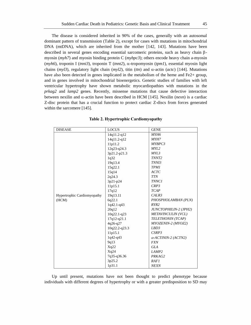
The disease is considered inherited in 90% of the cases, generally with an autosomal
dominant pattern of transmission (Table 2), except for cases with mutations in mitochondrial
DNA (mtDNA), which are inherited from the mother [142, 143]. Mutations have been
described in several genes encoding essential sarcomeric proteins, such as heavy chain -
myosin (myh7) and myosin binding protein C (mybpc3); others encode heavy chain a-myosin
(myh6), troponin I (tnni3), troponin T (tnnt2), -tropomyosin (tpm1), essential myosin light
chains (myl3), regulatory light chain (myl2), titin (ttn) and -actin (actc) [144]. Mutations
have also been detected in genes implicated in the metabolism of the heme and Fe2+ group,
and in genes involved in mitochondrial bioenergetics. Genetic studies of families with left
ventricular hypertrophy have shown metabolic myocardiopathies with mutations in the
prkag2 and lamp2 genes. Recently, missense mutations that cause defective interaction
between nexilin and -actin have been described in HCM [145]. Nexilin (nexn) is a cardiac
Z-disc protein that has a crucial function to protect cardiac Z-discs from forces generated
within the sarcomere [145].
Table 2. Hypertrophic Cardiomyopathy
DISEASE LOCUS GENE
Hypertrophic Cardiomyopathy
(HCM)
14q11.2-q12
14q11.2-q12
11p11.2
12q23-q24.3
3p21.2-p21.3
1q32
19q13.4
15q22.1
15q14
2q24.3
3p21-p24
11p15.1
17q12
19q13.11
6q22.1
1q42.1-q43
20q12
10q22.1-q23
17q12-q21.1
4q26-q27
10q22.2-q23.3
11p15.1
1q42-q43
9q13
Xq22
Xq24
7q35-q36.36
3p25.2
1p31.1
MYH6
MYH7
MYBPC3
MYL2
MYL3
TNNT2
TNNI3
TPM1
ACTC
TTN
TNNC1
CRP3
TCAP
CALR3
PHOSPHOLAMBAN (PLN)
RYR2
JUNCTOPHILIN-2 (JPH2)
METAVINCULIN (VCL)
TELETHONIN (TCAP)
MYOZENIN-2 (MYOZ2)
LBD3
CSRP3
-ACTININ-2 (ACTN2)
FXN
GLA
LAMP2
PRKAG2
RAF1
NEXN
Up until present, mutations have not been thought to predict phenotype because
individuals with different degrees of hypertrophy or with a greater predisposition to SD may
Georgia Sarquella-Brugada, Oscar Campuzano, Paola Berne et al. 46
be present in the same family and have the same mutation. This is due to the intervention of
modifying genes and polymorphisms, which require more exhaustive studies to achieve a full
understanding. It is assumed that interruption of mitochondrial energy metabolism in the heart
is the cause of HCM in patients with problems of sarcomeric contraction; this sheds some
light on several clinical observations such as heterogeneity, variability in clinical
presentation, and asymmetry in hypertrophy. The identification of the genotype may
contribute to risk stratification, but future genotype-phenotype studies need to be done to
confirm whether this is useful.
How to Follow a Child with Hypertrophic Cardiomyopathy
The sport is clearly contraindicated and any isometric exercise (lifting weights). In all
patients we recommend the administration of beta-blockers in high doses. The dose is
adjusted according to the medical tolerance to these drugs [146-149].
The implantation of an ICD will depend on the presence of symptoms, septal thickness,
presence of family history of arrhythmias and SD.
Study of the Relatives of a Patient with Hypertrophic Cardiomyopathy
In patients with HCM is important to perform echocardiography and ECG to their
relatives. It is advisable that the genetic study of these patients. In the case of a known
mutation, family members should be studied to rule out that they are carriers.
Arrhythmogenic Right Ventricular Dysplasia/Cardiomyopathy
Arrhythmogenic right ventricular dysplasia/cardiomyopathy (ARVD/C) is an inherited
cardiomyopathy characterized by right ventricular dysfunction and ventricular arrhythmias.
Patients with ARVD/C show fibrofatty replacement of right ventricular infarction. However,
ARVD/C may involve both ventricles and in very isolated cases only the left ventricle. The
ECG is not always altered, but the presence of epsilon waves and T negative ε, from V1 to
V3, may provide a pathological diagnosis [150]. About 50% of ARVD/C cases are familiar
with variable penetrance. It affects approximately 1/5000 individuals although the prevalence
is higher in men (80%), particularly young athletes. Most cases are diagnosed before the age
of 40 years. Usually, the individuals affected have symptomatic ventricular arrhythmias that
originate in the right ventricle, with syncope and a high risk of SD. This entity is responsible
for 5% of all SCD.
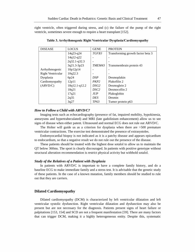
The disease has 2 different patterns of transmission: autosomal dominant pattern (the
most common) and an autosomal recessive pattern (Table 3). The recessive pattern has been
reported on the Greek island (Naxos), giving rise to the Naxos syndrome. This syndrome
comprises ARVD/C, palmoplantar keratoderma and typically curly hair. To date, several
genes are described as capable of inducing disease (http://www.arvcdatabase.info/). Most
cases involve genes encoding proteins responsible for the junctions of intercalated discs,
especially in desmosome, which led to the appointment of ARVD/C as a desmosomal disease
with a disorder of the connections between myocytes [151].
The clinical picture may include (a) a subclinical stage hidden structural defects, during
which the affected person may have a cardiac arrest/SCD as the first manifestation of the
disease, (b) an electrical disorder with palpitations and syncope tachyarrhythmias arising the
Sudden Cardiac Death in Pediatrics: Genetic Basis and Clinical Treatment 47
right ventricle, often triggered during stress, and (c) the failure of the pump of the right
ventricle, sometimes severe enough to require a heart transplant [152].
Table 3. Arrhythmogenic Right Ventricular Dysplasia/Cardiomyopathy
DISEASE LOCUS GENE PROTEIN
Arrhythmogenic
Right Ventricular
Dysplasia
Cardiomyopathy
(ARVD/C)
14q23-q24
14q12-q22
2q32.1-q32.3
3q21.3-3p23
10p12p14
10q22.3
6p24
12p11
18q12.1-q12.2
18q21
17q21
2q35
3q27
TGFΒ3
-
-
TMEM43
-
-
DSP
PKP2
DSG2
DSC2
JUP
DES
TP63
Transforming growth factor beta 3
-
-
Transmembrain protein 43
-
-
Desmoplakin
Plakofilin 2
Desmoglein 2
Desmocollin 2
Plakoglobin
Desmin
Tumor protein p63
How to Follow a Child with ARVD/C?
Imaging tests such as echocardiography (presence of fat, impaired mobility, hypokinesia,
aneurysms and hypertrabeculated) and MRI (late gadolinium enhancement) allow us to see
signs of disease when either patent. Ultrasound and normal ECG does not rule out ARVD/C.
The Holter will guide us as a criterion for dysplasia when there are >500 premature
ventricular contractions. The exercise test demonstrated the presence of extrasystoles.
Endomyocardial biopsy is not indicated as it is a patchy disease and appears epicardium
to endocardium, so that a negative result we do not rule out the presence of the disease.
These patients should be treated with the highest dose sotalol to allow us to maintain the
QT below 360ms. The sport is clearly discouraged. In patients with positive genotype without
structural alteration recommendation is restrict physical activity but withhold sotalol.
Study of the Relatives of a Patient with Dysplasia
In patients with ARVD/C is important to have a complete family history, and do a
baseline ECG to make immediate family and a stress test. It is advisable that the genetic study
of these patients. In the case of a known mutation, family members should be studied to rule
out that they are carriers.
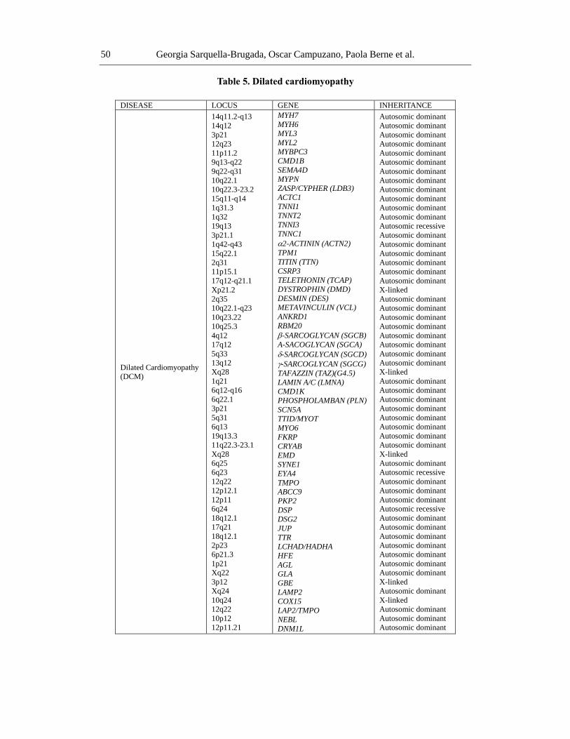
Dilated Cardiomyopathy
Dilated cardiomyopathy (DCM) is characterized by left ventricular dilatation and left
ventricular systolic dysfunction. Right ventricular dilatation and dysfunction may also be
present but are not necessary for the diagnosis. Patients present signs of heart failure or
palpitations [153, 154] and SCD are not a frequent manifestation [59]. There are many factors
that can trigger DCM, making it a highly heterogeneous entity. Despite this, systematic
Georgia Sarquella-Brugada, Oscar Campuzano, Paola Berne et al. 48
studies show that at least 35% of cases are hereditary. Arrhythmias that occur in patients with
familial DCM are usually the same as in the acquired forms, with defects in atrioventricular
and intraventricular conduction, ventricular arrhythmias and AF. Normally, a progressive
decline in ventricular function occurs in these patients and they die of either heart failure or
arrhythmic events.
In adults, the prevalence is 1/2500 individuals, with an incidence of 7/100000 per year
(but it could be underdiagnosed). This disorder develops at any age, in either sex, and in
people of any ethnic origin. In adults, DCM arises more commonly in men than in women. In
children, the yearly incidence is 0,57 cases per 100000 per year overall, but is higher in boys
than in girls (0·66 vs. 0·47 cases per 100000, p<0·006), in black people than in white people
(0,98 vs. 0,46 cases per 100000, p<0·001), and in babies younger than 1 year than in children
(4,40 vs. 0,34 cases per 100000, p<0·001). Two-thirds of children are thought to have
idiopathic disease.
DCM is an extremely complex disease, and so the clinical usefulness of genetic analysis
is limited. Even so, systematic studies of relatives of patients with DCM indicate that at least
35% of the cases are hereditary (Table 5). In 1994, the first locus of DCM with
atrioventricular block was identified on chromosome 1. To date, there are mutations in genes
that code for proteins of the cytoskeleton, cell nucleus and sarcomere [155, 156]. In 30% of
the cases, the mutation occurs in the lamin A/C gene (lmna), which encodes a protein that is
expressed in almost all cell types and whose function is to contribute to the integrity of the
nucleus by providing mechanical support. Other genes, such as myh7, mybpc3, tnni3, tnnc1,
tcap, vcl, csrp3, pln, ttn, tpm1, actc and tnnt2 can also cause DCM. Moreover, a mutation in
the scn5a gene was identified in a large family with DCM [157]. A recent new addition to the
long list of DCM candidate genes is nebulette (nebl), which encodes a 107 kDa protein that
aligns thin filaments and connects them with the myocardial Z-disk [158].
Another is dynamin-1-like (dnm1l) gene, which is known to be critical for mitochondrial
fission and has also been implicated in causing DCM by reducing levels of mitochondrial
enzyme complexes and cardiac ATP depletion [159].
It is estimated that 20%-50% of the DCM cases are heredity. Three patterns of
transmission are described in the inherited forms:
a) Autosomal dominant disease, a locus for which has been reported in several genes
(actin, desmin, lamin A/C, -sarcoglycan and -sarcoglycan);
b) X-linked disease, associated to mutations in dystrophin and tafazin. Although
mutations in the dystrophin gene are not a common cause of DCM, direct mutations
in this gene give rise to Duchene or Becker-type muscular dystrophy, which affect
both cardiac and skeletal muscle;
c) Mitochondrial diseases, which typically affect other tissues besides the myocardium;
so far, 2 loci of DCM have been reported in association with primary arrhythmias,
one not being the cause of the other. These families had autosomal dominant disease.
DCM is associated with complex remodeling of one or both ventricles, resulting in a
change of the ventricle shape and the architecture of the myocardium fibers. In the most
severe cases, affected individuals present signs and symptoms of heart failure—diaphoresis,
breathlessness at rest or with exertion, orthopnoea, exercise intolerance, early onset fatigue,
Sudden Cardiac Death in Pediatrics: Genetic Basis and Clinical Treatment 49
abdominal pain, and pallor. Heart failure symptoms can be exercise-induced or persistent at
rest.
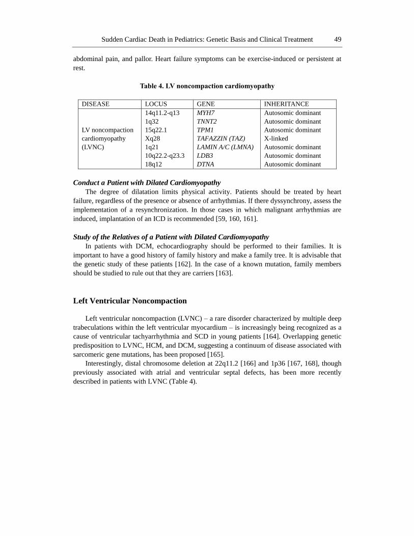
Table 4. LV noncompaction cardiomyopathy
DISEASE LOCUS GENE INHERITANCE
LV noncompaction
cardiomyopathy
(LVNC)
14q11.2-q13
1q32
15q22.1
Xq28
1q21
10q22.2-q23.3
18q12
MYH7
TNNT2
TPM1
TAFAZZIN (TAZ)
LAMIN A/C (LMNA)
LDB3
DTNA
Autosomic dominant
Autosomic dominant
Autosomic dominant
X-linked
Autosomic dominant
Autosomic dominant
Autosomic dominant
Conduct a Patient with Dilated Cardiomyopathy
The degree of dilatation limits physical activity. Patients should be treated by heart
failure, regardless of the presence or absence of arrhythmias. If there dyssynchrony, assess the
implementation of a resynchronization. In those cases in which malignant arrhythmias are
induced, implantation of an ICD is recommended [59, 160, 161].
Study of the Relatives of a Patient with Dilated Cardiomyopathy
In patients with DCM, echocardiography should be performed to their families. It is
important to have a good history of family history and make a family tree. It is advisable that
the genetic study of these patients [162]. In the case of a known mutation, family members
should be studied to rule out that they are carriers [163].
Left Ventricular Noncompaction
Left ventricular noncompaction (LVNC) – a rare disorder characterized by multiple deep
trabeculations within the left ventricular myocardium – is increasingly being recognized as a
cause of ventricular tachyarrhythmia and SCD in young patients [164]. Overlapping genetic
predisposition to LVNC, HCM, and DCM, suggesting a continuum of disease associated with
sarcomeric gene mutations, has been proposed [165].
Interestingly, distal chromosome deletion at 22q11.2 [166] and 1p36 [167, 168], though
previously associated with atrial and ventricular septal defects, has been more recently
described in patients with LVNC (Table 4).
Georgia Sarquella-Brugada, Oscar Campuzano, Paola Berne et al. 50
Table 5. Dilated cardiomyopathy
DISEASE LOCUS GENE INHERITANCE
Dilated Cardiomyopathy
(DCM)
14q11.2-q13
14q12
3p21
12q23
11p11.2
9q13-q22
9q22-q31
10q22.1
10q22.3-23.2
15q11-q14
1q31.3
1q32
19q13
3p21.1
1q42-q43
15q22.1
2q31
11p15.1
17q12-q21.1
Xp21.2
2q35
10q22.1-q23
10q23.22
10q25.3
4q12
17q12
5q33
13q12
Xq28
1q21
6q12-q16
6q22.1
3p21
5q31
6q13
19q13.3
11q22.3-23.1
Xq28
6q25
6q23
12q22
12p12.1
12p11
6q24
18q12.1
17q21
18q12.1
2p23
6p21.3
1p21
Xq22
3p12
Xq24
10q24
12q22
10p12
12p11.21
MYH7
MYH6
MYL3
MYL2
MYBPC3
CMD1B
SEMA4D
MYPN
ZASP/CYPHER (LDB3)
ACTC1
TNNI1
TNNT2
TNNI3
TNNC1
2-ACTININ (ACTN2)
TPM1
TITIN (TTN)
CSRP3
TELETHONIN (TCAP)
DYSTROPHIN (DMD)
DESMIN (DES)
METAVINCULIN (VCL)
ANKRD1
RBM20
-SARCOGLYCAN (SGCB)
A-SACOGLYCAN (SGCA)
-SARCOGLYCAN (SGCD)
-SARCOGLYCAN (SGCG)
TAFAZZIN (TAZ)(G4.5)
LAMIN A/C (LMNA)
CMD1K
PHOSPHOLAMBAN (PLN)
SCN5A
TTID/MYOT
MYO6
FKRP
CRYAB
EMD
SYNE1
EYA4
TMPO
ABCC9
PKP2
DSP
DSG2
JUP
TTR
LCHAD/HADHA
HFE
AGL
GLA
GBE
LAMP2
COX15
LAP2/TMPO
NEBL
DNM1L
Autosomic dominant
Autosomic dominant
Autosomic dominant
Autosomic dominant
Autosomic dominant
Autosomic dominant
Autosomic dominant
Autosomic dominant
Autosomic dominant
Autosomic dominant
Autosomic dominant
Autosomic dominant
Autosomic recessive
Autosomic dominant
Autosomic dominant
Autosomic dominant
Autosomic dominant
Autosomic dominant
Autosomic dominant
X-linked
Autosomic dominant
Autosomic dominant
Autosomic dominant
Autosomic dominant
Autosomic dominant
Autosomic dominant
Autosomic dominant
Autosomic dominant
X-linked
Autosomic dominant
Autosomic dominant
Autosomic dominant
Autosomic dominant
Autosomic dominant
Autosomic dominant
Autosomic dominant
Autosomic dominant
X-linked
Autosomic dominant
Autosomic recessive
Autosomic dominant
Autosomic dominant
Autosomic dominant
Autosomic recessive
Autosomic dominant
Autosomic dominant
Autosomic dominant
Autosomic dominant
Autosomic dominant
Autosomic dominant
Autosomic dominant
X-linked
Autosomic dominant
X-linked
Autosomic dominant
Autosomic dominant
Autosomic dominant
Sudden Cardiac Death in Pediatrics: Genetic Basis and Clinical Treatment 51
OTHER ANOMALIES WITH GENETIC DEFECTS
Anomalous Origin of the Coronary Arteries
Several congenital coronary artery malformations are the second cause of SD in young
athletes, accounting for 12% to 20% of SCD cases [169-172]. The anomalous origin of the
left main coronary artery from the right (anterior) sinus of Valsalva, with the left coronary
artery coursing between the aorta and the pulmonary trunk to reach the left atrioventricular
groove, is the most common type found at autopsies of subjects with SD or death due to
anatomical coronary artery anomalies [173]. During exertion, increased stroke volume causes
expansion of both the aorta and the pulmonary trunk, which can lead to further compression
of the already slit like ostial lumen of the anomalous artery [174]. Decreased blood flow and
resultant decreased O2 supply, in combination with increased O2 demand during exercise, can
lead to myocardial ischemia. This ischemia can trigger ventricular arrhythmias and
subsequent SD [175].
Most patients with coronary artery anomalies are asymptomatic, and those who are
symptomatic rarely complain of typical angina, reporting nonspecific symptoms such as
syncope, dyspnea on exertion and palpitations [174]. Transthoracic echocardiography is
utilized in diagnosis although highly trained ultrasound technicians using high-quality
imaging systems are required for accurate and consistent identification of the origin of the
coronary arteries [174]. Genetic predisposition for these defects has been identified, but
diagnostic is made by echocardiographic findings and if exist, it should be corrected
surgically.
Marfan Syndrome
Marfan syndrome (MS) has an increased risk for SD due to aortic dissection [176]. Aortic
root dilation and dissection are secondary to pathological changes in the aorta, namely, cystic
medial necrosis [177]. The disease is an autosomal dominant genetic disease with variable
penetrance, with an estimated incidence of 1 in 7000. It induce particular characteristics as
tall thin children, with arachnid fingers, long face, associated with strong myopic defects,
cardiac valve prolepses and other connective tissue anomalies [176, 178]. More than 100
mutations on the fibrillin-1 gene have been identified as causes of MS. These mutations lead
to defective fibrillin in the extracellular matrix, resulting in abnormalities of the ocular,
cardiovascular, skeletal, pulmonary, and integumentary systems [176]. The Berlin nosology
or the more recent “Ghent criteria”, which consider family history of MS, skeletal and
cardiovascular features, as well as other phenotypic expressions, are used to aid in diagnosis
[179]. These characteristics may be subtle, and body habitus measurements may be useful.
Echocardiography can be used to measure and monitor the degree of aortic root dilation. Non-
Marfan cases of familial aortic root aneurysms are also now being recognized and studied
[177].
Georgia Sarquella-Brugada, Oscar Campuzano, Paola Berne et al. 52
Loeys-Dietz Syndrome
Loeys-Dietz syndrome (LDS) is a rare autosomal dominant disorder showing the
involvement of cutaneous, cardiovascular, craniofacial, and skeletal systems. In particular,
LDS patients show arterial tortuosity with widespread vascular aneurysm and dissection, and
have a high risk of aortic dissection or rupture at an early age and at aortic diameters that
ordinarily are not predictive of these events [180, 181]. The LDS is a severe form of
extracellular matrix disease, with more premature SD. It has an autosomal dominant
inheritance pattern, related to a mutation in either the tgfbr1 or tgfbr2 genes (transforming
growth factor beta receptor 1 or 2). The LDS has been subdivided in type I (LDSI) and type II
(LDSII) on the basis of the presence or the absence of cranio-facial involvement, respectively.
Furthermore, LDSII patients display at least two of the major signs of vascular Ehlers-Danlos
syndrome (EDS) [182]. These genes encode the receptors for a molecule that plays a role in
extracellular signaling. A wide variety of mutations have been identified in both genes,
however, there is no significant correlation between the mutation, the location, the gene, and
clinical presentation [183-185]. There is no way to predict the severity of vascular, skeletal or
skin findings that may occur in an offspring.
REFERENCES
[1] Krous HF, Beckwith JB, Byard RW, et al. Sudden infant death syndrome and
unclassified sudden infant deaths: a definitional and diagnostic approach. Pediatrics.
Jul 2004;114(1):234-238.
[2] Byard RW, Krous HF. Research and sudden infant death syndrome: definitions,
diagnostic difficulties and discrepancies. J. Paediatr. Child Health. Aug
2004;40(8):419-421.
[3] Papadakis M, Sharma S, Cox S, et al. The magnitude of sudden cardiac death in the
young: a death certificate-based review in England and Wales. Europace. Oct
2009;11(10):1353-1358.
[4] Wisten A, Forsberg H, Krantz P, et al. Sudden cardiac death in 15-35-year olds in
Sweden during 1992-99. J. Intern. Med. Dec 2002;252(6):529-536.
[5] Vaartjes I, Hendrix A, Hertogh EM, et al. Sudden death in persons younger than 40
years of age: incidence and causes. Eur. J. Cardiovasc. Prev. Rehabil. Oct
2009;16(5):592-596.
[6] Cote A, Russo P, Michaud J. Sudden unexpected deaths in infancy: what are the
causes? J. Pediatr. Oct 1999;135(4):437-443.
[7] Wren C. Sudden death in children and adolescents. Heart. Oct 2002;88(4):426-431.
[8] Arnestad M, Vege A, Rognum TO. Evaluation of diagnostic tools applied in the
examination of sudden unexpected deaths in infancy and early childhood. Forensic. Sci.
Int. Feb 18 2002;125(2-3):262-268.
[9] Liberthson RR. Sudden death from cardiac causes in children and young adults. N.
Engl. J. Med. Apr 18 1996;334(16):1039-1044.
[10] Guntheroth WG, Spiers PS. The triple risk hypotheses in sudden infant death syndrome.
Pediatrics. Nov 2002;110(5):e64.
Sudden Cardiac Death in Pediatrics: Genetic Basis and Clinical Treatment 53
[11] Liberthson RR, Dinsmore RE, Fallon JT. Aberrant coronary artery origin from the
aorta. Report of 18 patients, review of literature and delineation of natural history and
management. Circulation. Apr 1979;59(4):748-754.
[12] Ferrero-Miliani L, Holst AG, Pehrson S, et al. Strategy for clinical evaluation and
screening of sudden cardiac death relatives. Fundam. Clin. Pharmacol. Oct
2010;24(5):619-635.
[13] Mohler PJ, Wehrens XH. Mechanisms of human arrhythmia syndromes: abnormal
cardiac macromolecular interactions. Physiology (Bethesda). Oct 2007;22:342-350.
[14] Priori SG, Napolitano C. Role of genetic analyses in cardiology: part I: mendelian
diseases: cardiac channelopathies. Circulation. Feb 28 2006;113(8):1130-1135.
[15] Schwartz PJ. Cardiac sympathetic innervation and the sudden infant death syndrome. A
possible pathogenetic link. Am. J. Med. Feb 1976;60(2):167-172.
[16] Maron BJ, Clark CE, Goldstein RE, et al. Potential role of QT interval prolongation in
sudden infant death syndrome. Circulation. Sep 1976;54(3):423-430.
[17] Klaver EC, Versluijs GM, Wilders R. Cardiac ion channel mutations in the sudden
infant death syndrome. Int. J. Cardiol. Jan 5 2011.
[18] Tester DJ, Ackerman MJ. Cardiomyopathic and channelopathic causes of sudden
unexplained death in infants and children. Annu. Rev. Med. 2009;60:69-84.
[19] Moss AJ, Kass RS. Long QT syndrome: from channels to cardiac arrhythmias. J. Clin.
Invest. Aug 2005;115(8):2018-2024.
[20] Goldenberg I, Moss AJ. Long QT syndrome. J. Am. Coll Cardiol. Jun 17
2008;51(24):2291-2300.
[21] Viskin S, Halkin A. Treating the long-QT syndrome in the era of implantable
defibrillators. Circulation. Jan 20 2009;119(2):204-206.
[22] Schimpf R, Veltmann C, Wolpert C, et al. Channelopathies: Brugada syndrome, long
QT syndrome, short QT syndrome, and CPVT. Herz. Jun 2009;34(4):281-288.
[23] Schimpf R, Veltmann C, Wolpert C, et al. Arrhythmogenic hereditary syndromes:
Brugada Syndrome, long QT syndrome, short QT syndrome and CPVT. Minerva
Cardioangiol. Dec 2010;58(6):623-636.
[24] Arnestad M, Crotti L, Rognum TO, et al. Prevalence of long-QT syndrome gene
variants in sudden infant death syndrome. Circulation. Jan 23 2007;115(3):361-367.
[25] Otagiri T, Kijima K, Osawa M, et al. Cardiac ion channel gene mutations in sudden
infant death syndrome. Pediatr. Res. Nov 2008;64(5):482-487.
[26] Roden DM, Viswanathan PC. Genetics of acquired long QT syndrome. J. Clin. Invest.
Aug 2005;115(8):2025-2032.
[27] Tester DJ, Benton AJ, Train L, et al. Prevalence and spectrum of large deletions or
duplications in the major long QT syndrome-susceptibility genes and implications for
long QT syndrome genetic testing. Am. J. Cardiol. Oct 15 2010;106(8):1124-1128.
[28] Kapplinger JD, Tester DJ, Salisbury BA, et al. Spectrum and prevalence of mutations
from the first 2,500 consecutive unrelated patients referred for the FAMILION long QT
syndrome genetic test. Heart Rhythm. Sep 2009;6(9):1297-1303.
[29] Chen S, Zhang L, Bryant RM, et al. KCNQ1 mutations in patients with a family history
of lethal cardiac arrhythmias and sudden death. Clin. Genet. Apr 2003;63(4):273-282.
[30] Bellocq C, van Ginneken AC, Bezzina CR, et al. Mutation in the KCNQ1 gene leading
to the short QT-interval syndrome. Circulation. May 25 2004;109(20):2394-2397.
Georgia Sarquella-Brugada, Oscar Campuzano, Paola Berne et al. 54
[31] Curran ME, Splawski I, Timothy KW, et al. A molecular basis for cardiac arrhythmia:
HERG mutations cause long QT syndrome. Cell. Mar 10 1995;80(5):795-803.
[32] January CT, Gong Q, Zhou Z. Long QT syndrome: cellular basis and arrhythmia
mechanism in LQT2. J. Cardiovasc. Electrophysiol. Dec 2000;11(12):1413-1418.
[33] Bianchi L, Shen Z, Dennis AT, et al. Cellular dysfunction of LQT5-minK mutants:
abnormalities of IKs, IKr and trafficking in long QT syndrome. Hum. Mol. Genet. Aug
1999;8(8):1499-1507.
[34] Tsuboi M, Antzelevitch C. Cellular basis for electrocardiographic and arrhythmic
manifestations of Andersen-Tawil syndrome (LQT7). Heart Rhythm. Mar
2006;3(3):328-335.
[35] Yang Y, Yang Y, Liang B, et al. Identification of a Kir3.4 Mutation in Congenital Long
QT Syndrome. Am. J. Hum. Genet. Jun 2 2010.
[36] Schwartz PJ, Priori SG, Locati EH, et al. Long QT syndrome patients with mutations of
the SCN5A and HERG genes have differential responses to Na+ channel blockade and
to increases in heart rate. Implications for gene-specific therapy. Circulation. Dec 15
1995;92(12):3381-3386.
[37] Wang Q, Shen J, Splawski I, et al. SCN5A mutations associated with an inherited
cardiac arrhythmia, long QT syndrome. Cell. Mar 10 1995;80(5):805-811.
[38] Medeiros-Domingo A, Kaku T, Tester DJ, et al. SCN4B-encoded sodium channel beta4
subunit in congenital long-QT syndrome. Circulation. Jul 10 2007;116(2):134-142.
[39] Ueda K, Valdivia C, Medeiros-Domingo A, et al. Syntrophin mutation associated with
long QT syndrome through activation of the nNOS-SCN5A macromolecular complex.
Proc. Natl. Acad. Sci. USA. Jul 8 2008;105(27):9355-9360.
[40] Wu G, Ai T, Kim JJ, et al. alpha-1-syntrophin mutation and the long-QT syndrome: a
disease of sodium channel disruption. Circ. Arrhythm Electrophysiol. Aug
2008;1(3):193-201.
[41] Vatta M, Ackerman MJ, Ye B, et al. Mutant caveolin-3 induces persistent late sodium
current and is associated with long-QT syndrome. Circulation. Nov 14
2006;114(20):2104-2112.
[42] Splawski I, Timothy KW, Sharpe LM, et al. Ca(V)1.2 calcium channel dysfunction
causes a multisystem disorder including arrhythmia and autism. Cell. Oct 1
2004;119(1):19-31.
[43] Yarotskyy V, Gao G, Peterson BZ, et al. The Timothy syndrome mutation of cardiac
CaV1.2 (L-type) channels: multiple altered gating mechanisms and pharmacological
restoration of inactivation. J. Physiol. Feb 1 2009;587(Pt 3):551-565.
[44] Kauferstein S, Kiehne N, Erkapic D, et al. A novel mutation in the cardiac ryanodine
receptor gene (RyR2) in a patient with an unequivocal LQTS. Int. J. Cardiol. Jan 21
2011;146(2):249-250.
[45] Chen L, Marquardt ML, Tester DJ, et al. Mutation of an A-kinase-anchoring protein
causes long-QT syndrome. Proc. Natl. Acad. Sci. USA. Dec 26 2007;104(52):20990-
20995.
[46] Mohler PJ. Ankyrins and human disease: what the electrophysiologist should know. J.
Cardiovasc. Electrophysiol. Oct 2006;17(10):1153-1159.
[47] Mohler PJ, Bennett V. Ankyrin-based cardiac arrhythmias: a new class of
channelopathies due to loss of cellular targeting. Curr. Opin. Cardiol. May
2005;20(3):189-193.
Sudden Cardiac Death in Pediatrics: Genetic Basis and Clinical Treatment 55
[48] Mohler PJ, Schott JJ, Gramolini AO, et al. Ankyrin-B mutation causes type 4 long-QT
cardiac arrhythmia and sudden cardiac death. Nature. Feb 6 2003;421(6923):634-639.
[49] Moss AJ, Zareba W, Hall WJ, et al. Effectiveness and limitations of beta-blocker
therapy in congenital long-QT syndrome. Circulation. Feb 15 2000;101(6):616-623.
[50] Goldenberg I, Moss AJ, Peterson DR, et al. Risk factors for aborted cardiac arrest and
sudden cardiac death in children with the congenital long-QT syndrome. Circulation.
Apr 29 2008;117(17):2184-2191.
[51] Hobbs JB, Peterson DR, Moss AJ, et al. Risk of aborted cardiac arrest or sudden cardiac
death during adolescence in the long-QT syndrome. Jama. Sep 13 2006;296(10):1249-
1254.
[52] Chatrath R, Bell CM, Ackerman MJ. Beta-blocker therapy failures in symptomatic
probands with genotyped long-QT syndrome. Pediatr. Cardiol. Sep-Oct
2004;25(5):459-465.
[53] Schwartz PJ, Priori SG, Cerrone M, et al. Left cardiac sympathetic denervation in the
management of high-risk patients affected by the long-QT syndrome. Circulation. Apr
20 2004;109(15):1826-1833.
[54] Pellizzon OA, Kalaizich L, Ptacek LJ, et al. Flecainide suppresses bidirectional
ventricular tachycardia and reverses tachycardia-induced cardiomyopathy in Andersen-
Tawil syndrome. J. Cardiovasc. Electrophysiol. Jan 2008;19(1):95-97.
[55] Schulze-Bahr E, Fenge H, Etzrodt D, et al. Long QT syndrome and life threatening
arrhythmia in a newborn: molecular diagnosis and treatment response. Heart. Jan
2004;90(1):13-16.
[56] Moltedo JM, Kim JJ, Friedman RA, et al. Use of a cardioselective beta-blocker for
pediatric patients with prolonged QT syndrome. Pediatr. Cardiol. Jan 2011;32(1):63-
66.
[57] Roden DM. Clinical practice. Long-QT syndrome. N. Engl. J. Med. Jan 10
2008;358(2):169-176.
[58] Schwartz PJ, Spazzolini C, Priori SG, et al. Who are the long-QT syndrome patients
who receive an implantable cardioverter-defibrillator and what happens to them?: data
from the European Long-QT Syndrome Implantable Cardioverter-Defibrillator (LQTS
ICD) Registry. Circulation. Sep 28 2010;122(13):1272-1282.
[59] Zipes DP, Camm AJ, Borggrefe M, et al. ACC/AHA/ESC 2006 guidelines for
management of patients with ventricular arrhythmias and the prevention of sudden
cardiac death--executive summary: A report of the American College of
Cardiology/American Heart Association Task Force and the European Society of
Cardiology Committee for Practice Guidelines (Writing Committee to Develop
Guidelines for Management of Patients with Ventricular Arrhythmias and the
Prevention of Sudden Cardiac Death) Developed in collaboration with the European
Heart Rhythm Association and the Heart Rhythm Society. Eur. Heart J. Sep
2006;27(17):2099-2140.
[60] Hofbeck M, Ulmer H, Beinder E, et al. Prenatal findings in patients with prolonged QT
interval in the neonatal period. Heart. Mar 1997;77(3):198-204.
[61] Garson A, Jr., Dick M, 2nd, Fournier A, et al. The long QT syndrome in children. An
international study of 287 patients. Circulation. Jun 1993;87(6):1866-1872.
Georgia Sarquella-Brugada, Oscar Campuzano, Paola Berne et al. 56
[62] Gorgels AP, Al Fadley F, Zaman L, et al. The long QT syndrome with impaired
atrioventricular conduction: a malignant variant in infants. J. Cardiovasc.
Electrophysiol. Nov 1998;9(11):1225-1232.
[63] Horigome H, Nagashima M, Sumitomo N, et al. Clinical characteristics and genetic
background of congenital long-QT syndrome diagnosed in fetal, neonatal, and infantile
life: a nationwide questionnaire survey in Japan. Circ. Arrhythm. Electrophysiol. Feb 1
2010;3(1):10-17.
[64] Barsheshet A, Peterson DR, Moss AJ, et al. Genotype-Specific QT Correction for Heart
Rate and the Risk of Life Threatening Cardiac Events in Adolescents with the
Congenital Long-QT Syndrome. Heart Rhythm. Mar 9 2011.
[65] Gussak I, Brugada P, Brugada J, et al. Idiopathic short QT interval: a new clinical
syndrome? Cardiology. 2000;94(2):99-102.
[66] Morita H, Wu J, Zipes DP. The QT syndromes: long and short. Lancet. Aug 30
2008;372(9640):750-763.
[67] Crotti L, Taravelli E, Girardengo G, et al. Congenital short QT syndrome. Indian
Pacing Electrophysiol J. 2010;10(2):86-95.
[68] Bjerregaard P, Nallapaneni H, Gussak I. Short QT interval in clinical practice. J.
Electrocardiol. Sep-Oct 2010;43(5):390-395.
[69] Triedman JK. Brugada and short QT syndromes. Pacing Clin. Electrophysiol. Jul
2009;32 Suppl 2:S58-62.
[70] Zareba W, Cygankiewicz I. Long QT syndrome and short QT syndrome. Prog
Cardiovasc Dis. Nov-Dec 2008;51(3):264-278.
[71] Patel C, Yan GX, Antzelevitch C. Short QT syndrome: from bench to bedside. Circ.
Arrhythm. Electrophysiol. Aug 1 2010;3(4):401-408.
[72] Patel U, Pavri BB. Short QT syndrome: a review. Cardiol. Rev. Nov-Dec
2009;17(6):300-303.
[73] Brugada R, Hong K, Dumaine R, et al. Sudden death associated with short-QT
syndrome linked to mutations in HERG. Circulation. Jan 6 2004;109(1):30-35.
[74] Hong K, Bjerregaard P, Gussak I, et al. Short QT syndrome and atrial fibrillation
caused by mutation in KCNH2. J. Cardiovasc. Electrophysiol. Apr 2005;16(4):394-
396.
[75] Hong K, Piper DR, Diaz-Valdecantos A, et al. De novo KCNQ1 mutation responsible
for atrial fibrillation and short QT syndrome in utero. Cardiovasc. Res. Dec 1
2005;68(3):433-440.
[76] Priori SG, Pandit SV, Rivolta I, et al. A novel form of short QT syndrome (SQT3) is
caused by a mutation in the KCNJ2 gene. Circ. Res. Apr 15 2005;96(7):800-807.
[77] Antzelevitch C, Pollevick GD, Cordeiro JM, et al. Loss-of-function mutations in the
cardiac calcium channel underlie a new clinical entity characterized by ST-segment
elevation, short QT intervals, and sudden cardiac death. Circulation. Jan 30
2007;115(4):442-449.
[78] Templin C, Ghadri JR, Rougier JS, et al. Identification of a novel loss-of-function
calcium channel gene mutation in short QT syndrome (SQTS6). Eur. Heart J. Mar 10
2011.
[79] Zipes DP, Camm AJ, Borggrefe M, et al. ACC/AHA/ESC 2006 guidelines for
management of patients with ventricular arrhythmias and the prevention of sudden
cardiac death: a report of the American College of Cardiology/American Heart
Sudden Cardiac Death in Pediatrics: Genetic Basis and Clinical Treatment 57
Association Task Force and the European Society of Cardiology Committee for
Practice Guidelines (Writing Committee to Develop Guidelines for Management of
Patients With Ventricular Arrhythmias and the Prevention of Sudden Cardiac Death). J.
Am. Coll Cardiol. Sep 5 2006;48(5):e247-346.
[80] Brugada P, Brugada J. Right bundle branch block, persistent ST segment elevation and
sudden cardiac death: a distinct clinical and electrocardiographic syndrome. A
multicenter report. J. Am. Coll Cardiol. Nov 15 1992;20(6):1391-1396.
[81] Benito B, Brugada J, Brugada R, et al. Brugada syndrome. Rev. Esp. Cardiol. Nov
2009;62(11):1297-1315.
[82] Vatta M, Dumaine R, Varghese G, et al. Genetic and biophysical basis of sudden
unexplained nocturnal death syndrome (SUNDS), a disease allelic to Brugada
syndrome. Hum. Mol. Genet. Feb 1 2002;11(3):337-345.
[83] Kapplinger JD, Tester DJ, Alders M, et al. An international compendium of mutations
in the SCN5A-encoded cardiac sodium channel in patients referred for Brugada
syndrome genetic testing. Heart Rhythm. Jan 2010;7(1):33-46.
[84] London B, Michalec M, Mehdi H, et al. Mutation in glycerol-3-phosphate
dehydrogenase 1 like gene (GPD1-L) decreases cardiac Na+ current and causes
inherited arrhythmias. Circulation. Nov 13 2007;116(20):2260-2268.
[85] Van Norstrand DW, Valdivia CR, Tester DJ, et al. Molecular and functional
characterization of novel glycerol-3-phosphate dehydrogenase 1 like gene (GPD1-L)
mutations in sudden infant death syndrome. Circulation. Nov 13 2007;116(20):2253-
2259.
[86] Watanabe H, Koopmann TT, Le Scouarnec S, et al. Sodium channel beta1 subunit
mutations associated with Brugada syndrome and cardiac conduction disease in
humans. J. Clin. Invest. Jun 2008;118(6):2260-2268.
[87] Hu D, Barajas-Martinez H, Burashnikov E, et al. A mutation in the beta 3 subunit of the
cardiac sodium channel associated with Brugada ECG phenotype. Circ. Cardiovasc.
Genet. Jun 2009;2(3):270-278.
[88] Delpon E, Cordeiro JM, Nunez L, et al. Functional effects of KCNE3 mutation and its
role in the development of Brugada syndrome. Circ. Arrhythm. Electrophysiol. Aug
2008;1(3):209-218.
[89] Ueda K, Hirano Y, Higashiuesato Y, et al. Role of HCN4 channel in preventing
ventricular arrhythmia. J. Hum. Genet. Feb 2009;54(2):115-121.
[90] Haissaguerre M, Chatel S, Sacher F, et al. Ventricular fibrillation with prominent early
repolarization associated with a rare variant of KCNJ8/KATP channel. J. Cardiovasc.
Electrophysiol. Jan 2009;20(1):93-98.
[91] Medeiros-Domingo A, Tan BH, Crotti L, et al. Gain-of-function mutation S422L in the
KCNJ8-encoded cardiac K(ATP) channel Kir6.1 as a pathogenic substrate for J-wave
syndromes. Heart Rhythm. Oct 2010;7(10):1466-1471.
[92] Kattygnarath D, Maugenre S, Neyroud N, et al. MOG1: A New Susceptibility Gene for
Brugada Syndrome. Circ. Cardiovasc. Genet. Mar 29 2011.
[93] Ohno S, Zankov DP, Ding WG, et al. KCNE5 (KCNE1L) Variants Are Novel
Modulators of Brugada Syndrome and Idiopathic Ventricular Fibrillation. Circ.
Arrhythm. Electrophysiol. Jun 1 2011;4(3):352-361.

Georgia Sarquella-Brugada, Oscar Campuzano, Paola Berne et al. 58
[94] Corrado D, Basso C, Pavei A, et al. Trends in sudden cardiovascular death in young
competitive athletes after implementation of a preparticipation screening program.
Jama. Oct 4 2006;296(13):1593-1601.
[95] Maron BJ. Distinguishing hypertrophic cardiomyopathy from athlete's heart
physiological remodelling: clinical significance, diagnostic strategies and implications
for preparticipation screening. Br. J. Sports Med. Sep 2009;43(9):649-656.
[96] Maron BJ, Doerer JJ, Haas TS, et al. Sudden deaths in young competitive athletes:
analysis of 1866 deaths in the United States, 1980-2006. Circulation. Mar 3
2009;119(8):1085-1092.
[97] Sumi S, Maruyama S, Shiga Y, et al. High efficacy of disopyramide in the management
of ventricular fibrillation storms in a patient with Brugada syndrome. Pacing Clin.
Electrophysiol. Jun 1 2010;33(6):e53-56.
[98] Schweizer PA, Becker R, Katus HA, et al. Successful acute and long-term management
of electrical storm in Brugada syndrome using orciprenaline and quinine/quinidine.
Clin. Res. Cardiol. Jul 2010;99(7):467-470.
[99] Mehrotra S, Juneja R, Naik N, et al. Successful Use of Quinine in the Treatment of
Electrical Storm in a Child with Brugada Syndrome. J. Cardiovasc. Electrophysiol. Oct
6 2010.
[100] Lenegre J, Moreau P. [Chronic Auriculo-Ventricular Block. Anatomical, Clinical and
Histological Study.]. Arch. Mal. Coeur. Vaiss. Aug 1963;56:867-888.
[101] Lev M. Anatomic Basis for Atrioventricular Block. Am. J. Med. Nov 1964;37:742-748.
[102] Brink PA, Ferreira A, Moolman JC, et al. Gene for progressive familial heart block type
I maps to chromosome 19q13. Circulation. Mar 15 1995;91(6):1633-1640.
[103] Tan HL, Bink-Boelkens MT, Bezzina CR, et al. A sodium-channel mutation causes
isolated cardiac conduction disease. Nature. Feb 22 2001;409(6823):1043-1047.
[104] Schott JJ, Alshinawi C, Kyndt F, et al. Cardiac conduction defects associate with
mutations in SCN5A. Nat. Genet. Sep 1999;23(1):20-21.
[105] Schott JJ, Benson DW, Basson CT, et al. Congenital heart disease caused by mutations
in the transcription factor NKX2-5. Science. Jul 3 1998;281(5373):108-111.
[106] Liu H, El Zein L, Kruse M, et al. Gain-of-Function Mutations in TRPM4 Cause
Autosomal Dominant Isolated Cardiac Conduction Disease. Circ. Cardiovasc. Genet.
Jun 19 2010.
[107] Reid DS, Tynan M, Braidwood L, et al. Bidirectional tachycardia in a child. A study
using His bundle electrography. Br. Heart J. Mar 1975;37(3):339-344.
[108] Viskin S, Belhassen B. Polymorphic ventricular tachyarrhythmias in the absence of
organic heart disease: classification, differential diagnosis, and implications for therapy.
Prog. Cardiovasc. Dis. Jul-Aug 1998;41(1):17-34.
[109] Hayashi M, Denjoy I, Extramiana F, et al. Incidence and risk factors of arrhythmic
events in catecholaminergic polymorphic ventricular tachycardia. Circulation. May 12
2009;119(18):2426-2434.
[110] Ylanen K, Poutanen T, Hiippala A, et al. Catecholaminergic polymorphic ventricular
tachycardia. Eur. J. Pediatr. May 2010;169(5):535-542.
[111] Laitinen PJ, Swan H, Piippo K, et al. Genes, exercise and sudden death: molecular basis
of familial catecholaminergic polymorphic ventricular tachycardia. Ann. Med. 2004;36
Suppl 1:81-86.
Sudden Cardiac Death in Pediatrics: Genetic Basis and Clinical Treatment 59
[112] Medeiros-Domingo A, Iturralde-Torres P, Ackerman MJ. [Clinical and genetic
characteristics of long QT syndrome]. Rev. Esp. Cardiol. Jul 2007;60(7):739-752.
[113] Lahat H, Eldar M, Levy-Nissenbaum E, et al. Autosomal recessive catecholamine- or
exercise-induced polymorphic ventricular tachycardia: clinical features and assignment
of the disease gene to chromosome 1p13-21. Circulation. Jun 12 2001;103(23):2822-
2827.
[114] Tester DJ, Arya P, Will M, et al. Genotypic heterogeneity and phenotypic mimicry
among unrelated patients referred for catecholaminergic polymorphic ventricular
tachycardia genetic testing. Heart Rhythm. Jul 2006;3(7):800-805.
[115] Tester DJ, Ackerman MJ. Genetic testing for potentially lethal, highly treatable
inherited cardiomyopathies/channelopathies in clinical practice. Circulation. Mar 8
2011;123(9):1021-1037.
[116] Katz G, Arad M, Eldar M. Catecholaminergic polymorphic ventricular tachycardia
from bedside to bench and beyond. Curr. Probl. Cardiol. Jan 2009;34(1):9-43.
[117] Leenhardt A, Lucet V, Denjoy I, et al. Catecholaminergic polymorphic ventricular
tachycardia in children. A 7-year follow-up of 21 patients. Circulation. Mar 1
1995;91(5):1512-1519.
[118] Wilde AA, Bhuiyan ZA, Crotti L, et al. Left cardiac sympathetic denervation for
catecholaminergic polymorphic ventricular tachycardia. N. Engl. J. Med. May 8
2008;358(19):2024-2029.
[119] Gopinathannair R, Olshansky B, Iannettoni M, et al. Delayed maximal response to left
cardiac sympathectomy for catecholaminergic polymorphic ventricular tachycardia.
Europace. Jul 2010;12(7):1035-1039.
[120] Kang G, Giovannone SF, Liu N, et al. Purkinje cells from RyR2 mutant mice are highly
arrhythmogenic but responsive to targeted therapy. Circ. Res. Aug 20 2010;107(4):512-
519.
[121] Watanabe H, Chopra N, Laver D, et al. Flecainide prevents catecholaminergic
polymorphic ventricular tachycardia in mice and humans. Nat. Med. Apr
2009;15(4):380-383.
[122] Hilliard FA, Steele DS, Laver D, et al. Flecainide inhibits arrhythmogenic Ca2+ waves
by open state block of ryanodine receptor Ca2+ release channels and reduction of Ca2+
spark mass. J. Mol. Cell Cardiol. Feb 2010;48(2):293-301.
[123] Knollmann BC. Power and pitfalls of using transgenic mice to optimize therapy for
CPVT: a need for prospective placebo-controlled clinical trials in genetic arrhythmia
disorders. Heart Rhythm. Nov 2010;7(11):1683-1685.
[124] Biernacka EK, Hoffman P. Efficacy of flecainide in a patient with catecholaminergic
polymorphic ventricular tachycardia. Europace. Jan 2011;13(1):129-130.
[125] van der Werf C, van Langen IM, Wilde AA. Sudden death in the young: what do we
know about it and how to prevent? Circ. Arrhythm. Electrophysiol. Feb 1 2010;3(1):96-
104.
[126] Survivors of out-of-hospital cardiac arrest with apparently normal heart. Need for
definition and standardized clinical evaluation. Consensus Statement of the Joint
Steering Committees of the Unexplained Cardiac Arrest Registry of Europe and of the
Idiopathic Ventricular Fibrillation Registry of the United States. Circulation. Jan 7
1997;95(1):265-272.
Georgia Sarquella-Brugada, Oscar Campuzano, Paola Berne et al. 60
[127] van der Werf C, Hofman N, Tan HL, et al. Diagnostic yield in sudden unexplained
death and aborted cardiac arrest in the young: the experience of a tertiary referral center
in The Netherlands. Heart Rhythm. Oct 2010;7(10):1383-1389.
[128] Alders M, Koopmann TT, Christiaans I, et al. Haplotype-sharing analysis implicates
chromosome 7q36 harboring DPP6 in familial idiopathic ventricular fibrillation. Am. J.
Hum. Genet. Apr 2009; 84(4):468-476.
[129] Radicke S, Cotella D, Graf EM, et al. Expression and function of dipeptidyl-
aminopeptidase-like protein 6 as a putative beta-subunit of human cardiac transient
outward current encoded by Kv4.3. J. Physiol. Jun 15 2005;565(Pt 3):751-756.
[130] Haissaguerre M, Derval N, Sacher F, et al. Sudden cardiac arrest associated with early
repolarization. N. Engl. J. Med. May 8 2008; 358(19):2016-2023.
[131] Nam GB, Kim YH, Antzelevitch C. Augmentation of J waves and electrical storms in
patients with early repolarization. N. Engl. J. Med. May 8 2008;358(19):2078-2079.
[132] Rosso R, Kogan E, Belhassen B, et al. J-point elevation in survivors of primary
ventricular fibrillation and matched control subjects: incidence and clinical
significance. J. Am. Coll Cardiol. Oct 7 2008;52(15):1231-1238.
[133] Burashnikov E, Pfeiffer R, Barajas-Martinez H, et al. Mutations in the cardiac L-type
calcium channel associated with inherited J-wave syndromes and sudden cardiac death.
Heart Rhythm. Dec 2010;7(12):1872-1882.
[134] Miyazaki S, Shah AJ, Haissaguerre M. Early repolarization syndrome - a new electrical
disorder associated with sudden cardiac death. Circ. J. Oct 2010;74(10):2039-2044.
[135] Mitten MJ, Maron BJ, Zipes DP. Task Force 12: legal aspects of the 36th Bethesda
Conference recommendations. J. Am. Coll Cardiol. Apr 19 2005;45(8):1373-1375.
[136] Basso C, Calabrese F, Corrado D, et al. Postmortem diagnosis in sudden cardiac death
victims: macroscopic, microscopic and molecular findings. Cardiovasc. Res. May
2001;50(2):290-300.
[137] Ho CY. Hypertrophic cardiomyopathy. Heart Fail Clin. Apr 2010;6(2):141-159.
[138] Pelliccia A. Athlete"s heart and hypertrophic cardiomyopathy. Curr. Cardiol. Rep. Mar
2000;2(2):166-171.
[139] Sharma S, Maron BJ, Whyte G, et al. Physiologic limits of left ventricular hypertrophy
in elite junior athletes: relevance to differential diagnosis of athlete's heart and
hypertrophic cardiomyopathy. J. Am. Coll Cardiol. Oct 16 2002;40(8):1431-1436.
[140] Ostman-Smith I, Wettrell G, Keeton B, et al. Age- and gender-specific mortality rates
in childhood hypertrophic cardiomyopathy. Eur. Heart J. May 2008;29(9):1160-1167.
[141] Ostman-Smith I, Wettrell G, Keeton B, et al. Echocardiographic and
electrocardiographic identification of those children with hypertrophic cardiomyopathy
who should be considered at high-risk of dying suddenly. Cardiol. Young. Dec
2005;15(6):632-642.
[142] Wang L, Seidman JG, Seidman CE. Narrative review: harnessing molecular genetics
for the diagnosis and management of hypertrophic cardiomyopathy. Ann. Intern. Med.
Apr 20 2010;152(8):513-520, W181.
[143] Marian AJ. Hypertrophic cardiomyopathy: from genetics to treatment. Eur. J. Clin.
Invest. Apr 2010;40(4):360-369.
[144] Konno T, Chang S, Seidman JG, et al. Genetics of hypertrophic cardiomyopathy. Curr.
Opin. Cardiol. Jan 30 2010.
Sudden Cardiac Death in Pediatrics: Genetic Basis and Clinical Treatment 61
[145] Wang H, Li Z, Wang J, et al. Mutations in NEXN, a Z-disc gene, are associated with
hypertrophic cardiomyopathy. Am. J. Hum. Genet. Nov 12 2010;87(5):687-693.
[146] Corrado D, Basso C, Schiavon M, et al. Screening for hypertrophic cardiomyopathy in
young athletes. N. Engl. J. Med. Aug 6 1998;339(6):364-369.
[147] Maron BJ, Shirani J, Poliac LC, et al. Sudden death in young competitive athletes.
Clinical, demographic, and pathological profiles. Jama. Jul 17 1996;276(3):199-204.
[148] Ackerman MJ. Cardiac causes of sudden unexpected death in children and their
relationship to seizures and syncope: genetic testing for cardiac electropathies. Semin.
Pediatr. Neurol. Mar 2005;12(1):52-58.
[149] Ackerman MJ. Genetic testing for risk stratification in hypertrophic cardiomyopathy
and long QT syndrome: fact or fiction? Curr. Opin. Cardiol. May 2005;20(3):175-181.
[150] Ellinor PT, MacRae CA, Thierfelder L. Arrhythmogenic right ventricular
cardiomyopathy. Heart Fail Clin. Apr 2010;6(2):161-177.
[151] Awad MM, Calkins H, Judge DP. Mechanisms of disease: molecular genetics of
arrhythmogenic right ventricular dysplasia/cardiomyopathy. Nat. Clin. Pract.
Cardiovasc. Med. May 2008;5(5):258-267.
[152] Azaouagh A, Churzidse S, Konorza T, et al. Arrhythmogenic right ventricular
cardiomyopathy/dysplasia: a review and update. Clin. Res. Cardiol. Mar 1 2011.
[153] Hamilton RM, Azevedo ER. Sudden cardiac death in dilated cardiomyopathies. Pacing
Clin. Electrophysiol. Jul 2009;32 Suppl 2:S32-40.
[154] Luk A, Ahn E, Soor GS, et al. Dilated cardiomyopathy: a review. J. Clin. Pathol. Mar
2009;62(3):219-225.
[155] Fatkin D, Otway R, Richmond Z. Genetics of dilated cardiomyopathy. Heart Fail Clin.
Apr 2010;6(2):129-140.
[156] Dellefave L, McNally EM. The genetics of dilated cardiomyopathy. Curr. Opin.
Cardiol. Feb 24 2010.
[157] Moller DV, Andersen PS, Hedley P, et al. The role of sarcomere gene mutations in
patients with idiopathic dilated cardiomyopathy. Eur. J. Hum. Genet. Oct
2009;17(10):1241-1249.
[158] Purevjav E, Varela J, Morgado M, et al. Nebulette mutations are associated with dilated
cardiomyopathy and endocardial fibroelastosis. J. Am. Coll Cardiol. Oct 26
2010;56(18):1493-1502.
[159] Ashrafian H, Docherty L, Leo V, et al. A mutation in the mitochondrial fission gene
Dnm1l leads to cardiomyopathy. PLoS Genet. Jun 2010;6(6):e1001000.
[160] Priori SG, Aliot E, Blomstrom-Lundqvist C, et al. Task Force on Sudden Cardiac Death
of the European Society of Cardiology. Eur. Heart J. Aug 2001;22(16):1374-1450.
[161] Priori SG, Aliot E, Blomstrom-Lundqvist C, et al. Task Force on Sudden Cardiac
Death, European Society of Cardiology. Europace. Jan 2002;4(1):3-18.
[162] Hershberger RE, Lindenfeld J, Mestroni L, et al. Genetic evaluation of
cardiomyopathy--a Heart Failure Society of America practice guideline. J. Card Fail.
Mar 2009;15(2):83-97.
[163] Meune C, Van Berlo JH, Anselme F, et al. Primary prevention of sudden death in
patients with lamin A/C gene mutations. N. Engl. J. Med. Jan 12 2006;354(2):209-210.
[164] Captur G, Nihoyannopoulos P. Left ventricular non-compaction: genetic heterogeneity,
diagnosis and clinical course. Int. J. Cardiol. Apr 15 2010;140(2):145-153.
Georgia Sarquella-Brugada, Oscar Campuzano, Paola Berne et al. 62
[165] Baldi M, Sgalambro A, Nistri S, et al. [Clinical and genetic features of left ventricular
noncompaction: a continuum in cardiomyopathies]. G. Ital. Cardiol. (Rome). May
2010;11(5):377-385.
[166] Madan S, Madan-Khetarpal S, Park SC, et al. Left ventricular non-compaction on MRI
in a patient with 22q11.2 distal deletion. Am. J. Med. Genet A. May
2010;152A(5):1295-1299.
[167] Cremer K, Ludecke HJ, Ruhr F, et al. Left-ventricular non-compaction (LVNC): a
clinical feature more often observed in terminal deletion 1p36 than previously expected.
Eur. J. Med. Genet. Nov-Dec 2008;51(6):685-688.
[168] Gajecka M, Saitta SC, Gentles AJ, et al. Recurrent interstitial 1p36 deletions: Evidence
for germline mosaicism and complex rearrangement breakpoints. Am. J. Med. Genet. A.
Dec 2010;152A(12):3074-3083.
[169] Fuller CM. Cost effectiveness analysis of screening of high school athletes for risk of
sudden cardiac death. Med. Sci. Sports Exerc. May 2000;32(5):887-890.
[170] Iskandar EG, Thompson PD. Exercise-related sudden death due to an unusual coronary
artery anomaly. Med. Sci. Sports Exerc. Feb 2004;36(2):180-182.
[171] Maron BJ. Sudden death in young athletes. N. Engl. J. Med. Sep 11 2003;349(11):1064-
1075.
[172] Trusty JM, Beinborn DS, Jahangir A. Dysrhythmias and the athlete. AACN Clin. Issues.
Jul-Sep 2004;15(3):432-448.
[173] Maron BJ. Risk profiles and cardiovascular preparticipation screening of competitive
athletes. Cardiol. Clin. Aug 1997;15(3):473-483.
[174] Zeppilli P, dello Russo A, Santini C, et al. In vivo detection of coronary artery
anomalies in asymptomatic athletes by echocardiographic screening. Chest. Jul
1998;114(1):89-93.
[175] Loeys B, De Paepe A. New insights in the pathogenesis of aortic aneurysms. Verh. K.
Acad. Geneeskd. Belg. 2008;70(2):69-84.
[176] Kinoshita N, Mimura J, Obayashi C, et al. Aortic root dilatation among young
competitive athletes: echocardiographic screening of 1929 athletes between 15 and 34
years of age. Am. Heart J. Apr 2000;139(4):723-728.
[177] Bader RS, Goldberg L, Sahn DJ. Risk of sudden cardiac death in young athletes: which
screening strategies are appropriate? Pediatr. Clin. North Am. Oct 2004;51(5):1421-
1441.
[178] Munoz L, Norgan G, Rauschhuber M, et al. An exploratory study of cardiac health in
college athletes. Appl. Nurs. Res. Nov 2009;22(4):228-235.
[179] Nienaber CA, Eagle KA. Aortic dissection: new frontiers in diagnosis and management:
Part I: from etiology to diagnostic strategies. Circulation. Aug 5 2003;108(5):628-635.
[180] Loeys BL, Schwarze U, Holm T, et al. Aneurysm syndromes caused by mutations in
the TGF-beta receptor. N Engl J Med. Aug 24 2006;355(8):788-798.
[181] Loeys BL, Chen J, Neptune ER, et al. A syndrome of altered cardiovascular,
craniofacial, neurocognitive and skeletal development caused by mutations in TGFBR1
or TGFBR2. Nat. Genet. Mar 2005;37(3):275-281.
[182] Drera B, Ritelli M, Zoppi N, et al. Loeys-Dietz syndrome type I and type II: clinical
findings and novel mutations in two Italian patients. Orphanet J. Rare Dis. 2009;4:24.
[183] Gutman G, Baris HN, Hirsch R, et al. Loeys-Dietz syndrome in pregnancy: a case
description and report of a novel mutation. Fetal Diagn Ther. 2009;26(1):35-37.

Sudden Cardiac Death in Pediatrics: Genetic Basis and Clinical Treatment 63
[184] Jamsheer A, Henggeler C, Wierzba J, et al. A new sporadic case of early-onset Loeys-
Dietz syndrome due to the recurrent mutation p.R528C in the TGFBR2 gene
substantiates interindividual clinical variability. J. Appl. Genet. 2009;50(4):405-410.
[185] Singh KK, Rommel K, Mishra A, et al. TGFBR1 and TGFBR2 mutations in patients
with features of Marfan syndrome and Loeys-Dietz syndrome. Hum. Mutat. Aug
2006;27(8):770-777.